Practice Essentials
Cervical disc disorders encountered in physiatric practice include herniated nucleus pulposus (HNP), degenerative disc disease (DDD), and internal disc disruption (IDD). HNP (seen in the image below) is defined as localized displacement of nucleus, cartilage, fragmented apophyseal bone, or fragmented anular tissue beyond the intervertebral disc space. [1] Most of the herniation is made up of the annulus fibrosus. DDD involves degenerative annular tears, loss of disc height, and nuclear degradation. IDD describes annular fissuring of the disc without external disc deformation. Cervical radiculopathy can result from nerve root injury in the presence of disc herniation or stenosis, most commonly foraminal stenosis, leading to sensory, motor, or reflex abnormalities in the affected nerve root distribution. [2, 3, 4]
Plain cervical spine radiographs are used to evaluate chronic degenerative changes, metastatic disease, infection, spinal deformity, and stability, while magnetic resonance imaging (MRI) remains the imaging modality of choice to assess cervical HNP. For most cervical disc disorders, studies support conservative treatment, such as the McKenzie approach and cervicothoracic stabilization programs, combined with aerobic conditioning.
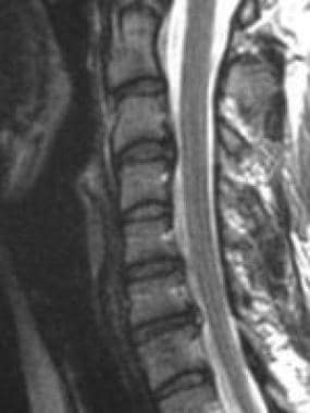 Sagittal magnetic resonance imaging (MRI) scan demonstrating cervical intervertebral disc protrusions at C3-C4 and C7-T1.
Understanding cervical disc disease requires basic knowledge of anatomy and biomechanics. The intervertebral disc is a functional unit connecting 2 vertebral bodies of the spine. The disc absorbs shock, accommodates movement, provides support, and separates vertebral bodies to lend height to intervertebral foramina. The disc consists of 3 structural components; an eccentrically located nucleus pulposus, a surrounding lamellar annulus fibrosus, and 2 cartilaginous endplates, separating each segmental level between the C2-T1 vertebrae. No disc exists between C1 and C2, and only ligaments and joint capsules resist excessive motion. Disc degeneration and/or herniation can injure the spinal cord or nerve roots and result in stenosis [5] and/or myofascial pain.
Symptoms of cervical disc disease
Discogenic pain without nerve root involvement is typically vague, diffuse and distributed axially. Depending on whether primarily motor or sensory involvement is present, radicular pain is deep, dull, and achy or sharp, burning, and electric. Such radicular pain follows a dermatomal or myotomal pattern into the upper limb. Cervical radicular pain most commonly radiates to the interscapular region, although pain can be referred to the occiput, shoulder, or arm as well. Neck pain does not necessarily accompany radiculopathy and frequently is absent.
Diagnosis of cervical disc disease
Consider performing rheumatologic workup to evaluate for possible rheumatoid arthritis, ankylosing spondylitis, Reiter syndrome, and polymyalgia rheumatica. In addition, consider performing infection workup to evaluate for possible discitis, epidural abscess, and vertebral osteomyelitis.
Plain cervical spine radiographs are used to evaluate chronic degenerative changes, metastatic disease, infection, spinal deformity, and stability.
Computed tomography (CT) scans delineate cervical spine fracture and are used extensively in trauma cases.
A myelogram followed by a CT scan may be obtained prior to cervical decompressive spinal cord or nerve root surgery. This study evaluates the spinal canal, its relationship to the spinal cord, and nerve root impingement from disc, spur, or foraminal encroachment.
Magnetic resonance imaging (MRI) remains the imaging modality of choice to evaluate cervical HNP, due to its low morbidity. [6, 7] Advantages include soft-tissue definition (eg, cervical discs, spinal cord), cerebrospinal fluid visualization, noninvasiveness, and lack of patient radiation exposure.
Provocative discography is the only procedure that can determine whether a disc serves as the pain generator. Discomfort and invasiveness render this procedure less desirable than cervical MRI, which provides much of the anatomical information that provocative discography does.
Electrodiagnostic studies continue to be standard for evaluating neurologic function of the cervical spine. Advantages of these tests include limited expense and low morbidity.
Management
For most cervical disc disorders, studies support conservative treatment, such as the McKenzie approach and cervicothoracic stabilization programs, combined with aerobic conditioning.
Physical modalities should be used to reduce pain only in the acute phase. Once past the acute phase, modalities are used sparingly on an as-needed basis.
Cervical traction may relieve radicular pain from nerve root compression. Traction does not improve soft-tissue injury pain. Hot packs, massage, and/or electrical stimulation should be applied prior to traction to relieve pain and relax muscles.
A soft cervical collar is recommended only for acute soft-tissue neck injuries and for short periods of time (ie, not to exceed 3-4 days' continuous use).
Spinal manipulation and mobilization may restore normal range of motion (ROM) and decrease pain; however, no clear therapeutic mechanism of action is known.
Cervical epidural, spinal nerve (or root), Z-joint, and sympathetic injections serve diagnostic and therapeutic roles. These procedures can be instrumental in determining the anatomic pain generator (eg, nerve root, facet) and providing aggressive, conservative treatment.
An anesthetic and corticosteroid mixture may be injected into the epidural space (interlaminar) or along the nerve root (transforaminal) after precise radiologic, contrast-enhanced fluoroscopic localization. [8]
Studies indicate that cervical HNP with radiculopathy can be managed conservatively. Surgery is warranted when neurogenic bowel or bladder dysfunction, deteriorating neurologic function, or intractable radicular or discogenic neck pain exists. Specifically, cervical spine surgical outcomes are most favorable for radicular pain, spinal instability, progressive myelopathy, or upper extremity weakness.
Surgical outcomes for patients with myelopathy have been shown to be significantly greater with regard to motor recovery if surgical intervention is performed less than 1 year after the onset of symptoms. [9]
The literature has demonstrated favorable cervical spine fusion outcomes for chronic discogenic axial neck pain when the presurgical evaluation has incorporated provocative cervical discography. However, fusion can increase intradiscal pressure and other stress at adjacent unfused levels, thereby accelerating postsurgical spinal degeneration. [10, 11, 12, 13, 14]
The possibility of obtaining the goals of anterior cervical decompression and fusion (ACDF) while maintaining adjacent segment motion led to the advent of total disk replacement (TDR).
Pathophysiology
Manifestations of HNP are divided into subcategories by type (ie, protrusion, extrusion, sequestration). Disc bulge, is not a true herniation, per se. It is described as generalized symmetrical or asymmetrical circumferential extension of the disc margin beyond the margins of the adjacent vertebral endplates.
Disc protrusion describes herniation of nuclear material through a defect in the annulus, producing a focal extension of the disc margin; it can further be defined if the greatest distance, in any plane, between the edges of the disc material beyond the disc space is less than the distance between the edges of the base, in the same plane.
Extrusion applies to herniation of nuclear material when, in at least one plane, any one distance between the edges of the disc material beyond the disc space is greater than the distance between the edges of the base, or when no continuity exists between the disc material beyond the disc space and that within the disc space. Extrusion may be further specified as sequestration, if the displaced disc material has lost completely any continuity with the parent disc.
The term migration may be used to signify displacement of disc material away from the site of extrusion, regardless of whether sequestrated or not. Examples of disc herniation are seen in the images below.
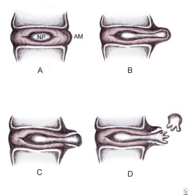 Disc herniation classification. A: Normal disc anatomy demonstrating nucleus pulposus (NP) and annular margin (AM). B: Disc protrusion, with NP penetrating asymmetrically through annular fibers but confined within the AM. C: Disc extrusion with NP extending beyond the AM. D: Disc sequestration, with nuclear fragment separated from extruded disc.
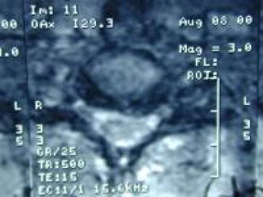 Axial magnetic resonance imaging (MRI) scan (C3-C4) demonstrating left-sided posterolateral protrusion of the nucleus pulposus with compression of the cerebrospinal fluid.
Sagittal magnetic resonance imaging (MRI) scan demonstrating cervical intervertebral disc protrusions at C3-C4 and C7-T1.
Axial magnetic resonance imaging (MRI) scan (C3-C4) demonstrating left-sided posterolateral protrusion of the nucleus pulposus with compression of the cerebrospinal fluid.
Sagittal magnetic resonance imaging (MRI) scan demonstrating cervical intervertebral disc protrusions at C3-C4 and C7-T1.
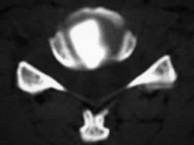 Postdiscography axial computed tomography (CT) scan demonstrating right posterolateral subligamentous protrusion.
Herniation typically occurs secondary to posterolateral annular stress. This is in part due to a thinner annulus fibrosus in this region as well as the absence of a supporting ligament posterolaterally. [15] There are 2 main types of herniation that are described in the literature: focal and broad-based. Focal herniation involves less than 25% of the disc circumference, whereas broad-based herniations involves between 25-50% of the disc circumference. [1] Herniation rarely results from a single traumatic incident. Acute traumatic cervical HNP serves as a major etiology of central cord syndrome. The C6-C7 disc herniates more frequently than discs at other levels.
Acute disc herniation causes radicular pain through chemical radiculitis in which proteoglycans and phospholipases released from the nucleus pulposus mediate chemical inflammation and/or direct nerve root compression. Interleukin 6 and nitric oxide are also released from the disc and play a role in the inflammatory cascade. Denda et al has also recently showed that chronic compression of the spinal canal can lead to higher than normal levels of nitric oxide (NO) in the cerebrospinal fluid (CSF). Excessive NO levels have been shown to be cytotoxic and can induce neuronal apoptosis. [16] Although high levels of NO have not been correlated to severity of pain or disease, this data may play a role in targets for future interventions.
The chemical radiculitis is a key element in the pain caused by HNP because nerve root compression alone is not always painful unless the dorsal root ganglion is also involved. Herniation may induce nerve demyelination with resulting neurologic symptoms. Cervical HNP maybe resorbed during the acute phase. Indeed, studies documenting frequent herniation resorption and correlating herniation regression with symptom resolution support conservative treatment of cervical radicular pain.
A rare trauma-induced high cervical (C2-C3) HNP syndrome manifests as nonspecific neck and shoulder pain, perioral hypesthesia, more radiculopathy than myelopathy, and more upper limb motor and sensory dysfunction than lower limb symptomatology. Decreased middle and/or lower cervical spine mobility from spondylosis, with consequent overload and hypermobility at higher segments, may precipitate high cervical disc lesions in older patients. A retro-odontoid disc may result from an upwardly migrating C2-C3 HNP. Some case reports describe cervical HNPs causing Brown-Séquard syndrome, as well as atypical nonradicular symptoms in patients with congenital insensitivity to pain. Although spondylosis may affect motion at adjacent levels, isolated disc herniation in the cervical spine does not seem to alter motion of adjacent levels, regardless of the degree of disc degeneration or the size of the disc herniation. [17]
A study by Nam et al indicated that in patients with cervical disc herniation, risk factors for motor weakness include decreased disc height, a percentage of the HNP in the spinal canal, and a signal intensity change in the spinal cord. A disc height cutoff value of 5.8 mm produced a sensitivity and specificity of 39.5% and 94.1%, respectively, while a cutoff value of 28.1% for HNP in the spinal canal produced a sensitivity and specificity of 57.9% and 82.4%, respectively. In addition to motor weakness, signal intensity change in the spinal cord also may predict delayed recovery. [18]
Cervical radiculopathy results from mechanical nerve root compression or intense inflammation (ie, chemical radiculitis). Specifically, nerve root compression may occur at the intervertebral foraminal entrance zone at the narrowest segment of the root sleeve anteriorly by disc protrusion and uncovertebral osteophytes and posteriorly by superior articulating process, ligamentum flavum, and periradicular fibrous tissue. [19] Decreased disc height, as well as age-related foraminal width decrease from inferior Z-joint hypertrophy, may impinge subsequently on nerve roots. The cervical region accounts for 5-36% of all radiculopathies encountered. Incidence of cervical radiculopathies by nerve root level is as follows: C7 (70%), C6 (19-25%), C8 (4-10%), and C5 (2%).
The most common cause of cervical radiculopathy is foraminal encroachment (70-75%). The cause is multifactorial, including degeneration of the discs and the uncovertebral joints of Luschka and the zygapophyseal joints. In contrast to lumbar spine disorders, HNP in the cervical spine is responsible for only 20-25% of radiculopathies.
Cervical DDD most commonly is due to age-related changes, but the condition also is affected by lifestyle, genetics, smoking, nutrition, and physical activity. Degenerative disc changes observed on radiographs may reflect simple aging and do not necessarily indicate a symptomatic process.
The disc begins to degenerate in the second decade of life. Degenerative disc disease is essentially a process disrupting homeostasis. This degenerative process of the less hydrated and more fibrous nucleus pulposus fails to withstand the compressive loading, resulting in uneven distribution of forces to the surrounding annulus, which leads to the formation of radial tears. Circumferential tears form in the posterolateral annulus after repetitive use. Several circumferential tears coalesce into radial tears, which progress into radial fissures. The disc then disrupts with tears passing throughout the disc.
Loss of disc height occurs with subsequent peripheral annular bulging. Proteoglycans and water escape through fissures formed from nuclear degradation, resulting in further thinning of the disc space. Changes in the cartilaginous endplates alter nutritional supply to the nucleus that contributes to preexisting dehydration of the disc, compounding the effects of the degenerative cascade. Vertebral sclerosis and osteophytic formation ultimately follow. [6]
IDD describes pathologic annular fissuring within the disc without external disc deformation. This disorder results from trauma-related nuclear degradation, cervical flexion/rotation-induced annular injury, or whiplash.
The intervertebral disc has few pain receptors and little innervation, except in the periphery of the disc. The intervertebral disc may not be irritated until the inflammation process becomes moderate or severe. The nucleus pulposus appears to be the first site of degeneration with the annulus fibrosis being the primary pain generator once injury or degeneration occurs. DDD ultimately may progress to IDD.
Epidemiology
Frequency
United States
HNP may be observed with magnetic resonance imaging (MRI) in 10% of asymptomatic individuals aged younger than 40 years and 5% of those older than 40 years. Degenerative disc disease (DDD) may be observed with MRI in 25% of asymptomatic individuals aged less than 40 years and 60% of those aged more than 40 years. The true incidence and prevalence of cervical radiculopathy is uncertain; however, studies have shown that 51-67% of adults experience neck and arm pain at some time, and 54% report pain present within the last 6 months. In a population-based study in Rochester, Minn published in 1994 the annual incidence of documented cervical radiculopathy for men and women from all causes was 107.3 and 63.5 cases per 100,000 population, respectively. [20] A separate study looking at a population at risk of more than 13,000,000 people in the military found an incidence of 1.79 per 1000 person-years. [21]
International
A study from Italy in 1996 reported the prevalence of cervical spondylotic radiculopathy to be 3.5 cases per 1000 people. [22]
Mortality/Morbidity
Occasionally, an acute HNP can herniate centrally and cause a myelopathy. This can manifest as hyperreflexia, positive pathologic reflexes (such as Babinski and Hoffman signs), and sphincter disturbances. If left untreated, the effects can be irreversible.
Sex
Kelley suggests that the male-to-female incidence of cervical disc herniation is approximately 1:1. [23] Marchiori and Henderson cite women as reporting higher disability with increasing levels of DDD than men. [24]
Schoenfeld et al found that female sex was a significant risk factor for developing cervical radiculopathy. [21]
Age
HNP typically affects younger patients (ie, < 40 y). DDD, part of natural aging, typically affects older patients (ie, >40 y).
Those older than 40 years were also found to have the greatest risk of cervical radiculopathy. [21]
-
Disc herniation classification. A: Normal disc anatomy demonstrating nucleus pulposus (NP) and annular margin (AM). B: Disc protrusion, with NP penetrating asymmetrically through annular fibers but confined within the AM. C: Disc extrusion with NP extending beyond the AM. D: Disc sequestration, with nuclear fragment separated from extruded disc.
-
Axial magnetic resonance imaging (MRI) scan (C3-C4) demonstrating left-sided posterolateral protrusion of the nucleus pulposus with compression of the cerebrospinal fluid.
-
Sagittal magnetic resonance imaging (MRI) scan demonstrating cervical intervertebral disc protrusions at C3-C4 and C7-T1.
-
Right C7 cervical transforaminal epidural steroid injection demonstrating epidural and radicular spread of radiologic contrast dye.
-
Cervical epidural steroid injection at the C7-T1 interlaminar space.
-
Cervical discography. Anteroposterior fluoroscopic image.
-
Cervical discography. Lateral fluoroscopic image.
-
Postdiscography axial computed tomography (CT) scan demonstrating right posterolateral subligamentous protrusion.


