You are in: eMedicine Specialties >
Physical Medicine and Rehabilitation > PLEXOPATHY
Thoracic Outlet Syndrome
Article Last Updated: Dec 13, 2006
AUTHOR AND EDITOR INFORMATION
Section 1 of 11
Author: Benjamin M Sucher, DO, FAAPMR, FAOCPMR, Medical Director, EMG Center of Arizona and Electrodiagnostic Medical Group
Benjamin M Sucher is a member of the following medical societies: American Academy of Physical Medicine and Rehabilitation, American Association of Neuromuscular and Electrodiagnostic Medicine, American Osteopathic Association, and American Osteopathic College of Physical Medicine and Rehabilitation
Editors: Robert E Windsor, MD, FAAPMR, FAAEM, FAAPM, President and Director, Georgia Pain Physicians, PC; Clinical Associate Professor, Department of Physical Medicine and Rehabilitation, Emory University School of Medicine; Francisco Talavera, PharmD, PhD, Senior Pharmacy Editor, eMedicine; Patrick M Foye, MD, FAAPMR, FAAEM, Associate Professor of Physical Medicine and Rehabilitation, Co-Director of Musculoskeletal Fellowship, Co-Director of Back Pain Clinic, Director of Coccyx Pain (Tailbone Pain, Coccydynia) Service, University of Medicine and Dentistry of New Jersey, New Jersey Medical School; Kelly L Allen, MD, Consulting Staff, Department of Physical Medicine and Rehabilitation, Lourdes Regional Rehabilitation Center, Our Lady of Lourdes Medical Center; Robert H Meier III, MD, Director, Amputee Services of America, Presbyterian St Luke's Hospital; Consulting Staff, North Valley Rehabilitation Hospital, Kindred Hospital, and North Suburban Hospital
Author and Editor Disclosure
Synonyms and related keywords:
thoracic outlet syndrome, brachial plexopathy, cervical rib syndrome, cervicobrachial myofascial pain syndrome, cervicobrachial pain syndrome, costoclavicular mass syndrome, costoclavicular syndrome, scalenus anticus syndrome, scalenus syndrome, thoracic outlet compression syndrome
Background
Thoracic outlet syndrome (TOS) is a controversial topic in the literature; many proponents support the existence of the condition, but some strongly vocal opponents doubt the validity of TOS as a medical entity. Even the name itself has been questioned because the actual site of pathology is technically the thoracic "inlet," not the "outlet." The primary controversy seems to center around the lack of objective criteria for diagnosis and the confusion with multiple types or clinical presentations.
Since TOS involves proximal neurovascular structures, symptoms often are confused with various distal compression neuropathies or cervical radiculopathies. In addition, surgical treatment has been known to have devastating complications, which further fuels debate with the opponents of recognition of this entity. Conservative treatment appears to be the most universally accepted approach, with even surgeons recommending a prolonged trial before any operative procedure.
Pathophysiology
TOS involves compression, injury, or irritation to the neurovascular structures at the root of the neck or upper thoracic region, bounded by the anterior and middle scalenes; between the clavicle and first rib (with possible enlargement/hypertrophy of the subclavius); or beneath the pectoralis minor muscle. Some authors define the thoracic outlet as an opening bordered by the first rib laterally, the vertebral column medially, and the claviculomanubrial complex anteriorly. The syndrome of compression at this site could be primarily neurologic, involving the brachial plexus, most often the lower trunk or medial cord; alternatively, it could involve compression of the subclavian artery, vein, or both. Thrombosis, embolus, or aneurysm of these vessels is a less likely possibility.
One proposed classification system has broken TOS into the following 3 categories:
- True neurogenic TOS: The brachial plexus is injured in these cases as documented by electromyography (EMG) and/or nerve conduction studies.
- True vascular TOS: The subclavian artery and/or vein is damaged or thrombosed, as documented by arteriogram or venogram.
- Nonspecific or disputed TOS: Patients have symptoms, but there are no abnormal tests to document the lesion clearly. This category is by far the most common type of this disorder seen in the clinical setting.
Many authors have discovered accessory cervical ribs associated with TOS; however, they have noted tough fibrous bands coming off the accessory ribs that are believed to be more responsible for the pathology. There have even been reports of bony fusion of variant cervical ribs, resulting in bifid ribs with attached fibrous bands. The bands cause tethering of the brachial plexus, which results in traction and, therefore, symptoms. Other authors report compression or irritation of the neurovascular bundle more distally under the pectoralis minor muscle or from anterior displacement of the humeral head.
Additionally, clavicle fractures can result in plexopathy from expanding hematomas or pseudoaneurysms that compress the plexus, with variable latent periods following the fracture. Delayed onset of symptoms may suggest exuberant callus from the healing fracture site. Nonunion of the fracture site also can result in direct compression by the lateral fragment, which is pulled inferiorly.
More recently, trapezius weakness due to spinal accessory nerve injury (following cervical lymph node biopsy) has been implicated as a cause of TOS. This results in "droopy shoulder" with secondary compression of the neurovascular bundle, particularly aggravated with arm elevation (abduction).
Frequency
United States
The inability to make a definitive and accurate diagnosis makes determination of the exact prevalence of this condition impossible. The prevalence of nonspecific TOS has been reported as high as 23% of soft tissue injuries of the cervical spine. TOS is overlooked or misdiagnosed commonly, especially upon presentation to the emergency department. The true or classical neurogenic or vascular TOS incidence is considered rare, with only 1 case per million population estimated for the neurogenic type. Some believe that TOS is the most common cause of acute arterial occlusion in the upper limbs of adults younger than 40 years. It is more common with occupations or activities that involve prolonged posturing of the neck, such as secretaries, cashiers, machine operators, surgeons, truck drivers, and overhead work or lifting. There is no increased incidence in athletes.
Mortality/Morbidity
No known mortality is associated directly with TOS. Morbidity often encompasses debilitating functional loss of the involved upper extremity, loss of livelihood or occupation, especially if the work involves overhead activity (eg, hairdressing, carpentry, painting). True neurogenic TOS causes neurologic deficit. Depending on the amount of nerve injury, there usually is weakness of the hand and sensory deficits in the lower trunk distribution. Devastating complications, usually to the brachial plexus, have been reported from surgical treatment for TOS.
Sex
Females are diagnosed more commonly with TOS than males, with some reports of a 9:1 female-to-male ratio. The shape of the chest wall is believed to predispose women by encouraging closure of the thoracic outlet. Large pendulous breasts have been particularly implicated, adding to the anterior forces on the chest, which leads to drooped shoulder posturing and further closing the outlet.
Age
Age of onset is from the second to the eighth decade, with a peak occurring in the fourth decade. In the younger age group, there appears to be a greater likelihood of anatomic or structural abnormality, such as cervical rib variants and fibrous bands.
History
- Pain, numbness and/or tingling, and heaviness of the involved upper extremity are common complaints reported by a patient with TOS. Often, the symptoms are vague and generalized. The entire extremity may be involved; additionally, neck pain and headaches are reported concomitantly.
- Symptoms may begin insidiously after repetitive or stressful activity, such as prolonged computer keyboard use or mechanical and overhead work. Trauma, such as an automobile accident with occurrence of a whiplash injury, also has been associated with onset of TOS with a frequency of up to 23%. Sports activities, especially throwing and swimming, have been implicated as well; symptoms may be similar to those of a clavicular fracture, with a delayed onset from hours to weeks.
- Autonomic phenomena (eg, cold hands, blanching, swelling) also may be reported. The proximity of the stellate ganglion to the first rib articulation, which is often dysfunctional or restricted in TOS, has been postulated as a cause.
Physical
A careful neurological and musculoskeletal examination is essential to diagnose TOS adequately. Often, the most important aspect of the physical examination is to diagnose or rule out other problems of the neck and arm.
- The mainstay of the physical examination diagnosis of TOS involves the so-called stress tests (or provocative maneuvers) (see Images 2-3). The sensitivity and specificity of these tests have been low in the studies on TOS completed to date. Different techniques for performing and interpreting these tests are discussed in the literature and vary even more in the bedside clinical situation.
- The most common tests are the Adson maneuvers, where the head is placed in extension and side bending while the patient takes a deep breath and holds it, followed by rotation to stretch or tether the plexus and/or artery by the anterior and middle scalenes. The maneuver is held for 15-30 seconds while the clinician observes for onset of symptoms and obliteration of the pulse. Symptoms have been reported both to the side of bending and, more commonly, to the side away from the bending. If the symptoms are reported on the side of bending, then this finding overlaps with the Spurling sign, commonly used to assist in the diagnosis of cervical radiculopathy. Some examiners ask the patient to pull the head forward while maintaining the test position, causing the anterior scalene to contract against the plexus to enhance the stress effect.
- Hyperabduction of the involved arm also can be used to stress the outlet; however, this maneuver often causes symptoms and loss of pulse even in normal individuals and may be misleading. The area of compression with this maneuver is considered more distal and frequently located at the anterior humeral head and plexus, with tethering under the pectoralis minor muscle.
- Costoclavicular bracing (military maneuver) closes the space between the clavicle and first rib and may reproduce symptoms.
- Focal stress tests involve applying pressure directly to the anterior scalene or upper segment of the pectoralis minor. These tests are considered positive if symptoms are reproduced within 15-30 seconds. In addition, some authors have noted a positive Tinel sign (percussing over the plexus) as diagnostic for TOS.
- The elevated arm stress test has been noted to be highly sensitive for TOS. The upper extremity is held in the "stick-em-up" position with the arms abducted and elbows flexed (both at 90°) for 3 minutes, while the patient simultaneously and vigorously flexes and extends the fingers (grasp and release). This test is considered positive if the patient cannot complete the full 3 minutes. Unfortunately, this test is challenging even for individuals without neurovascular symptoms to complete; thus, it may have limited practical usefulness in most clinical situations. In one study, over 80% of patients with carpal tunnel syndrome presenting to an electrodiagnostic medicine laboratory had a positive elevated arm stress test results.
- Careful observation for asymmetry of the upper chest wall may reveal clavicular irregularity consistent with prior fracture. A nontender hard mass over the middle third of the clavicle often is noted. Deformity from displaced fracture (with or without nonunion) or exuberant callus could be responsible for direct compression of the plexus. Pressure on the clavicle can reproduce or aggravate symptoms, especially when nonunion is present; motion can be detected between the fragments.
Causes
TOS most likely has multiple causes. The primary cause is believed to be mechanical or postural. Stress, depression, overuse, and habit all can lead to the forward head, droopy shoulder, and collapsed chest posture that allows the thoracic outlet to narrow and compress the neurovascular structures (see Image 1). Accessory ribs or fibrous bands also may be present, predisposing the site to narrowing and compression. Large breasts have been implicated as a contributing cause by pulling the chest wall forward (anterior and inferior); this theory has been supported by relief of discomfort following reduction mammoplasty. TOS has also been associated with breast implants, which may increase tension under the chest wall muscles and irritate the neurovascular tissue.
Trauma can lead to decompensation or shifting of structures in the shoulder and chest wall, leading to symptom onset. Additionally, trauma with fracture of the clavicle can result directly in compression of the plexus from bone fragments, exuberant callus, hematoma, or pseudoaneurysm. Following median sternotomy, brachial plexus injury can occur from displacement of the ribs, which usually involves the C8 fibers, distinguished from the typical pattern that primarily involves T1.
Primary vascular lesions, such as thrombus or aneurysm, may be present as well as secondary problems such as emboli. Tumors, such as upper lobe lung lesions (Pancoast tumor), are also possible causes.
- Many other problems may predispose an individual to TOS, some of which are classic perpetuators of myofascial pain syndrome, including the following:
- Sleep disorder
- Hormonal imbalance (estrogen, thyroid)
- Inflammatory disorder (rheumatoid arthritis)
- Fibromyalgia
- Nutritional insufficiency (B vitamins, folate, vitamin C)
- Anemia
- Infection
- Masses, tumors, axillary lymph nodes
- Mechanical disorders (short leg, hyperlordosis, hypolordosis)
- Psychologic (stress, depression)
- Nerve entrapment/impingement (other sites)
Adhesive Capsulitis
Carpal Tunnel Syndrome
Cervical Disc Disease
Cervical Myofascial Pain
Cervical Spondylosis
Cervical Sprain and Strain
Chronic Pain Syndrome
Complex Regional Pain Syndromes
Fibromyalgia
Radiation-Induced Brachial Plexopathy
Rheumatoid Arthritis
Rotator Cuff Disease
Traumatic Brachial Plexopathy
Workers' Compensation System
Other Problems to be Considered
Cervical myelopathy
Cervical radiculopathy
Double crush syndrome (thoracic outlet syndrome and compression at another distal or proximal site)
Paget-von Schroetter syndrome, effort syndrome (spontaneous venous thrombosis, primary deep venous thrombosis of the upper extremity)
Pancoast (apical lung) tumor
Shoulder tendonitis, bursitis, impingement
Shoulder (glenohumeral) instability
Raynaud syndrome
Ulnar neuropathy (cubital tunnel syndrome, Guyon canal syndrome)
Lab Studies
- Laboratory studies are used to rule out other systemic problems.
Imaging Studies
- In cases of nonspecific TOS, all studies have normal findings; however, testing to evaluate for other treatable problems or to establish true neurogenic or true vascular TOS usually is indicated.
- Radiographs
- Radiographs of the chest (with apical lordotic views) and cervical spine should be obtained.
- Rule out significant cervical spine degenerative changes that may be causing neck/shoulder pain or impingement of the spinal nerve roots; also eliminate the possibility of upper lobe lung masses.
- Radiographs can also help identify cervical ribs, which may be responsible for plexus injury or have associated fibrous bands.
- Radiographs may help identify elevated first ribs, caused by tight contracted anterior/middle scalene muscles. Displaced clavicle fractures, nonunion, and exuberant callus also can be identified.
- MRI and CT scans
- An MRI of the cervical spine and supraclavicular/brachial plexus area is useful to identify other causes, or a CT scan of the brachial plexus area and apical lung may be indicated.
- MRI and CT scans can identify cervical root injury from degenerative spurs, disc herniation, or other causes. Some authors have reported that MRI can identify distortion or displacement of the plexus in the thoracic outlet or supraclavicular space, usually from fibrous bands but also from clavicular abnormality (callus, fracture displacement). In addition, MRI has been reported to demonstrate differences in the costoclavicular measurements during provocative stress maneuvers, in patients with thoracic outlet compression.
- Doppler and noninvasive blood flow studies
- Doppler and plethysmography studies can identify interruption of blood flow to the involved extremity. A near-complete cut-off of flow during the stress maneuver with reproduction of the primary symptoms should be noted.
- Hyperabduction is somewhat controversial, since most normal individuals lose flow with the position and also may develop symptoms.
- Angiography/venography
- Angiography and venography can identify blockage of the vessels from thrombi or emboli, which result in vascular symptoms.
- Angiography can detect aneurysms that may be compressing the plexus and causing neurologic symptoms. This finding documents true vascular TOS.
- Autonomic assessment (thermography)
- Autonomic assessment has demonstrated thermal/temperature asymmetry consistent with nerve irritation or some interference with blood flow.
- Following successful treatment, thermal symmetry usually is restored and should correlate well with reduction or elimination of symptoms. This technique remains controversial, and, although the sensitivity has been considered high, the specificity is low and has not been well established.
Other Tests
- EMG and nerve conduction studies, also known as electrodiagnostic studies, are necessary to identify true neurogenic TOS and to rule out other sites of compression neuropathy. With true neurogenic TOS, reported abnormalities include the following:
- Low-amplitude ulnar sensory response. This finding may be variable, since the fifth digit is believed to be supplied primarily by C8 nerve fibers, whereas the T1 fibers are more deflected, stretched, or angulated by a cervical band (or rib) and thereby more likely to sustain injury in TOS.
- Low-amplitude median motor response. The thenar muscles are supplied more from the T1 root, whereas the hypothenar muscles receive primarily C8 innervation.
- Normal median sensory response.
- Neurogenic motor units within the C7-T1 or lower trunk distribution on needle EMG.
- Abnormal medial antebrachial cutaneous response (low-amplitude or unobtainable). Some authors believe that this finding can aid in the early diagnosis of neurogenic TOS. This test primarily assesses the T1 fibers.
- Abnormal C7-T1 root stimulation (prolonged latency).
- Abnormal F-wave study (prolonged latency, with either full F-wave or axillary F-loop)
- Absent or attenuated N13 response in ulnar-derived somatosensory evoked potentials
- The degree of nerve injury determines the abnormalities described above. In severe injuries with massive axon loss all of the above findings are seen. In cases where part of the lower plexus is spared, the findings are less dramatic. In these cases, the needle EMG is more likely to show abnormalities.
Procedures
- In most patients with nonspecific TOS, there are no indicated procedures. An anterior scalene block can be performed to anesthetize the anterior scalene muscle and to relieve pressure from the brachial plexus. Thus, this type of block is diagnostic but also may be therapeutic.
- The anterior scalene block is a potentially dangerous procedure because the plexus can be injured directly in the process; however, its reliability, accuracy, and safety may be improved with the use of electrophysiologic guidance to verify needle tip placement.
- Jordan and Machleder believe that a negative response to this block excludes a surgical treatment option, while a positive response predicts which patients should benefit from surgical decompression.
Histologic Findings
Biopsy results have not been reported systematically, and the procedure is not indicated unless some other condition is being considered.
Rehabilitation Program
Physical Therapy
Modalities with deep heat (eg, ultrasound), electric stimulation, superficial heat (eg, Hydrocollator packs), stretching exercises, postural correction exercises, and strength and endurance exercises are all useful or necessary components of TOS treatment. Ultrasound is the preferred modality as it is capable of heating deep muscular and soft tissue structures, which is essential to increase elasticity and facilitate effective stretching and/or manipulation, especially for the scalenes (see Image 6) and pectoralis minor muscles (see Image 7). Ideally, ultrasound should take immediately before the stretching or manual treatment, since the deep tissues cool (from 41-42°C back to 37°C) within 20-30 minutes.
Mobilization and manipulation procedures (often performed by an osteopathic physician) usually are indicated and necessary to release tight contracted/restricted vertebral segments and soft tissue (myofascial) regions, especially the anterior/middle scalenes (see Image 4) and pectoralis minor muscle (see Image 5) entrapment sites.
Spray and stretch with a vapo-coolant spray is an effective adjunct to the other modalities mentioned here. Note that the research-based scientific evidence for these modalities is limited.
Occupational Therapy
Work simplification and back protection techniques often are helpful. These educational tools are available from the occupational therapist, as well as from the physical therapist.
Medical Issues/Complications
Essentially, there are no randomized controlled trials demonstrating that specific treatment modalities are more effective than placebo. In general, research about TOS and its treatment can be difficult for the following several reasons:
- If the etiology of the problem cannot be agreed upon, then focusing treatment becomes difficult.
- The proposed areas of pathophysiology and diffuse nature of symptoms are not easily amenable to surgery or other treatment modalities.
- Many patients with TOS have coexistent chronic pain syndrome with disability and depression.
Treatment of the chronic pain and disability can be a much more complex problem and involves employment of multiple disciplines with consideration for a biopsychosocial model of treatment. Patients with nonspecific TOS and suggestion of chronic pain syndrome with disability may do best with chronic pain treatment and treatment of psychological issues.
Additional issues of concern include the following:
- More advanced/severe TOS can result in functional loss of the upper extremity.
- Concomitant cervical degenerative joint/disc disease limits the neck range of motion (ROM) that is necessary to stretch associated soft tissue restriction and limit conservative treatment options (eg, physical therapy).
- Shoulder arthritis/bursitis/tendonitis can interfere with stretching of the pectoral muscles and limit the effectiveness of conservative therapy approaches.
- Manual treatment to the pectoral/chest wall muscles must be sensitive to overlying breast tissue and implants (prostheses) in female patients.
- Manual treatment must consider the proximity of the involved muscles to the brachial plexus. Vigorous stretching or manipulation of these structures may not be tolerated well and can aggravate symptoms. The phrenic nerve and accessory phrenic nerve overlie the anterior scalene muscle and also must be considered.
- Scalene nerve block has been used as a treatment and as a diagnostic test; however, this procedure is dangerous due to the close proximity of the brachial plexus to the scalenes (see Procedures for more details).
Surgical Intervention
- First rib resection has been advocated by many surgeons to treat TOS. Many also use scalenectomy, in combination with rib removal, or as a second procedure, if the initial surgery is ineffective. Resection of accessory ribs and fibrous bands should be performed, especially if observed to be tethering the plexus.
- In cases where clavicular fracture is responsible for plexus compression, removal of hyperabundant callus may be necessary. Occasionally, resection of associated musculature is required, including the subclavius and infraclavicular soft tissue. Reduction of clavicular nonunion fragments and internal fixation (with pins) may be indicated.
- Complications from surgical treatment have been devastating, especially brachial plexopathy (eg, injury to the long thoracic nerve with scapular winging). Generally, surgery is used as a last resort after a prolonged trial (ie, months) of conservative treatment.
- Some authors have advocated breast reduction in extreme cases, where very large breasts obviously are adding uncontrollable weight loads to the anterior chest wall.
Consultations
- Physical medicine and rehabilitation specialists (physiatrists) are generally familiar with TOS and conservative treatment options. They usually are best positioned to implement and direct the therapy program, and they also have been trained extensively in electrodiagnosis, which may provide the best objective evidence for the disorder in its true neurogenic form. Neurologists are also capable diagnosticians for TOS.
- Some internists or cardiologists who specialize in peripheral cardiovascular disorders may be able to help with the diagnostic workup, especially when vascular or autonomic symptoms predominate. They also may be familiar with the use of medications to help control symptoms (eg, vasodilators, calcium-channel blockers). Rheumatologists can assist in ruling out connective tissue disorders or autoimmune diseases that might be associated with TOS or might be complicating the clinical picture.
- Thoracic and cardiovascular surgeons are the most likely surgical specialists to treat TOS; however, not all of these surgeons have an active interest or willingness to deal with the disorder. Occasionally, neurosurgeons can offer operative treatment for TOS patients.
- Plastic surgeons (or some general surgeons) may be used for reduction mammoplasty, when indicated.
- Anesthesiologists may be needed to perform diagnostic and therapeutic blocks.
- Orthopedists may be required to diagnose and treat concomitant shoulder joint pathology and other related disorders, such as complications from clavicular fractures.
Other Treatment
- Osteopathic manipulation has been shown to be highly effective in treating TOS, especially in combination with vigorous stretching exercise. The most useful procedures involve a vigorous myofascial release to the scalenes and pectoral muscles (especially pectoralis minor). These techniques are applied most easily immediately following modality treatment with ultrasound, which heats the deep muscular structures and renders them more elastic and responsive to manipulation and stretching.
- All other areas of restriction or dysfunction also should be addressed and treated with manipulation, since even pelvic/sacral abnormality can influence the thoracic outlet. This abnormality is most obvious with unleveling, or a sacral tilt that shifts the thorax and places skeletal and muscular strain on the shoulder girdle region. Some orthoses, such as a heel lift, may be a useful adjunct in such cases.
- Injection of trigger points in associated muscular structures may be indicated and necessary. An injection of deep muscular structures, such as a scalene block, however, is dangerous (close proximity to brachial plexus) and should be avoided or performed by an anesthesiologist (see Procedures).
Muscle relaxants (eg, Flexeril, Soma, Robaxin, Parafon forte) are options to help decrease muscle tightness and restriction, thereby facilitating conservative treatment with exercise and manipulation. Tizanidine (Zanaflex) is being used for muscle tension cephalgia and may hold promise for TOS.
Anti-inflammatory medication can decrease irritability and pain and enhance conservative treatment results. Long-acting preparations often are most effective and are tolerated better (eg, Relafen, Daypro, Celebrex). Voltaren or Arthrotec and Celebrex are relatively long-acting alternatives that are less irritating to the GI tract than Naproxen or Clinoril.
Some internists or rheumatologists may recommend vasodilators and calcium-channel blockers, if significant vascular and vasoconstrictive involvement exists. These agents are not primary drugs of choice for TOS but should be considered ancillary or secondary options to be considered and integrated into treatment as clinically indicated.
Drug Category: Muscle relaxants
Used to relax and loosen the tight musculature involved in TOS, facilitate stretching and manipulation treatments, relieve pain, and assist with sleep.
| Drug Name | Cyclobenzaprine (Flexeril) |
| Description | Muscle relaxer of moderate duration, centrally acting, related to TCAs chemically. This drug often produces a "hangover" effect, which can be minimized by taking the nighttime dose 2-3 h before going to sleep. |
| Adult Dose | 10 mg tid PO; 20-30 mg before hs may be necessary, and taking only 5 mg (half a tab) bid/tid (in addition to hs dose) may be less sedating and better tolerated during daytime; not to exceed 60 mg/d |
| Pediatric Dose | Not established |
| Contraindications | Documented hypersensitivity; have taken MAO inhibitors within the last 14 d; cardiac conditions; hyperthyroidism |
| Interactions | Coadministration with MAO inhibitors and TCAs may increase toxicity; cyclobenzaprine may have additive effect when used concurrently with anticholinergics; effects of alcohol, CNS depressants, and barbiturates may be enhanced with cyclobenzaprine; may block antihypertensive action of some medications, like guanethidine; may enhance seizure risk in patients taking tramadol (Ultram); atropinelike actions |
| Pregnancy | C - Safety for use during pregnancy has not been established.
|
| Precautions | Caution in patients with angle-closure glaucoma and urinary hesitance; use of anticholinergic medications; avoid operating machinery |
| Drug Name | Carisoprodol (Soma) |
| Description | Short-acting medication that works at spinal cord level. |
| Adult Dose | 350 mg PO tid/qid |
| Pediatric Dose | Not established |
| Contraindications | Documented hypersensitivity; acute intermittent porphyria |
| Interactions | Increases toxicity of alcohol, CNS depressants, MAO inhibitors, clindamycin, phenothiazines |
| Pregnancy | C - Safety for use during pregnancy has not been established.
|
| Precautions | Caution in renal and hepatic impairment; psychologic dependence, abuse, and possible withdrawal symptoms may occur |
| Drug Name | Methocarbamol (Robaxin) |
| Description | Short-acting muscle relaxer that probably works through CNS mechanisms. |
| Adult Dose | 750-1500 mg PO tid/qid; not to exceed 8 g/d |
| Pediatric Dose | Not established |
| Contraindications | Documented hypersensitivity; renal impairment |
| Interactions | May cause color interference with screening tests for 5-HIAA and VMA |
| Pregnancy | C - Safety for use during pregnancy has not been established.
|
| Precautions | Caution in patients with history of seizures |
| Drug Name | Chlorzoxazone (Flexaphen, Paraflex, Parafon Forte) |
| Description | Short-acting muscle relaxer, working via central pathways (spinal cord and subcortical). |
| Adult Dose | 250-500 mg PO tid/qid up to 750 mg PO tid/qid |
| Pediatric Dose | 20 mg/kg/d or 600 mg/m2/d PO tid/qid |
| Contraindications | Documented hypersensitivity; hepatic impairment |
| Interactions | Increases toxicity of CNS depressants |
| Pregnancy | C - Safety for use during pregnancy has not been established.
|
| Precautions | Rare idiosyncratic and unpredictable hepatocellular damage, possibly fatal; may cause tachycardia and tightness in chest |
Drug Category: Nonsteroidal anti-inflammatory drugs
Have analgesic, anti-inflammatory, and antipyretic activities. Their mechanism of action is not known, but may inhibit cyclo-oxygenase activity and prostaglandin synthesis. Other mechanisms may exist as well, such as inhibition of leukotriene synthesis, lysosomal enzyme release, lipoxygenase activity, neutrophil aggregation, and various cell-membrane functions.
| Drug Name | Nabumetone (Relafen) |
| Description | Nonacidic NSAID rapidly metabolized after absorption to a major active metabolite that inhibits cyclo-oxygenase enzyme, which in turn inhibits pain and inflammation. |
| Adult Dose | 1-2 g PO qd |
| Pediatric Dose | Not established |
| Contraindications | Documented hypersensitivity; active peptic ulceration, hepatic impairment |
| Interactions | Coadministration with aspirin increases risk of inducing serious NSAID-related adverse effects; probenecid may increase concentrations and, possibly, toxicity of NSAIDs; may decrease effect of hydralazine, captopril, and beta-blockers; may decrease diuretic effects of furosemide and thiazides; may increase PT when taking anticoagulants (instruct patients to watch for signs of bleeding); may increase risk of methotrexate toxicity; phenytoin levels may be increased when administered concurrently |
| Pregnancy | C - Safety for use during pregnancy has not been established.
|
| Precautions | Category D in third trimester of pregnancy; elderly patients may require lower doses; caution in hepatic and renal impairment |
| Drug Name | Oxaprozin (Daypro) |
| Description | For relief of mild to moderate pain. Inhibits inflammatory reactions and pain by decreasing activity of cyclo-oxygenase, which is responsible for prostaglandin synthesis. |
| Adult Dose | 600-1200 mg PO qd; not to exceed 1800 mg/d |
| Pediatric Dose | Not established |
| Contraindications | Documented hypersensitivity, history of GI disease, cardiac failure, renal or hepatic dysfunction, bleeding disorders |
| Interactions | Increases toxicity of anticoagulants, aspirin, and diuretics |
| Pregnancy | C - Safety for use during pregnancy has not been established.
|
| Precautions | May cause dizziness, indigestion, nausea, and abdominal cramps |
| Drug Name | Celecoxib (Celebrex) |
| Description | Inhibits primarily COX-2. COX-2 is considered an inducible isoenzyme, induced during pain and inflammatory stimuli. Inhibition of COX-1 may contribute to NSAID GI toxicity. At therapeutic concentrations, COX-1 isoenzyme is not inhibited thus GI toxicity may be decreased. Seek lowest dose of celecoxib for each patient. |
| Adult Dose | 200 mg/d PO qd; alternatively, 100 mg PO bid |
| Pediatric Dose | Not established |
| Contraindications | Documented hypersensitivity |
| Interactions | Coadministration with fluconazole may cause increase in celecoxib plasma concentrations because of inhibition of celecoxib metabolism; coadministration of celecoxib with rifampin may decrease celecoxib plasma concentrations |
| Pregnancy | B - Usually safe but benefits must outweigh the risks.
|
| Precautions | Category D in third trimester of pregnancy; may cause fluid retention and peripheral edema; caution in compromised cardiac function, hypertension, conditions predisposing to fluid retention; severe heart failure and hyponatremia, because may deteriorate circulatory hemodynamics; NSAIDs may mask usual signs of infection; caution in the presence of existing controlled infections; evaluate symptoms and signs suggesting liver dysfunction, or in abnormal liver lab results |
| Drug Name | Naproxen (Anaprox, Naprelan, Naprosyn) |
| Description | For relief of mild to moderate pain; inhibits inflammatory reactions and pain by decreasing activity of cyclo-oxygenase, which results in a decrease of prostaglandin synthesis. |
| Adult Dose | 500 mg PO followed by 250 mg q6-8h; not to exceed 1.25 g/d |
| Pediatric Dose | <2 years: Not established >2 years: 2.5 mg/kg/dose PO; not to exceed 10 mg/kg/d |
| Contraindications | Documented hypersensitivity; peptic ulcer disease; recent GI bleeding or perforation; renal insufficiency |
| Interactions | Coadministration with aspirin increases risk of inducing serious NSAID-related adverse effects; probenecid may increase concentrations and, possibly, toxicity of NSAIDs; may decrease effect of hydralazine, captopril, and beta-blockers; may decrease diuretic effects of furosemide and thiazides; may increase PT when taking anticoagulants (instruct patients to watch for signs of bleeding); may increase risk of methotrexate toxicity; phenytoin levels may be increased when administered concurrently |
| Pregnancy | B - Usually safe but benefits must outweigh the risks.
|
| Precautions | Category D in third trimester of pregnancy; acute renal insufficiency, interstitial nephritis, hyperkalemia, hyponatremia, and renal papillary necrosis may occur; patients with preexisting renal disease or compromised renal perfusion risk acute renal failure; leukopenia occurs rarely, is transient, and usually returns to normal during therapy; persistent leukopenia, granulocytopenia, or thrombocytopenia warrants further evaluation and may require discontinuation of drug |
| Drug Name | Sulindac (Clinoril) |
| Description | Decreases activity of cyclo-oxygenase and in turn inhibits prostaglandin synthesis. Results in a decreased formation of inflammatory mediators. |
| Adult Dose | 150-200 mg PO bid or 300-400 qd; not to exceed 400 mg/d |
| Pediatric Dose | Not established |
| Contraindications | Documented hypersensitivity; patients whom aspirin, iodides or other NSAIDs induce hypersensitivity; gastrointestinal (GI) bleed, and renal insufficiency |
| Interactions | Coadministration with aspirin increases risk of inducing serious NSAID-related adverse effects; probenecid may increase concentrations and, possibly, toxicity of NSAIDs; may decrease effect of hydralazine, captopril, and beta-blockers; may decrease diuretic effects of furosemide and thiazides; may increase PT when taking anticoagulants (instruct patients to watch for signs of bleeding); may increase risk of methotrexate toxicity; phenytoin levels may be increased when administered concurrently |
| Pregnancy | C - Safety for use during pregnancy has not been established.
|
| Precautions | Category D in third trimester of pregnancy; acute renal insufficiency, hyperkalemia, hyponatremia, interstitial nephritis, and renal papillary necrosis may occur; increases risk of acute renal failure in preexisting renal disease or compromised renal perfusion; low white blood cell counts occur rarely, and usually return to normal in ongoing therapy; discontinuation of therapy may be necessary if there is persistent leukopenia, granulocytopenia, or thrombocytopenia; caution in anticoagulation defects or are receiving anticoagulant therapy |
| Drug Name | Diclofenac (Voltaren, Cataflam) |
| Description | Inhibits prostaglandin synthesis by decreasing activity of enzyme cyclo-oxygenase which in turn decreases formation of prostaglandin precursors. |
| Adult Dose | 25 mg PO bid/tid If well tolerated, increase by 25 or 50 mg at weekly intervals until satisfactory response is obtained or total daily dose of 150-200 mg is reached Higher doses generally do not increase effectiveness |
| Pediatric Dose | <12 years: Not established >12 years: Administer as in adults |
| Contraindications | Documented hypersensitivity; do not administer into CNS or give to patients with peptic ulcer disease, recent GI bleeding or perforation, renal insufficiency, and those at high risk of bleeding |
| Interactions | Coadministration with aspirin increases risk of inducing serious NSAID-related adverse effects; probenecid may increase concentrations and, possibly, toxicity of NSAIDs; may decrease effect of hydralazine, captopril, and beta-blockers; may decrease diuretic effects of furosemide and thiazides; may increase PT when taking anticoagulants (instruct patients to watch for signs of bleeding) may increase risk of methotrexate toxicity; phenytoin levels may be increased when administered concurrently |
| Pregnancy | B - Usually safe but benefits must outweigh the risks.
|
| Precautions | Category D in third trimester of pregnancy; acute renal insufficiency, hyperkalemia, hyponatremia, interstitial nephritis, and renal papillary necrosis may occur; increases risk of acute renal failure in patients with preexisting renal disease or compromised renal perfusion; low white blood cell counts occur rarely, and usually return to normal in ongoing therapy; discontinuation of therapy may be necessary if there is persistent leukopenia, granulocytopenia, or thrombocytopenia |
| Drug Name | Diclofenac and misoprostol (Arthrotec) |
| Description | Diclofenac inhibits prostaglandin synthesis by decreasing activity of enzyme cyclo-oxygenase which in turn decreases formation of prostaglandin precursors. Misoprostol is prostaglandin analog that protects lining of GI tract by replacing depleted prostaglandin E1 in prostaglandin-inhibiting therapies. |
| Adult Dose | 1 tab PO bid/tid |
| Pediatric Dose | Not established |
| Contraindications | Documented hypersensitivity |
| Interactions | Coadministration with aspirin increases risk of inducing serious NSAID-related adverse effects; probenecid may increase concentrations and, possibly, toxicity of NSAIDs; may decrease effect of hydralazine, captopril, and beta-blockers; may decrease diuretic effects of furosemide and thiazides; may increase PT when taking anticoagulants (instruct patients to watch for signs of bleeding) may increase risk of methotrexate toxicity; phenytoin levels may be increased when administered concurrently |
| Pregnancy | X - Contraindicated in pregnancy
|
| Precautions | Acute renal insufficiency, hyperkalemia, hyponatremia, interstitial nephritis, and renal papillary necrosis may occur; increases risk of acute renal failure in patients with preexisting renal disease or compromised renal perfusion; low white blood cell counts occur rarely, and usually return to normal in ongoing therapy; discontinuation of therapy may be necessary if there is persistent leukopenia, granulocytopenia, or thrombocytopenia |
Further Inpatient Care
- No inpatient care is indicated for TOS, unless the patient is treated surgically.
Further Outpatient Care
- Outpatient care includes physical therapy, manipulation, and occupational therapy. See the Treatment section.
Deterrence
- In some cases, TOS may be prevented. Proper flexibility and strength, along with good posture, are very important for individuals who are at risk for developing this condition. One should avoid stressful positions and limit the amount of time completing repetitive or overhead activities. A workstation evaluation may be very beneficial to identify potential causes of TOS. If repetitive or overhead activities are required at work, one should take frequent rest breaks, change positions whenever possible, and ensure a proper workstation setup. Avoidance of working with outstretched arms and heavy lifting or carrying also can help to avoid developing TOS symptoms.
Complications
- Complications that may develop in individuals with TOS include the following:
- Chronic pain
- Disability and loss of functional ability with the upper extremity
- Depression
- Neurologic deficit
- Thrombosis
- Ischemia
- Pseudoaneurysm
Prognosis
- Prognosis is generally good for most cases of TOS, unless it is severe and requires surgery. Some patients may develop chronic pain and disability that can be complicated by legal and psychological issues. Recognition and prompt treatment of such psychological and disability issues can limit the complications.
- Most patients obtain relief of paresthesias and numbness with a return of strength or activity tolerance; however, recurrence is common, especially with resumption of the activity that led to symptom onset. Posture correction and strengthening usually is necessary to maintain improvement.
Patient Education
- Educating patients about the causes or perpetuating factors involved in TOS is essential. Offer training to minimize the likelihood of recurrence.
- Proper use of keyboards and adjustment of workstation ergonomics are useful. Specific techniques for exercise and motivation are necessary. Appropriate management of stress and depression also are helpful.
- For excellent patient education resources, visit eMedicine's Back, Ribs, Neck, and Head Center. Also, see eMedicine's patient education article Shoulder and Neck Pain.
Medical/Legal Pitfalls
- Surgery for TOS can result in serious nerve injury, such as long thoracic nerve damage with scapular winging. Resecting the first rib or scalene muscles are major operations with potential risk to the lung, phrenic nerve, brachial plexus, and surrounding tissues. Surgery should be avoided, except for the most severe cases of TOS.
Special Concerns
- Pregnancy often can lead to an increase in symptoms of TOS, because of the increasing abdominal girth that pulls the lumbar spine into hyperlordosis and aggravates postural strain. Increasing breast size during and after pregnancy also accentuates posture abnormality and shoulder girdle strain. Conservative treatment, especially exercise, can help control symptoms during this period.
| Media file 1:
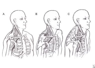
Progressive postural decompensation with neurovascular compression. A: Normal resting posture. B: Shoulder protraction beginning; the sternomastoid muscles are shortening, drawing the head anteriorly and inferiorly. C: Advanced deformity with adaptive shortening of scalene and pectoralis minor muscles. Also note narrowed costoclavicular space (ribs 1-5 have been relatively elevated). Neurovascular compression is evident at all 3 sites. |
 | View Full Size Image | |
Media type: Image
|
| Media file 2:
Scalene focal (left) and regional (right) stress tests for thoracic outlet syndrome. Both tests can be combined easily to enhance the stress effect (may be helpful in mild cases). |
 | View Full Size Image | |
Media type: Image
|
| Media file 3:
Pectoral focal (left) and regional (right) stress tests for thoracic outlet syndrome. |
 | View Full Size Image | |
Media type: Image
|
| Media file 4:
Myofascial release technique for the scalene muscles. Left: Side-lying approach. Right/top: Supine approach with pillow under thorax. Right/bottom: Supine approach with head extended off the table and supported by the operator's knees. Image courtesy of The Journal of the American Osteopathic Association |
 | View Full Size Image | |
Media type: Photo
|
| Media file 5:
Stretching technique for the scalene (anterior and middle) muscles. Left: The arm on the side to be stretched is secured down (hooked under the seat) to allow more control and effective stretch. Center: The opposite hand wraps partially around the head for good control to assist with the stretch. Right: After proceeding as far as tolerated, the patient leans the whole trunk away from the side being stretched, creating additional traction (downward) on the muscle by the arm that is secured. Image courtesy of The Journal of the American Osteopathic Association |
 | View Full Size Image | |
Media type: Photo
|
| Media file 6:
Stretching technique for the pectoralis minor muscle. Left: The patient has taken the slack out of the muscle. Center: He then rotates the body away from the side being stretched, increasing traction. Right: Maximum rotation and stretch effect are achieved slowly. Image courtesy of The Journal of the American Osteopathic Association |
 | View Full Size Image | |
Media type: Photo
|
| Media file 7:
Sagittal plane posture of patient with thoracic outlet syndrome before (left) and after (right) treatment. Notice the release of the pelvis that occurred with reduction in hyperlordosis, which allowed shoulder girdle to drop back and open the thoracic outlet. Image courtesy of The Journal of the American Osteopathic Association |
 | View Full Size Image | |
Media type: Photo
|
| Media file 8:
Algorithm for treatment of nonprogressive thoracic outlet syndrome (TOS). Anti-inflammatory medication, muscle relaxants, and activity modifications all are used as indicated and tolerated in all cases. Values for very severe TOS are greater than those for severe TOS, and the patient probably should be referred for surgical evaluation. |
 | View Full Size Image | |
Media type: Image
|
- Abe M, Ichinohe K, Nishida J. Diagnosis, treatment, and complications of thoracic outlet syndrome. J Orthop Sci. 1999;4(1):66-9. [Medline].
- Al-Shekhlee A, Katirji B. Spinal accessory neuropathy, droopy shoulder, and thoracic outlet syndrome. Muscle Nerve. Sep 2003;28(3):383-5. [Medline].
- Aligne C, Barral X. Rehabilitation of patients with thoracic outlet syndrome. Ann Vasc Surg. Jul 1992;6(4):381-9. [Medline].
- Atasoy E. Thoracic outlet compression syndrome. Orthop Clin North Am. Apr 1996;27(2):265-303. [Medline].
- Cagli K, Ozcakar L, Beyazit M, et al. Thoracic outlet syndrome in an adolescent with bilateral bifid ribs. Clinical Anatomy. 2006;19:558-560.
- Cherington M, Happer I, Machanic B, Parry L. Surgery for thoracic outlet syndrome may be hazardous to your health. Muscle Nerve. Sep 1986;9(7):632-4. [Medline].
- Colon E, Westdorp R. Vascular compression in the thoracic outlet. Age dependent normative values in noninvasive testing. J Cardiovasc Surg (Torino). Mar-Apr 1988;29(2):166-71. [Medline].
- Connolly JF, Ganjianpour M. Thoracic outlet syndrome treated by double osteotomy of a clavicular malunion: a case report. J Bone Joint Surg Am. Mar 2002;84-A(3):437-40. [Medline].
- Cuetter AC, Bartoszek DM. The thoracic outlet syndrome: controversies, overdiagnosis, overtreatment, and recommendations for management. Muscle Nerve. May 1989;12(5):410-9. [Medline].
- DeLisa JA, Gans BM, Walsch NE. Physical Medicine and Rehabilitation Principles and Practice, 4th Ed. Philadelphia, Lippincott Williams & Wilkins. 2005.
- Demirbag D, Unlu E, Ozdemir F, et al. The relationship between magnetic resonance imaging findings and postural maneuver and physical examination tests in patients with thoracic outlet syndrome: results of a double-blind, controlled study. Arch Phys Med Rehabil. Jul 2007;88(7):844-51. [Medline].
- Dobrusin R. An osteopathic approach to conservative management of thoracic outlet syndromes. J Am Osteopath Assoc. Aug 1989;89(8):1046-50, 1053-7. [Medline].
- England JD, Tiel RL. AAEM case report 33: costoclavicular mass syndrome. American Association of Electrodiagnostic Medicine. Muscle Nerve. Mar 1999;22(3):412-8. [Medline].
- Felice KJ, Butler KB, Druckemiller WH. Cervical root stimulation in a case of classic neurogenic thoracic outlet syndrome. Muscle Nerve. Sep 1999;22(9):1287-92. [Medline].
- Ferrante MA. Brachial plexopathies: Classification, causes, and consequences. Muscle & Nerve. 2004;30:547-568.
- Jordan SE, Machleder HI. Diagnosis of thoracic outlet syndrome using electrophysiologically guided anterior scalene blocks. Ann Vasc Surg. May 1998;12(3):260-4. [Medline].
- Kenny RA, Traynor GB, Withington D, Keegan DJ. Thoracic outlet syndrome: a useful exercise treatment option. Am J Surg. Feb 1993;165(2):282-4. [Medline].
- Lee RK, Pollak EW. Radiological diagnosis. In: Thoracic Outlet Syndrome: Diagnosis and Treatment. Mount Kisco, NY:. Futura Publishing Co;1986:107-124.
- Leffert RD. Thoracic outlet syndromes. Hand Clin. May 1992;8(2):285-97. [Medline].
- Levin LS, Dellon AL. Pathology of the shoulder as it relates to the differential diagnosis of thoracic outlet compression. J Reconstr Microsurg. Jul 1992;8(4):313-7. [Medline].
- Lindgren KA. Conservative treatment of thoracic outlet syndrome: a 2-year follow-up. Arch Phys Med Rehabil. Apr 1997;78(4):373-8. [Medline].
- Lindgren KA, Leino E. Subluxation of the first rib: a possible thoracic outlet syndrome mechanism. Arch Phys Med Rehabil. Sep 1988;69(9):692-5. [Medline].
- Lindgren KA, Leino E, Manninen H. Cineradiography of the hypomobile first rib. Arch Phys Med Rehabil. May 1989;70(5):408-9. [Medline].
- Machleder HI. Thoracic outlet syndromes: new concepts from a century of discovery. Cardiovasc Surg. Apr 1994;2(2):137-45. [Medline].
- Mackinnon SE, Novak CB. Evaluation of the patient with thoracic outlet syndrome. Semin Thorac Cardiovasc Surg. Apr 1996;8(2):190-200. [Medline].
- Mackinnon SE, Patterson GA, Novak CB. Thoracic outlet syndrome: a current overview. Semin Thorac Cardiovasc Surg. Apr 1996;8(2):176-82. [Medline].
- Nishida T, Price SJ, Minieka MM. Medial antebrachial cutaneous nerve conduction in true neurogenic thoracic outlet syndrome. Electromyogr Clin Neurophysiol. Jul-Aug 1993;33(5):285-8. [Medline].
- Novak CB, Collins ED, Mackinnon SE. Outcome following conservative management of thoracic outlet syndrome. J Hand Surg [Am]. Jul 1995;20(4):542-8. [Medline].
- Oh SJ. Clinical Electromyography: Nerve Conduction Studies (3rd Ed). Philadelphia, Lippincott Williams & Wilkins. 2003.
- Palmer JB, Uematsu S, Jankel WR, Arnold WP. A cellist with arm pain: thermal asymmetry in scalenus anticus syndrome. Arch Phys Med Rehabil. Mar 1991;72(3):237-42. [Medline].
- Panegyres PK, Moore N, Gibson R, et al. Thoracic outlet syndromes and magnetic resonance imaging. Brain. Aug 1993;116 ( Pt 4):823-41. [Medline].
- Pang D, Wessel HB. Thoracic outlet syndrome. Neurosurgery. Jan 1988;22(1 Pt 1):105-21. [Medline].
- Plewa MC, Delinger M. The false-positive rate of thoracic outlet syndrome shoulder maneuvers in healthy subjects. Acad Emerg Med. Apr 1998;5(4):337-42. [Medline].
- Press JM, Young JL. Vague upper-extremity symptoms? Consider thoracic outlet syndrome. The Physician and Sportsmedicine. 1994;22(7):57-64.
- Preston DC, Shapiro BE. Electromyography and Neuromuscular Disorders: Clinical-Electrophysiologic Correlations, 2nd Ed. Philadelphia, Elsevier Butterworth Heinemann. 2005.
- Rayan GM. Thoracic outlet syndrome. J Shoulder Elbow Surg. Jul-Aug 1998;7(4):440-51. [Medline].
- Redenbach DM, Nelems B. A comparative study of structures comprising the thoracic outlet in 250 human cadavers and 72 surgical cases of thoracic outlet syndrome. Eur J Cardiothorac Surg. Apr 1998;13(4):353-60. [Medline].
- Richardson AB. Thoracic outlet syndrome in aquatic athletes. Clin Sports Med. Apr 1999;18(2):361-78. [Medline].
- Roos DB. Congenital anomalies associated with thoracic outlet syndrome. Anatomy, symptoms, diagnosis, and treatment. Am J Surg. Dec 1976;132(6):771-8. [Medline].
- Roos DB. Thoracic outlet syndrome is underdiagnosed. Muscle Nerve. Jan 1999;22(1):126-9; discussion 137-8. [Medline].
- Sucher BM. Palpatory diagnosis and manipulative management of carpal tunnel syndrome: Part 2. ''Double crush'' and thoracic outlet syndrome. J Am Osteopath Assoc. Aug 1995;95(8):471-9. [Medline].
- Sucher BM. Thoracic outlet syndrome--a myofascial variant: Part 1. Pathology and diagnosis. J Am Osteopath Assoc. Aug 1990;90(8):686-96, 703-4. [Medline].
- Sucher BM. Thoracic outlet syndrome--a myofascial variant: Part 2. Treatment. J Am Osteopath Assoc. Sep 1990;90(9):810-2, 817-23. [Medline].
- Sucher BM, Glassman JH. Upper extremity syndromes. In: Mein E, Stanton D, ed. Manipulation. Philadelphia:. WB Saunders Co;1996:787-810.
- Sucher BM, Heath DM. Thoracic outlet syndrome--a myofascial variant: Part 3. Structural and postural considerations. J Am Osteopath Assoc. Mar 1993;93(3):334, 340-5. [Medline].
- Tilki HE, Stalberg E, Incesu L, Basoglu A. Bilateral neurogenic thoracic outlet syndrome. Muscle Nerve. Jan 2004;29(1):147-50.
- Vender MI, Truppa KL, Ruder JR, et al. Upper extremity compressive neuropathies. In: Derebery VJ, Kasdan ML, ed. Injuries and Rehabilitation of the Upper Extremity. 1998:243-262.
- Wilbourn AJ. Thoracic outlet syndrome is overdiagnosed. Muscle Nerve. Jan 1999;22(1):130-6; discussion 136-7. [Medline].
- Wilbourn AJ. Thoracic outlet syndrome surgery causing severe brachial plexopathy. Muscle Nerve. Jan 1988;11(1):66-74. [Medline].
- Wood VE, Biondi J. Double-crush nerve compression in thoracic-outlet syndrome. J Bone Joint Surg Am. Jan 1990;72(1):85-7. [Medline].
- Woods WW. Thoracic outlet syndrome after trauma, diagnosis is elusive. Modern Medicine. 1978;46:147.
Thoracic Outlet Syndrome excerpt Article Last Updated: Dec 13, 2006
|