Practice Essentials
Epiglottitis, also termed supraglottitis or epiglottiditis, is an inflammation of structures above the insertion of the glottis and is most often caused by bacterial infection. Before widespread Haemophilus influenzae type b (Hib) vaccination, H influenzae caused almost all pediatric cases of epiglottitis.
Affected structures include the epiglottis, aryepiglottic folds, arytenoid soft tissue, and, occasionally, the uvula. The epiglottis is the most common site of swelling. Acute epiglottitis and associated upper airway obstruction has significant morbidity and mortality and may cause respiratory arrest and death.
The following image illustrates the difference between a normal pediatric airway and one from a child with fatal epiglottitis.
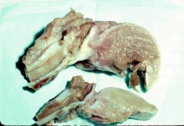 Comparison of a normal pediatric airway (bottom) and airway from a child who died from epiglottitis (top).
Signs and symptoms of pediatric epiglottitis
The clinical triad of drooling, dysphagia, and distress is the classic presentation. Fever with associated respiratory distress or air hunger occurs in most patients. Drooling occurs in up to 80% of cases.
An erythematous and classic swollen, cherry red epiglottis is often visible during careful examination of the oropharynx, although this examination should not be attempted if it may compromise respiratory effort.
See Presentation for more detail.
Diagnosis of pediatric epiglottitis
Laryngoscopy is the best way to confirm the diagnosis, but it is not advised to attempt any procedures without securing the airway.
Laboratory studies
Laboratory evaluation is nonspecific in patients with epiglottitis and should be performed once the airway is secured. The white blood cell (WBC) count may be elevated from 15,000-45,000 cells/µL with a predominance of bands.
Imaging studies
Classic cases of epiglottitis require no radiographic evaluation; however, radiography may be needed in some cases to confirm the diagnosis and to exclude other potential causes of acute airway obstruction.
See Workup for more detail.
Management of pediatric epiglottitis
Treatment in patients with epiglottitis is directed toward relieving the airway obstruction and eradicating the infectious agent.
See Treatment and Medication for more detail.
Anatomy
As with many other aspects of the pediatric airway, the epiglottis is significantly different in the child than in the adult. In the infant, the epiglottis is located more anteriorly and superiorly than in the adult, and it is at a greater angle with the trachea. The infant epiglottis is also more omega shaped and floppy than the more rigid, U-shaped epiglottis in the adult.
Pathophysiology
Haemophilus influenzae type b (Hib) or Streptococcus pneumoniae (see Etiology) can colonize the pharynges of otherwise healthy children through respiratory transmission from intimate contact. These bacteria may penetrate the mucosal barrier, invading the bloodstream and causing bacteremia and seeding of the epiglottis and surrounding tissues. Bacteremia may also lead to infection of the meninges, skin, lungs, ears, joints, and other structures.
Hib infection of the epiglottis leads to acute onset of inflammatory edema, beginning on the lingual surface of the epiglottis where the submucosa is loosely attached. Swelling significantly reduces the airway aperture. Edema rapidly progresses to involve the aryepiglottic folds, the arytenoids, and the entire supraglottic larynx. The tightly bound epithelium on the vocal cords halts edema spread at this level. Frank airway obstruction, aspiration of oropharyngeal secretions, or distal mucous plugging can cause respiratory arrest.
Noninfectious inflammation of any of the structures around the epiglottis may also result from thermal or chemical injury or from local trauma, including blunt trauma to the neck. [1]
Etiology
Historically, Haemophilus influenzae type b (Hib) was the predominant organism (>90%) in pediatric epiglottitis cases. Since the widespread use of the Hib vaccine, the incidence and causative agents of epiglottitis have changed; however, even vaccinated children can develop epiglottitis due to non – type b H influenzae. [2] Clearly, epiglottitis due to Hib persists in parts of the world where Hib vaccination is not used.
The following are other known bacterial causes of pediatric epiglottitis:
-
Streptococcus pneumoniae
-
Group A and group C (ie, beta-hemolytic) streptococci
-
Staphylococcus aureus
-
Moraxella catarrhalis
-
Haemophilus parainfluenzae
-
Neisseria meningitidis
-
Pseudomonas species
-
Candida albicans, especially in immunocompromised patients
-
Klebsiella pneumoniae
-
Pasteurella multocida
Although viruses normally do not cause epiglottitis, a previous viral infection may allow bacterial superinfection to occur. Viral agents may include herpes simplex virus (HSV), parainfluenzae virus, varicella-zoster virus (VZV), human immunodeficiency virus (HIV), [3] and Epstein-Barr virus (EBV). Varicella can cause a primary or secondary infection often with group A beta-hemolytic streptococci.
Noninfectious etiologies include thermal injuries, trauma-causing blind finger sweeps to remove a foreign body from the pharynx, angioneurotic edema, hemophagocytic lymphohistiocytosis, [4] and acute leukemia. Lymphoproliferative diseases may also cause epiglottic swelling.
Epidemiology
Historically, acute epiglottitis was most common in children aged 2-4 years. The use of the Haemophilus influenzae type b (Hib) vaccine has reduced incidence of epiglottitis in the United States, making this a rare condition in children. [5] Introduction of the polysaccharide vaccine in 1985, followed by the highly effective conjugate vaccine, dramatically reduced the incidence of epiglottitis, with concomitant declines in hospital admissions. Studies have shown an annual incidence rate of 0.63 cases per 100,000 persons, [6] and studies of children of all ages with epiglottitis report a seasonal variation in incidence.
A comparison made between a large US children's hospital's chart review from 1995 to 2003 and a previous report from the same hospital completed 27 years earlier, showed a 10-fold decline in acute epiglottitis admissions, with streptococci becoming the major pathogens. [7] Epiglottitis incidence in adults has remained constant.
A retrospective case series of 107 patients admitted to a pediatric hospital's intensive care unit (ICU) from 1997 to 2006 concluded that bacterial tracheitis is now 3 times more likely to be the cause of pediatric respiratory failure compared with viral croup and epiglottitis combined. The authors attributed this change in the epidemiology of acute infectious upper airway disease to Hib vaccine as well as the use of corticosteroids for the treatment of viral croup. [8]
The international incidence of epiglottitis widely varies, with a significantly greater prevalence in countries without universal immunization. Among countries with mandatory immunization, the reported incidences are 0.9 cases per 100,000 persons in Sweden and 0.6 cases per 100,000 in the United Kingdom, for example. However, in recent years, epiglottitis has been increasing in frequency in the United Kingdom. [9] The reason for this is unclear and may be due to the administration of 3 vaccines rather than 4. Other studies suggest that bacterial tracheitis is now the most common serious airway infection in children. [8, 10]
A retrospective review of a Danish population demonstrated a mean national incidence of epiglottitis in children of 4.9 cases per 100,000 per year in the decade before Hib vaccination. From 1996 to 2005, with the introduction of widespread Hib vaccination, an incidence of only 0.02 cases per 100,000 per year was seen. During this period, the incidence of acute epiglottitis in adults remained constant, at 1.9 cases per 100,000 per year. [9]
Racial, sexual, and age differences in incidence
Most studies show no racial predominance for epiglottitis, although a recent study showed higher incidence among Black and Hispanic individuals. There also appears to be a 60% male predominance, which has remained true even with the changing epidemiology of epiglottitis.
Epiglottitis was once believed to occur exclusively in children. In the past, this condition occurred most commonly in children aged 2-4 years; however, it may occur at any age. Adult cases have been reported in recent years, and some evidence suggests the incidence in adults is increasing.
Prognosis
The prognosis is good for patients with epiglottitis whose airways have been secured. The mortality rate is less than 1% in these patients. However, mortality rates as high as 10% can occur in children whose airways are not protected by endotracheal intubation.
An analysis by Allen et al of US mortality trends from 1979 to 2017 showed that deaths from acute epiglottitis decreased following the widespread use of the Hib vaccine. Of a total 1187 epiglottitis-related deaths during the 39-year period, 443 occurred among children and adolescents. Mortality rates in this age group fell from 0.064 per 100,000 individuals (41 deaths) in 1979 to 0.001 per 100,000 individuals (1 death) in 2017. [11]
Complications
During the bacteremic phase of the disease, other foci of infection are possible. Pneumonia is the most commonly cited associated illness, followed by otitis media. Meningitis has also been reported in association with epiglottitis.
As with other causes of upper airway obstruction, pulmonary edema can be observed after the airway has been secured. Accidental extubation and respiratory arrest are the 2 most common complications, and accidental extubation can cause additional complications. Cervical adenitis, tonsillitis, and otitis media have also been documented.
In summary, complications associated with a swollen epiglottis and surrounding tissues include airway obstruction, which can lead to respiratory arrest and death from hypoxia as well the following:
-
Aspiration
-
Endotracheal tube dislodgement
-
Extubation
-
Tracheal stenosis
-
Pneumothorax or pneumomediastinum
-
Epiglottic abscess
-
Adenitis
-
Cervical cellulitis
-
Septic shock
-
Pulmonary edema (rare)
-
Cerebral anoxia
-
Death from asphyxia
In classic cases involving bacteremia with Haemophilus influenzae, other structures may have concomitant infectious processes. These may include the following:
-
Meningitis
-
Pneumonia
-
Septicemia
-
Cellulitis
-
Septic arthritis
-
Otitis media
-
Pericarditis (rare)
Patient Education
For patient education information, see Cold & Flu Center. Also see the WebMD patient education resource Epiglottis.
-
Swollen epiglottis with characteristic thumbprint sign.
-
Comparison of a normal pediatric airway (bottom) and airway from a child who died from epiglottitis (top).
-
Child assuming the sniffing position with upper airway obstruction.
-
Swollen epiglottis with characteristic thumbprint sign.
-
Radiograph of a child with acute epiglottitis; note the hypopharyngeal dilatation, obliteration of the vallecula, and thickened aryepiglottic folds—a positive thumb sign.
-
Correct positioning for a cricothyroid needle insertion
-
Child with acute epiglottitis after intubation. Note cherry red epiglottis. This image was taken in 2008 and the child was completely immunized and grew HiB from surface culture.


