Practice Essentials
Lymphadenitis is the inflammation or enlargement of a lymph node. Lymph nodes are small, ovoid nodules normally ranging in size from a few millimeters to 2 cm. (See the image below.) They are distributed in clusters along the course of lymphatic vessels located throughout the body. The primary function of lymph nodes is to filter out microorganisms and abnormal cells that have collected in lymph fluid. [1]
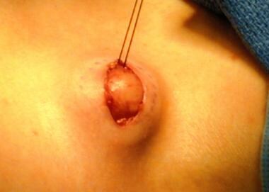 A lymph node biopsy is performed. Note that a marking pen has been used to outline the node before removal and that a silk suture has been used to provide traction to assist the removal.
Lymph node enlargement is a common feature in a variety of diseases and may serve as a focal point for subsequent clinical investigation of diseases of the reticuloendothelial system or regional infection. The majority of cases represent a benign response to localized or systemic infection. Most children with lymphadenitis exhibit small, palpable cervical, axillary, and inguinal lymph nodes. Less common is enlargement of the suboccipital or postauricular nodes. Palpable supraclavicular, epitrochlear, and popliteal lymph nodes are uncommon, as are enlarged mediastinal and abdominal nodes.
Lymphadenitis may affect a single node or a group of nodes (regional adenopathy) and may be unilateral or bilateral. The onset and course of lymphadenitis may be acute, subacute, or chronic.
Pathophysiology
Increased lymph node size may be caused by the following:
-
Multiplication of cells within the node, including lymphocytes, plasma cells, monocytes, or histiocytes
-
Infiltration of cells from outside the node, such as malignant cells or neutrophils
-
Draining of an infection (eg, abscess) into local lymph nodes
Etiology
Infectious agents/causes and lymphadenitis characteristics are as follows [2] :
-
Bartonella henselae (catscratch disease) – Single-node involvement determined by scratch site; discrete, mobile, nontender
-
Coccidioides immitis (coccidioidomycosis) – Mediastinal
-
Cytomegalovirus – Generalized
-
Dental caries/abscess – Submaxillary
-
Epstein-Barr virus (mononucleosis) - Anterior cervical, mediastinal, bilateral; discrete, firm, nontender
-
Francisella tularensis (tularemia) - Cervical, mediastinal, or generalized; tender
-
Histoplasma capsulatum (histoplasmosis) – Mediastinal
-
Mycobacterium tuberculosis - Mediastinal, mesenteric, anterior cervical, localized disease (discrete, firm, mobile, tender); generalized hematogenous spread (soft, fluctuant, matted, and adhere to overlying, erythematous skin)
-
Parvovirus - Posterior auricular, posterior cervical, occipital
-
Rubella - Posterior auricular, posterior cervical, occipital
-
Salmonella - Generalized
-
Seborrheic dermatitis, scalp infections - Occipital, postauricular
-
Staphylococcus aureus adenitis - Cervical, submandibular; unilateral, firm, tender
-
Group A streptococcal (GAS) pharyngitis - Submandibular and anterior cervical; unilateral, firm, tender
-
Toxoplasma gondii - Generalized, often nontender
-
Viral pharyngitis - Bilateral postcervical; firm, tender
-
Yersinia enterocolitica - Cervical or abdominal
-
Yersinia pestis (plague) - Axillary, inguinal, femoral, cervical; extremely tender with overlying erythema
Immunologic or connective tissue disorders causing lymphadenitis are as follows:
Primary diseases of lymphoid or reticuloendothelial tissue causing lymphadenitis are as follows:
-
Lymphosarcoma
-
Reticulum cell sarcoma
-
Malignant histocytosis or histocytic lymphoma
-
Nonendemic Burkitt tumor
-
Nasopharyngeal rhabdomyosarcoma
-
Thyroid carcinoma, chronic lymphocytic thyroiditis
-
Histiocytosis X
-
Kikuchi disease
-
Benign sinus histiocytosis
-
Angioimmunoblastic or immunoblastic lymphadenopathy
-
Chronic pseudolymphomatous lymphadenopathy (chronic benign lymphadenopathy)
Immunodeficiency syndromes and phagocytic dysfunction causing lymphadenitis are as follows:
-
Chronic granulomatous disease of childhood
-
Acquired immunodeficiency syndrome
Metabolic and storage diseases causing lymphadenitis are as follows:
-
Cystinosis
Hematopoietic diseases causing lymphadenitis are as follows:
-
Congenital hemolytic anemia
-
Autoimmune hemolytic anemia
Miscellaneous disorders causing lymphadenitis are as follows:
-
PFAPA (periodic fever, aphthous stomatitis, pharyngitis, and adenitis) syndrome
-
Castleman disease (also known as benign giant lymph node hyperplasia)
Medications causing lymphadenitis are as follows:
-
Mesantoin - Most commonly causes cervical lymphadenitis
-
Hydantoin - Generalized lymphadenopathy
Prognosis
Prognosis depends on the etiology of the lymphadenopathy and timing of intervention.
Complications
The following complications may occur:
-
Cellulitis
-
Suppuration
-
Systemic involvement
-
Internal jugular vein thrombosis
-
Septic embolic phenomena
-
Carotid artery rupture
-
Mediastinal abscess
-
Purulent pericarditis
-
A lymph node biopsy is performed. Note that a marking pen has been used to outline the node before removal and that a silk suture has been used to provide traction to assist the removal.


