Practice Essentials
Inguinal hernia is a type of ventral hernia that occurs when an intra-abdominal structure, such as bowel or omentum, protrudes through a defect in the abdominal wall. Most hernias that are present at birth or in childhood are indirect inguinal hernias. Other less common types of ventral hernias include umbilical, epigastric, and incisional hernias.
Approximately 400 years ago, a French surgeon named Ambroise Pare described the reduction of an incarcerated pediatric hernia and the application of trusses. He recognized that inguinal hernias in children were probably congenital in nature and that they could be cured.
Unfortunately, despite the many historical descriptions of conservative medical management of inguinal hernias, no effective nonsurgical means of treating this condition is recognized. All pediatric inguinal hernias require operative treatment to prevent the development of complications, such as inguinal hernia incarceration or strangulation. Today, inguinal hernia repair is one of the most common pediatric operations performed.
In this article, the embryology, clinical presentation, and management of inguinal hernias are discussed in relation to the pediatric population. Because inguinal hernias are common, every clinician must be well versed in the subject and able to provide optimal care to patients and their families, especially because hernias can be organ-threatening or life-threatening if not expeditiously managed. Examples of hernias are shown in the images below.
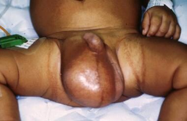 Typical appearance of an infant with a large right indirect inguinal hernia. The right scrotal sac is enlarged and contains palpable loops of bowel and fluid.
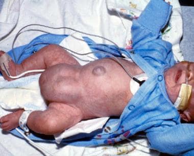 A premature baby boy with bilateral giant inguinoscrotal hernias. Because of the large size of the hernias, operative repair typically requires repair of the inguinal floor in addition to the high ligation of the indirect hernia sac.
Pathophysiology
The processus vaginalis is an outpouching of peritoneum attached to the testicle that trails behind as it descends retroperitoneally into the scrotum. When obliteration of the processus vaginalis fails to occur, inguinal hernia results. [1] A review of embryonic development of the inguinal region is important to understanding the pathophysiology and surgical management of inguinal hernias.
Although the sex of the embryo is determined at fertilization, the gonads do not begin to differentiate until 7 weeks' gestation. Primordial germ cells migrate along the dorsal mesentery of the gut. They arrive at the primitive gonads early in the fifth week of development and, during the sixth week, invade the genital ridges, which lie on the medial aspect of the mesonephros. The coelomic epithelium proliferates, and the underlying mesenchyme condenses, forming the primitive sex cords.
Under the influence of the Y chromosome, the cords in the male embryo proliferate to form the testes. Near the end of the second month, the testis and mesonephros are attached by the urogenital mesentery to the posterior abdominal wall. As the mesonephros degenerates, only the testis remains suspended. At its caudal end, the attachment is ligamentous and is known as the caudal genital ligament. The gubernaculum, a mesenchymal structure rich in extracellular matrices, also extends from the caudal pole of the testis. This structure attaches in the inguinal region between the differentiating internal and external oblique muscles prior to descent of the testes. As the testes begin to descend at about 28 weeks' gestation, an outgrowth of gubernaculum from the inguinal region grows toward the scrotal area, and as the testis passes through the inguinal canal, this portion of the gubernaculum comes in contact with the scrotal floor.
During this time, the peritoneum of the coelomic cavity is forming an evagination on each side of the midline into the ventral abdominal wall. This evagination, known as the processus vaginalis, follows the path of the gubernaculum testis into the scrotal swellings and forms, along with the muscle and fascia, the inguinal canal. The descent of the testes through the inguinal canal is thought to be regulated by both androgenic hormones produced by the fetal testis and mechanical factors resulting from increased abdominal pressure.
As each testis descends, the layers of the abdominal wall contribute to the layers of the spermatic cord. The internal spermatic fascia is a reflection of the transversalis fascia, the internal oblique muscle helps form the cremaster muscle, and the external spermatic fascia results from the external oblique aponeurosis. In addition, a reflected fold of the processus vaginalis covers each testis and becomes known as the visceral and parietal layers of the tunica vaginalis.
In the female embryo, the ovaries descend into the pelvis but do not leave the abdominal cavity. The upper portion of the gubernaculum becomes the ovarian ligament, and the lower portion becomes the round ligament, which travels through the inguinal ring into the labium majus. If the processus vaginalis remains patent, it extends into the labium majus and is known as the canal of Nuck.
Before birth, the layers of the processus vaginalis normally fuse, closing off the entrance into the inguinal canal from the abdominal cavity. In some individuals, the processus vaginalis remains patent through infancy, into childhood, and possibly even into adulthood. The precise cause of the obliteration of the processus vaginalis is unknown, but some studies indicate that calcitonin gene-related peptide (CGRP), released from the genitofemoral nerve, may have a role in the fusion.
When luminal obliteration fails to occur, a ready-made sac is present where abdominal contents may herniate. Even when the processus vaginalis is patent, the entrance may be adequately covered by the internal oblique and transverse abdominal muscles, preventing escape of abdominal contents for many years. Failure of fusion can result not only in an inguinal hernia, but also in a communicating or noncommunicating hydrocele.
In infants, the most common type of hydrocele is the communicating type. A communicating hydrocele results when the proximal portion of the processus vaginalis remains patent, allowing fluid from the abdominal cavity to freely enter the scrotal sac. When closure is present proximally but fluid remains trapped within the tunica distally, a noncommunicating hydrocele results.
Etiology
The cause of inguinal hernia in children can be termed an abnormality of embryologic development of the fetus. However, some children may present with an acquired form of inguinal hernia, also called a direct inguinal hernia. In this type of hernia, weakness of the inguinal floor is present, which allows for protrusion of viscera from the abdominal cavity. The hernia sac is composed of the peritoneal fold that contains the hernia.
Anatomically speaking, indirect and direct inguinal hernias differ in that the direct hernia bulges through the inguinal floor medial to the inferior epigastric vessels and the indirect hernia arises lateral to the inferior epigastric vessels. Either hernia may cause fullness or a palpable bulge in the inguinal region, and distinguishing between the two types on the basis of physical examination findings may be difficult. The clinician may assume, until proven otherwise, that the pediatric patient with an inguinal hernia has an indirect inguinal hernia.
-
The following are associated with an increased risk of inguinal hernia:
Prematurity and low birth weight (Incidence approaches 50%.)
Urologic conditions
Epispadias
Exstrophy of the bladder
Patent processus vaginalis, which may be present because of increased abdominal pressure due to ventriculoperitoneal shunts, peritoneal dialysis, or ascites
Abdominal wall defects
Omphalocele
Family history
Meconium peritonitis
Connective tissue disease
Mucopolysaccharidosis
Congenital dislocation of the hip
Cloacal exstrophy
Liver disease with ascites
Ventriculoperitoneal shunting for hydrocephalus
-
Figures regarding inguinal hernia incarceration indicate the following risk patterns:
Incarceration occurs in 17% of right-sided hernias and 7% of left-sided hernias.
More than 50% of cases of incarceration occur within the first 6 months of life; the risk gradually decreases after age 1 year.
Premature infants have twice the risk of incarceration than the general pediatric population.
More than two thirds of all incarcerations occur in children younger than 1 year.
Girls are more likely to develop incarceration of an inguinal hernia; the incidence in girls is 17.2%, whereas the incidence in boys is 12%.
Epidemiology
United States statistics
Although the exact incidence of indirect inguinal hernia in infants and children is unknown, the reported incidence ranges from 1-5%. Sixty percent of hernias occur on the right side. Premature infants are at increased risk for inguinal hernia, with incidence rates of 2% in females and 7-30% in males. Approximately 5% of all males develop a hernia during their lifetime.
A study that evaluated the incidence of inguinal hernia in almost 80,000 children found that the cumulative incidence of inguinal hernia from birth to 15 years of age was 6.62% in males and 0.74% in females. [2]
International statistics
International incidence rates are similar to those in the United States.
Race-, sex-, and age-related demographics
Inguinal hernia appears to occur equally among races. Umbilical hernias, on the other hand, appear to be more common in Blacks than in other races.
Inguinal hernias are much more common in males than in females. The male-to-female ratio is estimated to be 4-8:1.
Premature infants are at an increased risk for inguinal hernia, with the incidence ranging from 7-30%. Moreover, the associated risk of incarceration is more than 60% in this population. Most pediatric ventral and inguinal hernias are detected in the first year of life. Occasionally, hernias may remain asymptomatic and unnoticed by the parents until later in life. Finding an adult patient with an indirect inguinal hernia that has been present since birth is not unusual.
Prognosis
Overall prognosis is excellent; most patients do extremely well after operative repair of their inguinal hernia. Mortality is extremely rare but, unfortunately, continues to be reported as a consequence of delayed recognition of an incarcerated and strangulated inguinal hernia.
Morbidity/mortality
An incarcerated or strangulated inguinal hernia can result in severe complications and even death. An incarcerated or strangulated inguinal and/or femoral hernia may also result in significant sequelae, depending on which visceral structure is involved in the hernia sac. Such sequelae can range from life-threatening complications to gonadal dysfunction, including intestinal necrosis and perforation, intestinal obstruction, intestinal stricture, testicular necrosis, testicular atrophy, ovarian necrosis, ovarian atrophy, and tubal stricture.
Complications
Few complications result from operative repair of an inguinal hernia. Possible consequences of hernia repair include decreased testicular size (≤ 20% of patients), testicular atrophy (1-2%), [3] vas injury (< 1%), and development of sperm-agglutinating antibodies. The risk of gonadal injury in females is low. Fortunately, in the hands of pediatric surgeons, such complications are quite rare.
The incidence of wound infection is 1-2%.
Hernia recurrence rates are around 1% when experienced pediatric surgeons perform the operation. Factors associated with recurrence of inguinal hernia include an unrecognized tear in the sac, failure to repair an enlarged inguinal ring, damage to the canal and inguinal floor, infection, history of incarceration, connective tissue disorder, and conditions producing increased intra-abdominal pressure (eg, chronic respiratory problems, constipation). The hernia recurrence rate with the laparoscopic technique has been reported to be higher if the surgeon is still in the "learning curve." However, in the hands of an experienced surgeon, the recurrence rate for the laparoscopic technique should be similar to the one reported for the open technique.
A study that included data from 9993 pediatric patients who underwent inguinal hernia repair reported a recurrence rate of 1.4% with an incidence of recurrence 3.46 per 1000 person-years. The study also found that the majority of recurrence occurred within a year and children with multiple comorbidities had a greater risk of recurrence. [4] A systematic review by Obayashi et al showed that the main comorbidities associated with a higher risk of recurrence are increased intra-abdominal pressure and weakness of the anterior abdominal wall. [5]
The vas deferens and ilioinguinal nerve occasionally may be injured and should be repaired with 7-0 or 8-0 Maxon sutures. This may be technically difficult because of the extremely small vas lumen not traversed by semen. One infertility expert advises marking the ends of the vas with permanent suture and performing vasovasotomy after puberty with a 2-layer closure. It is also important to remember that the finding of vas or epididymis on the surgical pathology report does not necessarily imply injury because embryonal müllerian remnants have been recognized in 1-6% of surgical specimens. Specific histologic features of the remnant include a smaller diameter and failure to show a prominent muscular wall with Masson trichrome staining.
Patient Education
Instruct parents and caretakers on the signs and symptoms of inguinal hernia incarceration. Delayed recognition of incarceration is likely to result in significant morbidity and mortality for the child.
-
Typical appearance of an infant with a large right indirect inguinal hernia. The right scrotal sac is enlarged and contains palpable loops of bowel and fluid.
-
A premature baby boy with bilateral giant inguinoscrotal hernias. Because of the large size of the hernias, operative repair typically requires repair of the inguinal floor in addition to the high ligation of the indirect hernia sac.
-
Illustration of the technique for intraoperative diagnostic laparoscopy to evaluate for the presence of an asymptomatic contralateral inguinal hernia at the time of elective repair of an indirect inguinal hernia.
-
Laparoscopic view of a left indirect inguinal hernia at the time of surgery for laparoscopic needle-assisted repair.
-
Laparoscopic needle-assisted repair of a left indirect inguinal hernia. Note the passage of a Prolene suture through a small 22G spinal needle; this is used for creation of the purse-string suture that closes the open inguinal ring.
-
Laparoscopic view of the repaired left indirect inguinal hernia with the closed Prolene purse-string suture around the internal inguinal ring.


