Background
Total anomalous pulmonary venous connection (TAPVC) consists of an abnormality of blood flow in which all four pulmonary veins drain into systemic veins or the right atrium with or without pulmonary venous obstruction. Systemic and pulmonary venous blood mix in the right atrium. An atrial defect or foramen ovale (part of the complex) is important in left ventricular output both in fetal and in newborn circulation. See the image below.
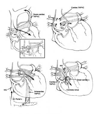 Types of total anomalous pulmonary venous connection.
Embryology
Early in the formation of the lungs, the blood coming from the lung buds drains to the splanchnic plexus, which connects to the paired common cardinal and umbilicovitelline veins. The right common cardinal system later evolves into the right sinus venosus, which, in turn, becomes the right superior vena cava and azygos vein. The left common cardinal vein evolves into the left sinus venosus, which, in turn, becomes the left superior vena cava and coronary sinus. The umbilicovitelline system becomes the inferior vena cava, ductus venosus, and portal vein.
At 25-27 days' gestation, the developing pulmonary venous plexus retains connections to the right superior vena cava, left superior vena cava, and portal system. No direct communication to the left atrium exists.
At 27-29 days' gestation, the primitive pulmonary vein appears as an endothelial out-pouching from either the posterior superior left atrial wall or from the central part of the sinus venosus proximal to the primordial lung venous plexus. Connection between the primitive pulmonary vein and pulmonary venous plexus occurs by 30 days' gestation. The common pulmonary vein enlarges and incorporates into the left atrium, and, normally, the pulmonary venous part of the splanchnic plexus gradually loses its connection with the cardinal and umbilicovitelline veins.
Knowledge of the normal development of pulmonary venous pathways facilitates an understanding of how the various types of anomalous pulmonary venous return might occur. Failure of the common pulmonary vein to connect with the pulmonary venous plexus leads to persistence of one or more earlier venous connections to the right superior vena cava, to the left vertical vein/innominate vein, or to the umbilicovitelline vein/portal vein. Failure of the septum primum to normally form or abnormal septation of the sinus venosus can lead to direct connection of the pulmonary veins to the right atrium. Late obstruction of the common pulmonary vein after earlier venous channels have disappeared can lead to isolated pulmonary vein atresia, a rare and usually fatal condition. Failure of incorporation of the common pulmonary vein may lead to a left atrial shelf or membrane of cor triatriatum (ie, stenosis of the common pulmonary vein). [1]
Because all pulmonary venous return connects to the systemic venous system, right atrial and right ventricular enlargement occurs, and, if significant pulmonary venous obstruction develops, right ventricular hypertrophy occurs. Total anomalous pulmonary venous connection occurs alone in two thirds of patients and occurs as part of a group of heart defects (eg, heterotaxy syndromes) in approximately one third of patients.
An atrial septal defect or patent foramen ovale, considered part of the complex, serves a vital function in this condition for maintaining left ventricular output. Because diagnosis of most patients occurs in early infancy, a ductus arteriosus is frequently found as well
Darling proposed the most commonly used classification system for total anomalous pulmonary venous connection based on the site of pulmonary venous drainage. In type I (ie, supracardiac connection), the four pulmonary veins drain via a common vein into the right superior vena cava, left superior vena cava, or their tributaries. In type II (ie, cardiac connection), the pulmonary veins connect directly to the right heart (eg, coronary sinus or directly to the right atrium). In type III (ie, infradiaphragmatic connection), the common pulmonary vein travels down anterior to the esophagus through the diaphragm to connect to the portal venous system. In type IV (ie, mixed connections), the right and left pulmonary veins drain to different sites (eg, left pulmonary veins into the left vertical vein to the left innominate, right pulmonary veins directly into the right atrium or coronary sinus).
Pulmonary venous obstruction may occur in all types of anomalous connections, and, in all cases, clinicians must identify any sites of obstruction and treat the obstruction whenever possible at the time of surgical repair. In supracardiac connections, obstruction may occur at the origin of the ascending (vertical) vein or its attachment to the innominate vein, or the vertical vein may be obstructed as it crosses between the left pulmonary artery and the left bronchus. In cardiac connections, obstruction to pulmonary veins seldom develops but may occur at the junction of the common vein to the coronary sinus.
In infradiaphragmatic connections, severe obstruction almost always inhibits pulmonary venous flow with obstruction of the common pulmonary vein. This obstruction occurs either as it travels through the diaphragm, at its junction with the portal vein system, or as an obstruction of pulmonary venous flow as the ductus venosus closes and pulmonary vein flow is forced to cross the liver portal sinusoid system. Finally, in all types, obstruction may occur because of restrictive atrial septal defect size and because of small left atrial size.
Recurrent pulmonary vein stenosis remains a problem in some patients with total anomalous pulmonary venous connection. Recent studies suggest missing myocardial muscle “sleeve” layer in the abnormal-draining pulmonary vein walls and in left atrial walls of patients with total anomalous pulmonary venous connection; this might help explain the increased risk of developing stenosis. [2]
Pathophysiology
As a result of the mixture of pulmonary and systemic venous flow, right atrial and right ventricular volume loading develops in all patients with total anomalous pulmonary venous connection. Whether right heart pressure loading is also present depends primarily on whether restriction to flow occurs at the atrial septum or an obstruction to pulmonary venous flow develops. If the foramen ovale is restrictive, right atrial pressure elevates, and systemic and pulmonary venous congestion both occur. Pulmonary blood flow increases, and pulmonary artery hypertension may occur. The left atrium and left ventricle receive less than the normal flow and pump less than the normal volume, with some decrease in the cardiac index.
Most patients with isolated total anomalous pulmonary venous connection have a patent foramen ovale with some degree of restriction to transatrial flow. If no pulmonary venous obstruction is present, pulmonary blood flow increases (eg, 3-5 times the systemic volume) in early infancy, and arterial oxygen saturation is maintained, usually at 90% or higher. Signs of right heart volume load or right heart failure are evident.
If obstruction of pulmonary venous flow is present, then pulmonary venous congestion occurs with increased pulmonary lymphatic flow and increased flow through available alternate pulmonary venous pathways. Reflex pulmonary arterial vasoconstriction may also occur. Increase in pulmonary vascular resistance leads to decrease in pulmonary blood flow and a lower volume of saturated blood in the venous mixture. Decrease in systemic oxygen saturation along with a decrease in the cardiac index may lead to a severe decrease in oxygen delivery.
Etiology
Sociodemographic findings in patients with total anomalous pulmonary venous connection were similar to those in control subjects. [3] Family history showed no other family members with total anomalous pulmonary venous connection. Noncardiac malformations were present in 9 patients (22%); however, other cardiac and noncardiac malformations were present in 6 first-degree relatives and 7 second-degree relatives of patients with isolated cases (41%). Altogether, a genetic etiology was suspected to contribute to a "failure of targeted pulmonary vein growth" because of the number of multiplex families. In addition, total anomalous pulmonary venous connection has been reported in siblings in other series.
Exposure histories showed possible association of total anomalous pulmonary venous connection with lead or pesticide exposure and raised questions of familial susceptibility to certain environmental teratogens.
Total anomalous pulmonary venous connection frequently occurs in association with asplenia and pulmonary atresia. Overall, one third of patients with total anomalous pulmonary venous connection have a major associated cardiovascular malformation and two thirds of patients have isolated total anomalous pulmonary venous connection.
Epidemiology
United States data
Total anomalous pulmonary venous connection occurred in 41 of 2659 cases with cardiovascular abnormalities in the Baltimore-Washington Infant Study (1981-1987) or in 1.5% of all patients with cardiovascular malformations. Regional prevalence was 6.8 per 100,000 live births. [3] A total of 68% of these patients were diagnosed as neonates.
Sex-related demographics
In the Baltimore-Washington Infant Study, the male-to-female ratio was 18:23. [3] In other reports, a strong male preponderance of 3:1 was observed in patients with infradiaphragmatic drainage.
-
Types of total anomalous pulmonary venous connection.


