Overview of Tuberculosis
Tuberculosis (TB) is the most common cause of infection-related death worldwide. In 1993, the World Health Organization (WHO) declared TB to be a global public health emergency. The image below depicts typical radiographic findings on a patient with tuberculosis.
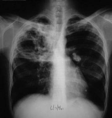 This radiograph shows a patient with typical radiographic findings of tuberculosis.
See Tuberculosis: Diagnostic Imaging and Treatment Challenges, a Critical Images slideshow, to help determine the best approach for patients with this multisystemic disease.
Tubercle bacilli belong to the order Actinomycetales and family Mycobacteriaceae. Mycobacterium tuberculosis is the most common cause of this disease, and it is seen in the image below. Other rare causes include M bovis and M africanum.
The acid-fast characteristic of the mycobacteria is their unique feature. M tuberculosis is an aerobic, non-spore-forming, nonmotile, slow-growing bacillus with a curved and beaded rod-shaped morphology. It is a very hardy bacillus that can survive under adverse environmental conditions. Humans are the only known reservoirs for M tuberculosis.
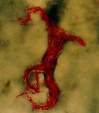 Acid-fast bacillus smear showing characteristic cording in Mycobacterium tuberculosis.
Most persons infected with M tuberculosis do not develop active disease. In healthy individuals, the lifetime risk of developing disease is 5-10%. In certain instances, such as extremes of age or defects in cell-mediated immune (CMI) response (eg, human immunodeficiency virus [HIV] infection, malnutrition, administration of chemotherapy, prolonged steroid use), TB may develop. For patients with HIV infection, the risk of developing TB is 7-10% per year.
For patient education information, see the Infections Center and Tuberculosis.
See Medscape Drugs & Diseases articles Tuberculosis, Miliary Tuberculosis, Primary Tuberculosis Imaging, Pediatric HIV Infection, and HIV Disease for more information on these topics.
TB Risk Factors
Risk factors for the acquisition of tuberculosis (TB) are usually exogenous to the patient. Thus, likelihood of being infected depends on the environment and the features of the index case. However, the development of TB disease depends on inherent immunologic status of the host.
Tuberculosis has been reported in patients treated for arthritis, inflammatory bowel disease, and other conditions with tumor necrosis factor (TNF)-alpha blockers/antagonists.
Factors in acquiring TB infection
The number of bacilli in the inoculum and the relative virulence of the organism are the major factors determining transmission of the disease. TB is transmitted by inhaling the tubercle bacilli.
The infectiousness of the source case is of vital importance in determining likelihood of transmission. Bacillary population of TB lesions varies and depends on the morphology of the lesion. Nodular lesions have 100-10,000 organisms, whereas cavitary lesions have 10 million to 1 billion bacilli. Thus, persons with cavitary lesions are highly infectious. Also, contacts of persons with sputum-positive smears have an increased prevalence of infection as opposed to contacts of those with sputum-negative smears.
Persons who have received anti-TB drugs are much less infectious than those who have not received any treatment. This decline in infectiousness is due primarily to reduction in the bacillary population in the lungs.
Environmental factors also contribute to the likelihood of acquiring the infection. The concentration of bacilli depends on the ventilation of the surroundings and exposure to ultraviolet light. Thus, overcrowding, congregation in prison settings, poor housing, and inadequate ventilation predispose individuals to the development of TB.
Factors in acquiring TB disease
Defects in cell-mediated immunity (CMI) and level of immunocompetence are major determinants for development of disease. In fact, infection with human immunodeficiency virus (HIV) is one of the most significant risk factors for TB infection. Case rates for persons who are dually infected with HIV and M tuberculosis exceed the lifetime risk of persons with TB infection who are not infected with HIV.
Steroid therapy, cancer chemotherapy, and hematologic malignancies increase the risk of TB. In addition, malnutrition interferes with the CMI response and therefore accounts for much of the increased frequency of TB in impoverished patients.
Non-TB infections, such as measles, varicella, and pertussis, may activate quiescent TB.
Individuals with certain human leukocyte antigen (HLA) types have a predisposition to TB. Hereditary factors, including the presence of a Bcg gene, have been implicated in susceptibility to acquisition of this disease.
Mechanism of TB Infection
Tuberculosis (TB) occurs when individuals inhale bacteria aerosolized by infected persons. The organism is slow growing and tolerates the intracellular environment, where it may remain metabolically inert for years before reactivation and disease. The main determinant of the pathogenicity of TB is its ability to escape host defense mechanisms, including macrophages and delayed hypersensitivity responses.
Virulence factors and infective droplets
Among the several virulence factors in the mycobacterial cell wall are the cord factor, lipoarabinomannan (LAM), and a highly immunogenic 65-kd M tuberculosis heat shock protein. Cord factor is a surface glycolipid present only in virulent strains that causes M tuberculosis to grow in serpentine cords in vitro. LAM is a heteropolysaccharide that inhibits macrophage activation by interferon (IFN)-gamma and induces macrophages to secrete TNF-alpha, which causes fever, weight loss, and tissue damage.
The infective droplet nucleus is very small, measuring 5 µm or less, and may contain approximately 1-10 bacilli. Although a single organism may cause disease, 5-200 inhaled bacilli are usually necessary for infection. The small size of the droplets allows them to remain suspended in the air for a prolonged time period. Primary infection of the respiratory tract occurs as a result of inhalation of these aerosols. The risk of infection is increased in small enclosed areas and in areas with poor ventilation. Upon inhalation, the bacilli are deposited (usually in the midlung zone) into the distal respiratory bronchiole or alveoli, which are subpleural in location. Subsequently, the alveolar macrophages phagocytose the inhaled bacilli. However, these naïve macrophages are unable to kill the mycobacteria, and the bacilli continue to multiply unimpeded.
Seeding
Transportation of the infected macrophages to the regional lymph nodes then occurs. Lymphohematogenous dissemination of the mycobacteria travels to other lymph nodes, the kidney, epiphyses of long bones, vertebral bodies, juxtaependymal meninges adjacent to the subarachnoid space, and, occasionally, to the apical posterior areas of the lungs. In addition, chemotactic factors released by the macrophages attract circulating monocytes to the site of infection, leading to differentiation of the monocytes into macrophages and ingestion of free bacilli. Logarithmic multiplication of the mycobacteria occurs within the macrophage at the primary site of infection.
Immune response
A cell-mediated immune (CMI) response terminates the unimpeded growth of the M tuberculosis 2-3 weeks after initial infection. CD4 helper T cells activate the macrophages to kill the intracellular bacteria with resultant epithelioid granuloma formation. CD8 suppressor T cells lyse the macrophages infected with the mycobacteria, resulting in the formation of caseating granulomas. Mycobacteria cannot continue to grow in the acidic extracellular environment, so most infections are controlled.
TNF is a potent inflammatory cytokine that plays an important role in immune defense against M tuberculosis. TNF-mediated innate immune responses, including phagolysosomal maturation and cell-mediated responses (eg, IFN-gamma secretion by memory T cells, complement-mediated lysis of M tuberculosis –reactive CD8+ T cells) are important immune responses in M tuberculosis infection.
Evidence of infection includes a positive tuberculin skin test (TST) result (see Tuberculin Skin Test) or a positive IFN-gamma release assay (IGRAs) finding. However, the initial pulmonary site of infection and its adjacent lymph nodes (ie, primary complex or Ghon focus) sometimes reach sufficient size to develop necrosis and subsequent radiographic calcification.
Disease progression
Progression of the primary complex may lead to enlargement of hilar and mediastinal nodes with resultant bronchial collapse. Progressive primary TB may develop when the primary focus cavitates and organisms spread through contiguous bronchi.
Lymphohematogenous dissemination, especially in young patients, may lead to miliary TB when caseous material reaches the bloodstream from a primary focus or a caseating metastatic focus in the wall of a pulmonary vein (Weigert focus). TB meningitis may also result from hematogenous dissemination. Bacilli may remain dormant in the apical posterior areas of the lung for several months or years, with later progression of disease resulting in the development of reactivation-type TB (ie, endogenous reinfection TB).
Go to Miliary Tuberculosis, and Tuberculous Meningitis for more information on these topics.
TB Incidence and Prevalence
Globally, the World Health Organization (WHO) reports more than 9 million new cases of tuberculosis (TB) occur each year, [1] and an estimated, 19-43.5% of the world's population is infected with M tuberculosis. [2] This disease occurs disproportionately among disadvantaged populations, such as homeless individuals, malnourished individuals, and those living in crowded areas. Most cases of TB occur in the South-East Asia (35%), African (30%), and Western Pacific (20%) regions. [1]
In the United States, approximately 15 million people are infected with M tuberculosis. The number of US cases reported annually dropped 74% between 1953 and 1985 (84,304 to 22,201), but there was a subsequent resurgence, peaking at 26,673 cases in 1992. Unfortunately, although the incidence of TB increased by approximately 13% in all ages from 1985-1994, the rate among children younger than 15 years increased by 33%.
This resurgence was attributed to the human immunodeficiency virus (HIV) epidemic, which increased the risk of developing active TB among persons infected with HIV and those latent TB infection. Other contributory factors included emigration from developing countries and transmission in settings such as endemic hospitals and prisons. In addition, the development of multidrug-resistant (MDR) organisms and deterioration of the public health infrastructure for TB services further contributed to the rise in the number of cases.
Go to Tuberculosis, HIV Infection, and HIV Disease for more information on these topics.
Factors contributing to decline in US cases
Since 1992, the US case numbers of TB has declined. Increased awareness of the disease, the institution of more aggressive preventive measures, improvement in healthcare strategies (eg, prompt identification and treatment of patients with TB), and highly active antiretroviral therapy (HAART) for individuals with HIV infection have contributed to this decline. However, a huge reservoir of individuals who are infected with M tuberculosis remains.
According to the Centers for Disease Control and Prevention (CDC), a total of 12,898 new cases of TB were reported in the United States in 2008, [3] representing a rate of 4.2 cases per 100,000 population. This was the lowest rate recorded since national reporting began in 1953. However, the rate of the decline has slowed since 2000, [4, 5] influenced by the rate of TB of foreign origin, which increased 5% from 1993-2004, whereas the rate of affected US-born individuals declined 62% over the same period.
ATS Staging Criteria of Pediatric TB
Although the natural history of tuberculosis (TB) in children follows a continuum, the American Thoracic Society (ATS) definition of stages is useful. [6, 7, 8, 9, 10]
Stage 1
Exposure has occurred, implying that the child has had recent contact with an adult who has contagious TB. The child has no physical signs or symptoms and has a negative tuberculin skin test (TST) result (see Tuberculin Skin Test). Chest radiography does not reveal any changes at this stage.
Not all patients who are exposed become infected, and the TST result may not be positive for 3 months. Unfortunately, children younger than 5 years may develop disseminated TB in the form of miliary disease or TB meningitis before the TST result becomes positive. Thus, a very high index of suspicion is required when a young patient has a history of contact.
Go to Miliary Tuberculosis, and Tuberculous Meningitis for more information on these topics.
Stage 2
This second stage is TB infection heralded by a positive TST result. No signs and symptoms occur, although an incidental chest radiograph may reveal the primary complex.
Stage 3
In stage 3, TB disease occurs and is characterized by the appearance of signs and symptoms depending on the location of the disease. Radiographic abnormalities may also be seen.
Stage 4
Stage 4 is defined as TB with no current disease. This implies that the patient has a history of previous episodes of TB or abnormal, stable radiographic findings with a significant reaction to the TST and negative bacteriologic studies. No clinical findings suggesting current disease are present.
Stage 5
TB is suspected, and the diagnosis is pending.
Overview of Pediatric TB Evaluation
Any patient with pneumonia, pleural effusion, or a cavitary or mass lesion in the lung that does not improve with standard antibacterial therapy should be evaluated for tuberculosis (TB). Also, patients with fever of unknown origin, failure to thrive, significant weight loss, or unexplained lymphadenopathy should be evaluated for TB.
Congenital TB
Congenital disease is rare. Symptoms typically develop during the second or third week of life and include poor feeding, poor weight gain, cough, lethargy, and irritability. Other symptoms include fever, ear discharge, and skin lesions.
Signs of congenital TB include failure to thrive, icterus, hepatosplenomegaly, tachypnea, and lymphadenopathy.
Asymptomatic infection
Patients with asymptomatic infection have a positive tuberculin skin test (TST) result, but they do not have any clinical or radiographic manifestations. Children with asymptomatic infection may be identified on a routine well-child physical examination, or they may be identified subsequent to TB diagnosis in household or other contacts (eg, children who recently have immigrated, adopted children).
Primary TB is characterized by the absence of any signs on clinical evaluation. As discussed above, these patients are identified by a positive TST result. Tuberculin hypersensitivity may be associated with erythema nodosum and phlyctenular conjunctivitis.
Evaluation of Pediatric Pulmonary TB
Pulmonary tuberculosis (TB) may manifest itself in several forms, including endobronchial TB with focal lymphadenopathy, progressive pulmonary disease, pleural involvement, and reactivated pulmonary disease. Symptoms of primary pulmonary disease in the pediatric population are often meager. Fever, night sweats, anorexia, nonproductive cough, failure to thrive, and difficulty gaining weight may occur. Signs of disease depend on the site involved (pulmonary or extrapulmonary).
Endobronchial TB with lymphadenopathy
Endobronchial disease with enlargement of lymph nodes is the most common variety of pulmonary TB. Symptoms are the result of impingement on various structures by the enlarged lymph nodes. Enlargement of lymph nodes and persistent cough may result in signs suggestive of bronchial obstruction or hemidiaphragmatic paralysis, whereas difficulty swallowing may result from esophageal compression. Vocal cord paralysis may be suggested by hoarseness or difficulty breathing and may occur as a result of local nerve compression. Dysphagia due to esophageal compression may also be observed.
TB pleural effusion
Pleural effusions due to TB usually occur in older children and are rarely associated with miliary disease. The typical history reveals an acute onset of fever, chest pain that increases in intensity on deep inspiration, and shortness of breath. Fever usually persists for 14-21 days.
Signs include tachypnea, respiratory distress, dullness to percussion, decreased breath sounds, and, occasionally, features of mediastinal shift.
Progressive primary TB
Progression of the pulmonary parenchymal component leads to enlargement of the caseous area and may lead to pneumonia, atelectasis, and air trapping. This is more likely to occur in young children than in adolescents. The child usually appears ill with symptoms of fever, cough, malaise, and weight loss.
This condition presents with classic signs of pneumonia, including tachypnea, nasal flaring, grunting, dullness to percussion, egophony, decreased breath sounds, and crackles.
Reactivation TB
Reactivation of TB disease usually has a subacute presentation with weight loss, fever, cough, and, rarely, hemoptysis. This condition typically occurs in older children and adolescent and is more common in patients who acquire TB at age 7 years and older.
Physical examination results may be normal or may reveal posttussive crackles.
Evaluation of Pediatric Extrapulmonary TB
Extrapulmonary tuberculosis (TB) includes peripheral lymphadenopathy, TB meningitis, miliary TB, skeletal TB, and other organ involvement. Other unusual sites for TB include the middle ear, gastrointestinal (GI) tract, skin, kidneys, and ocular structures.
Go to Scrofula, Tuberculosis of the Genitourinary System, Miliary Tuberculosis, and Tuberculous Meningitis for more information on these topics.
Lymphadenopathy
Patients with lymphadenopathy (ie, scrofula) may have a history of enlarged nodes. Fever, weight loss, fatigue, and malaise are usually absent or minimal. Lymph node involvement typically occurs 6-9 months following initial infection by the tubercle bacilli. More superficial lymph nodes commonly are involved. Frequent sites of involvement include the anterior cervical, submandibular, and supraclavicular nodes. TB of the skeletal system may lead to involvement of the inguinal, epitrochlear, or axillary lymph nodes.
Typically, infected lymph nodes are firm and nontender with nonerythematous overlying skin. The nodes are initially nonfluctuant. Suppuration and spontaneous drainage of the lymph nodes may occur with caseation and the development of necrosis.
A study reported on the accuracy and safety of endobronchial ultrasound (EBUS) transbronchial needle aspiration (TBNA) for the diagnosis of tuberculous mediastinal lymphadenitis. The study concluded that EBUS-TBNA is a safe and well tolerated procedure in the assessment of patients with suspected isolated mediastinal lymphadenitis. The authors add that EBUS-TBNA should be considered the procedure of choice for patients in whom TB is suspected. [11]
TB meningitis
One of the most severe complications of TB is TB meningitis, which develops in 5-10% of children younger than 2 years; thereafter, the frequency drops to less than 1%. A very high index of suspicion is required to make a timely diagnosis because of the insidious onset of the disease.
A subacute presentation usually occurs within 3-6 months after the initial infection. Nonspecific symptoms such as anorexia, weight loss, and fever may be present. After 1-2 weeks, patients may experience vomiting and seizures or alteration in the sensorium. Deterioration of mental status, coma, and death may occur despite prompt diagnosis and early intervention.
Three stages of TB meningitis have been identified. Stage 1 is defined by the absence of focal or generalized neurologic signs. Possibly, only nonspecific behavioral abnormalities are found.
Stage 2 is characterized by the presence of nuchal rigidity, altered deep tendon reflexes, lethargy, and/or cranial nerve palsies. TB meningitis most often affects the sixth cranial nerve due to the pressure of the thick basilar inflammatory exudates on the cranial nerves or to hydrocephalus; this results in lateral rectus palsy. The third, fourth, and seventh cranial nerves may also be affected. Funduscopic changes may include papilledema and the presence of choroid tubercles, which should be carefully sought.
Stage 3, the final stage, comprises major neurologic defects, including coma, seizures, and abnormal movements (eg, choreoathetosis, paresis, paralysis of one or more extremities). In the terminal phase, decerebrate or decorticate posturing, opisthotonus, and/or death may occur. Patients with tuberculomas or TB brain abscesses may present with focal neurologic signs. Spinal cord disease may result in the acute development of spinal block or a transverse myelitis–like syndrome. A slowly ascending paralysis may develop over several months to years.
Miliary TB
This is a complication of primary TB in young children. Miliary TB may manifest subacutely with low-grade fever, malaise, weight loss, and fatigue. A rapid onset of fever and associated symptoms may also be observed. History of cough and respiratory distress may be obtained.
Physical examination findings include lymphadenopathy, hepatosplenomegaly, and systemic signs including fever. Respiratory signs may evolve to include tachypnea, cyanosis, and respiratory distress. Other signs, which are subtle and should be carefully sought in the physical examination, include papular, necrotic, or purpuric lesions on the skin or choroidal tubercles in the retina.
Bone or joint TB
Skeletal TB may present acutely or subacutely. Vertebral disease may go unrecognized for months to years because of its indolent nature. Common sites involved include the large weightbearing bones or joints, including the vertebrae (50%), hip (15%), and knee (15%).
Destruction of the bones with deformity is a late sign of TB. Manifestations may include angulation of the spine (gibbus deformity) and/or Pott disease (severe kyphosis with destruction of the vertebral bodies). Cervical spine involvement may result in atlantoaxial subluxation, which may lead to paraplegia or quadriplegia.
Diagnostic Overview
Making the diagnosis of tuberculosis (TB) in children is extremely challenging because of the difficulty in isolating M tuberculosis. Definitive TB diagnosis depends on isolation of the organism from secretions or biopsy specimens. Despite innovations in rapid diagnosis, many of the classic diagnostic tools remain useful and continue to be used in the evaluation of patients with TB.
To make a diagnosis of congenital TB, the infant should have proven TB lesions and at least one of the following:
-
Skin lesions during the first week of life, including papular lesions or petechiae
-
Documentation of TB infection of the placenta or the maternal genital tract
-
Presence of a primary complex in the liver
-
Exclusion of the possibility of postnatal transmission
Differentials
The following conditions should also be considered in cases of suspected TB:
Tuberculin Skin Test
The tuberculin skin test (TST) is a widely used diagnostic test for evaluation of patients who have symptoms of tuberculosis (TB) or in whom infection with M tuberculosis is suspected. The TST is neither 100% sensitive nor 100% specific. Interferon gamma release assays (IGRA) are now replacing the TST as the preferred test for screening and testing for tuberculosis in children > 5 years old who have been vaccinated with Bacille Calmette-Guerin (BCG).
AAP guidelines for pediatric testing
According to the American Academy of Pediatrics (AAP), immediate skin testing is indicated for the following children [12] :
-
Those who have been in contact with persons with active or suspected TB
-
Immigrants from TB-endemic countries (eg, Asia, Middle East, Africa, Latin America) or children with travel histories to these countries
-
Those who have radiographic or clinical findings suggestive of TB
An annual TST is indicated for the following children [12] :
-
Children who are infected with human immunodeficiency virus (HIV) or those living in a household with persons infected with HIV
-
Incarcerated adolescents
Testing at 2-year to 3-year intervals is indicated if the child has been exposed to high-risk individuals including those who are homeless, institutionalized adults who are infected with HIV, users of illicit drugs, residents of nursing homes, and incarcerated adolescents or adults. [12]
Testing when children are aged 4-6 years and 11-16 years is indicated for the following children [12] :
-
Children without risk factors residing in high-prevalence areas
-
Children whose parents emigrated from regions of the world with a high prevalence of TB or who have continued potential exposure by travel to the endemic areas and/or household contact
Performing an initial TST before the initiation of immunosuppressive therapy is recommended in any patient. [12]
Administration of TST
The recommended TST is the Mantoux test. The dosage of 0.1 mL or 5 tuberculin units [TU] of purified protein derivative (PPD) should be injected intradermally into the volar aspect of the forearm using a 27-gauge needle. A detergent called Tween 80 to prevent loss of efficacy on contact and adsorption by glass stabilizes the PPD. A wheal should be raised and should measure approximately 6-10 mm in diameter.
Skilled personnel should always read the test 48-72 hours after administration. Measure the amount of induration and not erythema. This should be measured transverse to the long axis of the forearm.
Multiple puncture tests (eg, tine test, Heaf test) lack sensitivity and specificity and hence are not recommended.
Interpretation of TST results
The Centers for Disease Control and Prevention (CDC) and the AAP provided recommendations regarding the size of the induration created by the TST that is considered a positive result and indicative of disease. [12, 13] The TST is interpreted on the basis of 3 "cut points": 5 mm, 10 mm, and 15 mm.
Induration of 5 mm or more is considered a positive TST result in the following children [12, 13] :
-
Children having close contact with known or suspected contagious cases of the disease, including those with household contacts with active TB whose treatment cannot be verified before exposure
-
Children with immunosuppressive conditions (eg, HIV) or children who are on immunosuppressive medications
-
Children who have an abnormal chest radiograph finding consistent with active TB, previously active TB, or clinical evidence of the disease
Induration of 10 mm or more is considered a positive TST result in the following children [12, 13] :
-
Children who are at a higher risk of dissemination of TB disease, including those younger than 5 years or those who are immunosuppressed because of conditions such as lymphoma, Hodgkin disease, diabetes mellitus, and malnutrition
-
Children with increased exposure to the disease, including those who are exposed to adults in high-risk categories (eg, homeless, HIV infected, users of illicit drugs, residents of nursing homes, incarcerated or institutionalized persons); those who were born in or whose parents were born in high-prevalence areas of the world; and those with travel histories to high-prevalence areas of the world
Induration of 15 mm or more is considered a positive TST result in children aged 5 years or older without any risk factors for the disease. [12, 13]
False-positive and false-negative results
False-positive reactions and false-negative results can have various causes. False-positive reactions are often attributed to asymptomatic infection by environmental non-TB mycobacteria (due to cross-reactivity).
False-negative results may be due to vaccination with live-attenuated virus, anergy, immunosuppression, immune deficiency, or malnutrition. Other factors that may cause a false-negative result include improper administration (eg, subcutaneous injection, injection of too little antigen), improper storage, and contamination. PPD has been recognized to have an initial false-negative rate of 29%.
Previous BCG vaccination
Some important points regarding administering the TST to previous recipients of the bacille Calmette-Guérin (BCG) are briefly discussed.
Immunization with BCG is not a contraindication to the TST. BCG vaccination is used in many parts of the world, especially in developing countries.
Differentiating tuberculin reactions caused by vaccination with BCG versus reactions caused by infection with M tuberculosis is difficult. History of contact with a person with contagious TB or emigration from a country with a high prevalence of TB suggests that the positive results are due to infection with M tuberculosis. However, multiple BCG vaccinations may increase the likelihood that the positive TST result is due to the BCG vaccination. The positive reactivity caused by BCG vaccination generally wanes with the passage of time. With the administration of TST, this positive tuberculin reactivity may be boosted.
A previous BCG vaccination does not affect interpretation of a TST result for a person who is symptomatic or in whom TB is strongly suspected.
Specimen Collection for Analysis
The initial step in detection and isolation of the mycobacterium is to obtain appropriate specimens for bacteriologic examination. Examination of sputum, gastric lavage, bronchoalveolar lavage, lung tissue, lymph node tissue, bone marrow, blood, liver, cerebrospinal fluid (CSF), urine, and stool may be useful, depending on the location of the disease.
Decontamination of other microorganisms in the specimens obtained may be performed by the addition of sodium hydroxide, usually in combination with N -acetyl-L -cysteine. Other body fluids (eg, CSF, pleural fluid, peritoneal fluid) can also be centrifuged; the sediment can be stained and evaluated for presence of acid-fast bacilli (AFB). CSF smear results are positive in fewer than 10% of patients in some series. Enhancement of the yield may be possible by staining any clot that may have formed in standing CSF specimens, as well as using the sediment of a centrifuged specimen. Increased yield may also be obtained from cisternal or ventricular fluid.
Sputum specimens
Sputum specimens may be used in older children, but not in very young children (< 6 y), who usually do not have a cough deep enough to produce sputum for analysis. In those younger than 6 years, gastric aspirates are used.
Nasopharyngeal secretions and saliva are not acceptable. In older children, bronchial secretions may be obtained by the stimulation of cough by an aerosol solution of propylene glycol in 10% sodium chloride (see Bronchial secretions).
Gastric aspirates
Gastric aspirates are used in lieu of sputum in children younger than 6 years.
Using the correct technique for obtaining the gastric lavage is important because of the scarcity of the organisms in children compared with adults. An early morning sample should be obtained before the child has had a chance to eat or ambulate, because these activities dilute the bronchial secretions accumulated during the night.
Initially, the stomach contents should be aspirated, and then a small amount of sterile water is injected through the orogastric tube. This aspirate should also be added to the specimen.
Because gastric acidity is poorly tolerated by the tubercle bacilli, neutralization of the specimen should be performed immediately with 10% sodium carbonate or 40% anhydrous sodium phosphate. Even with careful attention to detail and meticulous technique, the tubercle bacilli can be detected in only 70% of infants and in 30-40% of children with disease.
Bronchial secretions
Bronchoalveolar lavage may be used in older children (6 y or older). Bronchial secretions may be obtained by the stimulation of cough by an aerosol solution of propylene glycol in 10% sodium chloride. This technique may also be used to provide bronchial secretions for detection of tubercle bacilli.
Urine specimens
Obtain overnight urine specimens in the early morning. Send immediately for analysis, because the tubercle bacilli poorly tolerate the acidic pH of urine.
AFB Staining
Because M tuberculosis is an acid-fast bacilli (AFB), AFB staining provides preliminary confirmation of the diagnosis. Conventional methods include the Ziehl-Neelsen staining method. The Kinyoun stain is modified to make heating unnecessary. Fluorochrome stains, such as auramine and rhodamine, are variations of the traditional stains. The major advantage of these methods is that slides can be screened faster, because the acid-fast material stands out against the dark, nonfluorescent background. However, fluorochrome-positive smears must be confirmed by Ziehl-Neelsen staining.
Staining can also give a quantitative assessment of the number of bacilli being excreted (eg, 1+, 2+, 3+). This can be of clinical and epidemiologic importance in estimating the infectiousness of the patient and in determining the discontinuation of respiratory isolation. However, for reliably producing a positive result, smears require approximately 10,000 organisms/mL. Therefore, in early stages of the disease or in children in whom the bacilli in the respiratory secretions are sparse, the results may be negative. A single organism on a slide is highly suggestive and warrants further investigation.
A significant drawback of AFB smears is that they cannot be used to differentiate M tuberculosis from other acid-fast organisms such as other mycobacterial organisms or Nocardia species.
Mycobacterium Cultures
Culture of mycobacterium is the definitive method to detect bacilli. It is also more sensitive than examination of the smear. Approximately 10 acid-fast bacilli (AFB) per millimeter of a digested concentrated specimen are sufficient to detect the organisms by culture.
Another advantage of culture is that it allows specific species identification and testing for recognition of drug susceptibility patterns. However, because M tuberculosis is a slow-growing organism, a period of 6-8 weeks is required for colonies to appear on conventional culture media.
Conventional growth techniques
Conventional solid media include the Löwenstein-Jensen medium, which is an egg-based medium, and the Middlebrook 7H10 and the 7H11 media, which are agar-based media. Liquid media (eg, Dubos oleic-albumin media) are also available, and they require incubation in 5-10% carbon dioxide for 3-8 weeks. These media usually have antibacterial antibiotics, which are slightly inhibitory for tubercle bacilli.
Rapid growth techniques
Because mycobacteria require 6-8 weeks for isolation from conventional media, automated radiometric culture methods (eg, BACTEC) are increasingly used for the rapid growth of mycobacteria. The methodology uses a liquid Middlebrook 7H12 medium that contains radiometric palmitic acid labeled with radioactive carbon-14 (14 C). Several antimicrobial agents are added to this medium to prevent the growth of nonmycobacterial contaminants. Production of14 CO2 by the metabolizing organisms provides a growth index for the mycobacteria. Growth is generally detected within 9-16 days.
Another rapid method for isolation of mycobacteria is SEPTICHEK. This nonradiometric approach has a biphasic broth-based system that decreases the mean recovery time versus conventional methods.
Mycobacterial growth indicator tubes (MGITs), which presently are used as a research tool, have round-bottom tubes with oxygen-sensitive sensors at the bottom. MGITs indicate microbial growth and provide a quantitative index of M tuberculosis growth.
Species Identification
M tuberculosis can be reliably differentiated from other species on the basis of culture characteristics, growth parameters, and other empiric tests. M tuberculosis produces heat-sensitive catalase, reduces nitrates, produces niacin, and grows slowly. Serpentine cording is demonstrated on smears prepared from the BACTEC system.
Addition of p -nitro-acetyl-amino-hydroxy-propiophenone (NAP) inhibits the growth of M tuberculosis complex (including M bovis and M africanum) but does not inhibit growth of other mycobacteria. This provides the basis for the NAP differentiation test.
Chromatographic analysis of mycobacterial cell wall lipids can provide further speciation. The most useful approaches include gas-liquid chromatography and high-performance liquid chromatography (HPLC). The unique mycolic acid pattern associated with the species can be detected by the chromatographic separation of the ester.
A significant drawback of these chromatographic methods is the requirement of bacterial colonies grown in conventional solid media, a process that takes at least 3 weeks. However, the recent combination of HPLC with fluorescence detection has made the method more sensitive; thus, BACTEC broth culture can be used instead of conventional solid media. This may make the method comparable to the NAP and AccuProbe tests (see Nucleic Acid Probes). The expense of the initial equipment limits the availability of HPLC.
Nucleic Acid Probes
Because biochemical methods are time-consuming and laborious, nucleic acid hybridization using molecular probes has become widely accepted. The basic principle is the use of a chemiluminescent, ester-labeled, single-strand DNA probe. A luminometer is used to assess the chemiluminescence.
Commercially available probes, including the AccuProbe technology, help advance identification of the M tuberculosis complex. Sensitivity and specificity approach 100% when at least 100,000 organisms are present.
Positive test results should be reported as M tuberculosis complex, because the probe does not reliably differentiate between M tuberculosis and other members of the complex (eg, M bovis). In addition, final identification to species level is required, because pyrazinamide should not be included in the treatment regimen if the isolate is M bovis.
Niacin production, nitrate reduction, pyrazinamidase, and susceptibility to thiophene-2-carboxylic acid hydrazide can help differentiate between M bovis and M tuberculosis.
Nucleic Acid Amplification Tests
Nucleic acid amplification techniques (eg, polymerase chain reaction [PCR]) allows the direct identification of M tuberculosis in clinical specimens, unlike the nucleic acid probes, which require substantial time for bacterial accumulation in broth culture.
The US Food and Drug Administration (FDA) has approved at least 2 tests, the amplified M tuberculosis direct test and the AMPLICOR M tuberculosis test. The amplified M tuberculosis direct test is an isothermal transcription-mediated amplification that targets RNA. The AMPLICLOR test targets the DNA. The most commonly used target sequence for the detection of M tuberculosis has been the insertion sequence IS6110.
Although amplification techniques are promising tools for the rapid diagnosis of tuberculosis (TB), several caveats remain. Contamination of samples by products of previous amplification and the presence of inhibitors in the sample may lead to false-positive or false-negative results.
Although the sensitivity and specificity of the nucleic acid techniques in smear-positive cases exceed 95%, the sensitivity of smear-negative cases varies from 40% to 70%. Thus, discordance between the acid-fast smear result and the nucleic acid amplification techniques requires careful clinical appraisal and judgment.
Immunoassays
IFN-gamma plays a critical role in regulating cell-mediated immune responses to M tuberculosis infection. This resulted in the development of IGRAs to aid clinicians in diagnosing M tuberculosis infection (latent infection and active infection).
IGRAs detect sensitization to M tuberculosis by measuring IFN-gamma release in response to antigens that represent M tuberculosis. Available assays include the QuantiFERON-TB test (QFT), the QuantiFERON-TB Gold test (QFT-G), the QuantiFERON-TB Gold In-Tube test (QFT-GIT), and the T-SPOT.TB test (T-Spot).
The use of IGRAs in children is subject to the following limitations:
-
Studies evaluating IGRAs performance in children are scant.
-
Indeterminate results for children are a potential limitation to implementing IGRAs into clinical practice. The frequencies of indeterminate IGRA results in children vary (range, 0–17%) and most are attributable to a low mitogen response as a result of a lack of immunologic maturity. A study of 761 children by Critselis et al confirmed that indeterminate results from the QFT-IT assay occur more frequently among younger children. [14]
-
Difficulties in collecting blood for these tests and the need for a relatively large volume of blood from small children (especially for infants) are also limitations.
Because of the above limitations, a TST is preferred for testing children younger than 5 years. Regardless, sensitivity of IGRAs in children is expected to be comparable to TST. Specificity of IGRAs in children is expected to be high. However, additional studies are needed to evaluate the performance of IGRAs in children.
Situations in which an IGRA is preferred but a TST is acceptable include the following:
-
Children > 5 years of age
-
Testing patients who have low rates of returning for TST
-
Testing persons who have received BCG as a vaccine or for cancer therapy to increase diagnostic specificity and improve acceptance of treatment for latent infection
M tuberculosis Drug Susceptibility
Because of the emergence of multidrug-resistant (MDR) organisms, determination of the drug susceptibility panel of an isolate is important so that appropriate treatment can be ensured.
Numerous chromosomal mutations are associated with drug resistance. Genotypic methods now being evaluated to identify these mutations include DNA sequencing, solid phase hybridization, and polymerase chain reaction (PCR)-single-strand combination polymorphism analysis.
Mutations of the catalase peroxidase gene katG, the inhA gene involved in fatty acid biosynthesis, the ahpc gene, and the oxyR gene have been identified as major determinants for isoniazid (INH) resistance.
Resistance to rifampin is determined by mutations in the rpoB gene encoding the beta subunit of the RNA polymerase.
Phenotypic susceptibility assays, which are still under investigation, use mycobacteriophages to type the mycobacteria grown in the presence of antituberculous agents.
Rapid molecular detection of TB and drug resistance using an automated molecular test for M tuberculosis and resistance to rifampin (Xpert MTB/RIF), by PCR assay to amplify an M tuberculosi– -specific sequence within the rifampin resistance–determining region has been studied in countries with a high TB burden. Overall, the findings suggest use of MTB/RIF test in low-resource countries may be feasible to allow to early diagnosis and treatment. This test can be performed using nasopharyngeal specimens in settings where induced sputum and culture are not practical. [15]
Serology
M tuberculosis increases the levels of antibody titers in the serum. However, there is no available serodiagnostic test for tuberculosis (TB) that has an adequate sensitivity and specificity for routine use in diagnosing TB in children.
Management Overview
The American Thoracic Society (ATS) and the Centers for Disease Control and Prevention (CDC) have provided standard guidelines for the treatment of tuberculosis (TB). The ultimate goal of treatment is to achieve sterilization of the TB lesion in the shortest possible time. The general rule is strict adherence to TB treatment regimens for a sufficient time period. To prevent the emergence of resistance, the regimens for the treatment of TB always should consist of multiple drugs.
Pharmacotherapy considerations
Anti-TB medications kill mycobacteria, thereby preventing further complications of early primary disease and progression of disease. However, disappearance of caseous or granulomatous lesions does not occur even with therapy. These drugs are classified as first-line and second-line drugs. First-line drugs have less toxicity with greater efficacy than second-line drugs. All first-line agents are bactericidal with the exception of ethambutol.
First-line agents include rifampin, isoniazid (INH), pyrazinamide, ethambutol, and streptomycin. Second-line agents are capreomycin, ciprofloxacin, cycloserine, ethionamide, kanamycin,ofloxacin, levofloxacin, and para-aminosalicylic acid.
INH and rifampin are effective against bacilli in necrotic foci and intracellular populations of mycobacteria. Streptomycin, aminoglycosides, and capreomycin have poor intracellular penetration. Multidrug-resistant (MDR) TB is defined as resistance to at least INH and rifampin (see Multidrug-Resistant TB). The emergence of drug-resistant strains has necessitated the use of second-line agents.
Naturally drug-resistant organisms occur with a frequency of approximately 10-6; however, individual resistances may vary. The resistance to streptomycin is 10-5, to INH is 10-6, and to rifampin is 10-8. The chance that an organism is naturally resistant to both INH and rifampin is on the order of 10-14. Because populations of this size do not occur in patients, organisms naturally resistant to 2 drugs are essentially nonexistent. If only a single medication is administered to a patient with TB, the subpopulations susceptible to that medication are destroyed, but the other categories continue to multiply. Thus, the use of multiple agents in the treatment of TB is essential.
Adverse drug effects
Adverse effects of isoniazid (INH) (eg, hepatitis) are rare in children; therefore, routine determination of serum aminotransferase levels is not necessary. Consider monthly monitoring of hepatic function tests in the following patients: (1) those with severe or disseminated TB; (2) those with concurrent or recent hepatic disease; (3) those receiving high daily doses of INH (10 mg/kg/d) in combination with rifampin, pyrazinamide, or both; (4) women who are pregnant or within the first 6 weeks postpartum; (5) those with clinical evidence of hepatotoxic effects; and (6) those with hepatobiliary tract disease from other causes.
Bed rest
The advisability of bed rest varies with the type and severity of the disease. No limitation of activity is required in patients with TB infection or asymptomatic primary pulmonary TB. Severely ill patients with miliary TB, TB meningitis, or disseminated TB may require complete bed rest; these individuals may also need transfer to the intensive care unit until their condition is stabilized.
Consultations
An infectious diseases consultation may be helpful in managing affected patients.
Treatment of Pulmonary TB
Recommendations for the treatment of pulmonary tuberculosis (TB) include a 6-month course of isoniazid (INH) and rifampin, supplemented during the first 2 months with pyrazinamide. Ethambutol (or streptomycin in children too young to be monitored for visual acuity) may need to be included in the initial regimen until the results of drug susceptibility studies are available.
Drug susceptibility studies may not be required if the risk of drug resistance is not significant. Significant risk factors include residence in a community with greater than 4% primary resistance to INH, history of previous treatment with anti-TB drugs, history of exposure to a drug-resistant case, and origin in a country with a high prevalence of drug resistance. The purpose of this recommendation is to decrease the development of multidrug-resistant (MDR) TB in areas in which primary INH resistance is increased.
Another treatment option is a 2-month regimen of INH, rifampin, and pyrazinamide daily, followed by 4 months of INH and rifampin twice a week. Effective treatment of hilar adenopathy when the organisms are fully susceptible is a 9-month regimen of INH and rifampin daily or a 1-month regimen of INH and rifampin once a day, followed by 8 months of INH and rifampin twice a week.
Because poor adherence to these regimens is a common cause of treatment failure, directly observed therapy (DOT) is recommended for treatment of TB. DOT means a healthcare provider or other responsible person must watch the patient ingest the medications. Intermittent regimens should be monitored by DOT for the duration of therapy, because poor compliance may result in inadequate drug delivery.
Another initiative recently launched by the World Health Organization (WHO) is the DOTS-plus strategy, which is based on finding appropriate treatment strategies for MDR TB and drug susceptibility testing, as well as judicious usage of second-line drugs. [16, 17] This initiative also focuses on community involvement and a good recording and reporting system.
Treating Extrapulmonary TB
Most cases of extrapulmonary tuberculosis (TB), including cervical lymphadenopathy, can be treated with the same regimens used to treat pulmonary TB. Exceptions include bone and joint disease, miliary disease, and meningitis. For these severe forms of drug-susceptible disease, the recommendation is a regimen of 2 months of isoniazid (INH), rifampin, pyrazinamide, and streptomycin once a day, followed by 7-10 months of INH and rifampin once a day.
Another recommended regimen is 2 months of INH, rifampin, pyrazinamide, and streptomycin, followed by 7-10 months of INH and rifampin twice a week. Streptomycin may be administered with initial therapy until drug susceptibility is known. Consider administering capreomycin or kanamycin instead of streptomycin in patients who may have acquired TB in areas in which resistance to streptomycin is common.
Managing TB With HIV Coinfection
Optimal therapy for tuberculosis (TB) in children with human immunodeficiency virus (HIV) infection has not been established. According to the guidelines provided by the Centers for Disease Control and Prevention (CDC), effective treatment of TB for patients infected with HIV should include directly observed therapy (DOT) and consultation with a specialist.
A regimen that uses rifabutin instead of rifampin has been advised when simultaneously treating HIV disease and TB. This situation may occur (1) when antiretroviral treatment is recommended for a newly diagnosed HIV infection in a patient with active TB or (2) when a patient with active TB has established HIV infection and continuation of antiretroviral therapy is recommended. This recommendation is based on the fact that the use of rifampin with protease inhibitors or nonnucleoside reverse transcriptase inhibitors is contraindicated.
The treatment regimen for TB should initially include at least 3 drugs and should be continued for at least 9 months. Isoniazid (INH), rifampin, and pyrazinamide with or without ethambutol or streptomycin should be administered for the first 2 months. Treatment of disseminated disease or drug-resistant TB may require the addition of a fourth drug (see Multidrug-Resistant TB).
Multidrug-Resistant TB
Infection caused by multidrug resistant (MDR) organisms, defined as organisms resistant to at least isoniazid (INH) and rifampin, has reached critical levels worldwide. The median prevalence of resistance to any of the 4 antituberculosis (TB) drugs in an update by the World Health Organization (WHO) and the International Union Against Tuberculosis and Lung Disease (IUATLD) was reported to be 10.2% (range 0-57.1%). [17, 18]
Categories of TB drug resistance
Primary and secondary resistance are 2 categories of drug resistance recognized. Primary resistance is defined as the occurrence of resistance to anti-TB treatment in an individual who has no history of previous treatment. Secondary resistance involves the emergence of resistance during the course of ineffectual anti-TB therapy.
In 2006, the WHO Global Task Force defined another category of MDR TB termed extensively drug-resistant (XDR) TB. [2] This is defined as resistance to first-line drugs, including resistance to at least rifampicin and isoniazid (INH), in addition to resistance to any fluoroquinolone and at least one of following second-line anti-TB drugs: capreomycin, kanamycin, and amikacin. This usually occurs as a result of mismanagement of MDR TB.
Risk factors for TB drug resistance
Risk factors for the development of primary drug resistance include patient contact with drug-resistant contagious TB, residence in areas with a high prevalence of drug-resistant M tuberculosis, birth outside the United States, ethnicity other than non-Hispanic white, young age, human immunodeficiency virus (HIV) infection, and the use of intravenous drugs. Secondary drug resistance reflects patient nonadherence to the regimen, inappropriate drug regimens, and/or interference with absorption of the drug.
MDR TB management principles
Guidelines endorsed by the Centers for Disease Control and Prevention (CDC) state that if a child is at risk of or has disease resistant to INH, then at least 2 drugs to which the isolate is susceptible should be administered. Another important management principle is to never add a single drug to an already failing regimen. The resistance pattern, toxicities of the drugs, and patients' responses to treatment determine duration and the regimen selected.
The initial treatment regimen for patients with MDR TB should include 4 drugs. At least 2 bactericidal drugs (eg, INH, rifampin), pyrazinamide, and either streptomycin or another aminoglycoside (also bactericidal) or high-dose ethambutol (25 mg/kg/d) should also be incorporated into the regimen.
Six-month treatment regimens are not advocated for patients with strains resistant to INH or rifampin. Intermittent therapy with twice-a-week regimens is also not recommended. In isolated INH resistance, the 4-drug, 6-month regimen should be initially started for the treatment of pulmonary TB. INH should be discontinued when resistance is documented. Continue pyrazinamide for the entire 6-month course of treatment.
In the 9-month regimen, INH should be discontinued upon the documentation of isolated INH resistance. If ethambutol was included in the initial regimen, continue treatment with rifampin and ethambutol for a minimum of 12 months. If ethambutol was not included, then repeating susceptibility tests is advocated, as are discontinuation of INH and the addition of 2 new drugs (eg, ethambutol and pyrazinamide).
Resistance to both INH and rifampin presents a complex problem that often necessitates consultation with a specialist. Continuing the initial drug regimen (with 2 drugs to which the organism is susceptible) until bacteriologic sputum conversion is documented is preferable; then administer at least 12 months of 2-drug therapy. The role of new agents such as quinolone derivatives and amikacin in MDR cases remains unclear.
The diarylquinoline antimycobacterial, bedaquiline (Sirturo), was approved by the FDA in December 2012 as part of a 24-week multidrug regimen for pulmonary MDR-TB. Approval was based on phase 2 data that showed bedaquiline significantly improved time to sputum culture conversion and included 2 consecutive negative sputum cultures collected at least 25 days apart during treatment. At week 24, sputum culture conversion was observed in 77.6% of patients in the bedaquiline treatment group compared with 57.6% of patients in the placebo treatment group. [19, 20]
In another phase 2 study, researchers found bedaquiline (TMC207) added to standard therapy for MDR-TB reduced the time to conversion to a negative sputum culture compared with placebo and increased the proportion of patients with conversion of sputum culture (48% vs 9%). [21]
Provisional guidelines from the Centers for Disease Control and Prevention (CDC) include use of bedaquiline for FDA-approved and off-label uses. In addition to the approved indication as part of at least a 4-drug regimen for treatment of MDR-TB, the guidelines include use on a case-by-case basis for children, HIV-infected persons, pregnant women, persons with extrapulmonary MDR-TB, and patients with comorbid conditions on concomitant medications when an effective treatment regimen cannot otherwise be provided. [22]
Bedaquiline gained FDA approval in August 2019 for adolescents aged 12 years or older and for children as young as 5 years in May 2020. This approval was based on evidence from a single-arm, open-label, phase 2 study that enrolled 15 pediatric patients with confirmed or probable MDR-TB infection. The patients were treated with the recommended dosage of bedaquiline for 24 weeks in combination with a background regimen. [23]
Neonates With Household Contacts With TB
The American Academy of Pediatrics (AAP) and Centers for Disease Control and Prevention (CDC) guidelines advocate avoidance of separation of the mother and infant, if possible. Authorities have endorsed recommendations regarding different clinical scenarios.
Mother with a positive TST result and no evidence of current disease
Because the positive tuberculin skin test (TST) result may be evidence of an unrecognized case of contagious tuberculosis (TB) within the household, careful screening and evaluation of the other members of the household should be performed. Perform a Mantoux test when the infant is aged 4-6 weeks and 3-4 months. Consider administration of isoniazid (INH) (10 mg/kg/d) to the infant if the family cannot be promptly tested.
Mother has current disease but is noncontagious at delivery
In this situation, separation of the mother and infant is not necessary, and the mother can breastfeed the infant. Evaluation of the infant includes chest radiography and Mantoux test at age 4-6 weeks; if the Mantoux test is negative, a repeat test is warranted at ages 3-4 months and 6 months. INH should be administered even if the TST result and chest radiography do not suggest TB, because sufficient cell-mediated immunity (CMI) to prevent progressive disease may not develop until age 6 months.
Mother has current disease and is contagious at delivery
In this situation, separation of the mother and infant is recommended until the mother is noncontagious. The rest of the management is the same as for the mother with current disease but who is noncontagious at delivery.
Mother with hematogenous spread
Congenital TB is possible in this scenario. Promptly perform a Mantoux test and chest radiography, and immediately begin treatment for the infant. INH should be administered until the infant is aged 6 months, at which time evaluation of the infant with a TST should be repeated. If the TST result is positive, the infant should be treated with INH for a total of 9 months.
Surgical Management of TB
Pulmonary resection in patients with tuberculosis (TB) may be required in drug-resistant cases because of the high likelihood of failure of the medication regimen. Surgical resection may also be required in patients with advanced disease with extensive caseation necrosis. Hemoptysis, although rare in children, may necessitate surgical intervention. TB abscesses and bronchopleural fistulae also should be surgically removed.
Complications of TB Disease
Miliary disease and tubercular (TB) meningitis are the earliest and most deadly complications of primary TB. A high index of suspicion is required for prompt diagnosis and management of these conditions. Pulmonary complications include the development of pleural effusions and pneumothorax. Complete obstruction of a bronchus can result if caseous material extrudes into the lumen. This can lead to atelectasis of the involved lung. Bronchiectasis, stenosis of the airways, bronchoesophageal fistula, and endobronchial disease caused by penetration through an airway wall are other catastrophes that may occur with primary TB.
Perforation of the small bowel, obstruction, enterocutaneous fistula, and the development of severe malabsorption may complicate TB of the small intestine.
Pericardial effusion can be an acute complication or can resemble chronic constrictive pericarditis.
Renal complications including hydronephrosis and autonephrectomy usually do not occur in children. Paraplegia may complicate Pott disease of the spine (ie, TB spondylitis) (see Pott Disease [Tuberculous Spondylitis]).
Outcomes of TB Disease
The prognosis of tuberculosis (TB) varies according to the clinical manifestation. Poor prognosis is associated with disseminated TB, miliary disease, and TB meningitis.
The prognosis of TB meningitis varies according to the stage of the disease at the time treatment is started (see TB Meningitis in Evaluation of Pediatric Extrapulmonary TB). Stage 1 has a good prognosis, whereas patients with stage 3 usually have sequelae such as deafness, blindness, paraplegia, intellectual disability, movement disorders, and diabetes insipidus.
The US mortality rate from TB is about 0.6 deaths per 100,000 individuals, which represents approximately 1,700 deaths per year and an annual mortality rate of approximately 7% per newly identified case. In 1953, the mortality rate was 12.5 deaths per 100,000 individuals. This decrease in mortality is attributed to improved health care and prompt initiation of therapy. However, multidrug-resistant TB cases have a reported fatality rate of greater than 70%. Worldwide, deaths due to TB are estimated to be 3 million per year.
Higher mortality rates occur in children younger than 5 years (20%) and in those with a illness lasting longer than 2 months (80%).
Patient Surveillance
Public health authorities should be notified of all cases of tuberculosis (TB).
Directly observed therapy (DOT) is mandatory for the treatment of patients with coexistent human immunodeficiency virus (HIV) disease, those with multidrug-resistant (MDR) TB, and those who may be noncompliant.
A regular follow-up appointment every 4-8 weeks should be scheduled to ensure compliance and to monitor the adverse effects of and response to the medications administered. Adherence to the regimen is of vital importance to its success. Therefore, every measure should be taken to provide language-specific and culturally appropriate material to ensure compliance. Clear and written instructions regarding the timing of medication and the quantity to be administered should be provided.
Monitoring of liver function test results is not indicated routinely. However, it may be required in the treatment of patients with miliary TB, TB meningitis, and coexistence of other hepatic disorders or with concomitant hepatotoxic drug therapy. In the rare event the patient has symptoms of hepatitis, discontinue the regimen and evaluate liver function. If the tests are normal or return to normal, then a decision to restart the medications may be made. Reintroduce the drugs one by one.
Follow-up chest radiography may be performed after 2-3 months of therapy to observe the response to treatment in patients with pulmonary TB. However, hilar lymphadenopathy may take several years to resolve. Thus, a normal chest radiography finding is not required for termination of therapy.
Prevention of TB Disease
The key method of preventing tuberculosis (TB) is prompt identification and treatment of patients with TB. Other strategies include patient education, treatment of latent infection, and vaccination.
The World Health Organization (WHO) launched the Stop TB strategy in 2006 (modelled after the directly observed therapy [DOT] strategy) and the core components include pursuing high-quality DOT expansion and enhancement; addressing TB and human immunodeficiency (HIV) infection, multidrug-resistant (MDR) TB, and other challenges; contributing to health system strengthening; engaging all care providers; empowering people with TB; and enabling and promoting research. [2]
Patient education
Thoroughly educate patients regarding compliance to therapy, adverse effects of medications, and follow-up care.
Treatment of latent TB infection
Recommendations for preventive therapy are based on a comparative analysis of the risk of administration of isoniazid (INH) versus the risk of acquiring the disease. Adults with a positive tuberculin skin test (TST) result and no clinical or radiographic manifestations who are receiving INH therapy have been demonstrated to have 54-88% protection against the development of the disease, whereas children have been shown to have 100% protection.
The risk of acquisition of TB is particularly high in very young children (< 5 y) and in the adolescent population. Thus, patients in these age groups with a positive TST result and no other manifestations should receive INH therapy. Active TB should be carefully excluded before the initiation of preventive therapy.
For recent contacts of patients with contagious TB (ie, in the past 3 mo), INH therapy is indicated even if the TST result is negative. This is especially true for contacts who are infected with HIV or for household contacts younger than 5 years. Household contacts of any age should be considered for INH therapy if they are from a high-prevalence area, even if the TST result is negative.
The recommendations from the American Academy of Pediatrics (AAP) are to administer 9 months of therapy. The drug of choice is INH. A treatment period of 12 months is recommended for patients with HIV infection. For the management of contacts of INH-resistant cases, rifampin is recommended for 6 months in children.
A newer regimen for latent TB is isoniazid plus rifapentine once-weekly for 12 weeks (administered as directly observed therapy [DOT]). [24, 25] This combination was approved by the FDA in November 2014 is recommended for patients 12 years of age and older with latent TB who are at high risk for developing active TB disease (including those in close contact with active TB patients, recent conversion to a positive tuberculin skin test, HIV-infected patients, or those with pulmonary fibrosis on radiograph). Dosing is weight-based. A study among children 0-18 years of age showed 12 weeks of once-weekly therapy with rifapentine plus isoniazid for treatment of TB infection is associated with fewer side effects with increased completion of treatment compared with traditional 9 months daily isoniazid. [26]
In case of a high probability of infection with MDR TB, observation is recommended, because none of the other drugs have been evaluated for preventive therapy. Several drugs have been used in these circumstances, including pyrazinamide, fluoroquinolones, and ethambutol, depending on the susceptibility patterns.
Vaccination
The bacille Calmette-Guérin (BCG) vaccine is available for the prevention of disseminated TB. BCG is a live vaccine prepared from attenuated strains of M bovis. The major role of BCG vaccination is the prevention of serious and life-threatening disease such as disseminated TB and TB meningitis in children. The BCG vaccine does not prevent infection with M tuberculosis.
Although the BCG vaccine has been in use since 1921 and approximately 3 billion doses have been administered, its efficacy continues to be debated. Several trials have been performed to assess the efficacy of the vaccine, and results vary. However, 2 meta-analyses of the various trials concluded that the vaccine is efficacious against miliary and meningeal TB. [27] Controversy surrounds the efficacy of BCG vaccination against pulmonary TB.
The WHO’s Expanded Program on Immunization recommends the administration of BCG at birth. The vaccine is used in more than 100 countries. In the United States, BCG vaccination is currently recommended only in certain situations, including the following:
-
Child has negative HIV and TST results, is exposed to persons with contagious MDR (resistant to INH and rifampin) pulmonary TB, and cannot be removed from the exposure
-
Child has negative HIV and TST results, is exposed to persons with untreated or ineffectively treated contagious pulmonary TB, and cannot be removed from the exposure or treated with anti-TB medication
From birth to age 2 months, administration of BCG does not require a previous TST. Thereafter, a TST is mandatory before vaccination.
Adverse reactions due to the vaccine include subcutaneous abscess formation and the development of lymphadenopathy. Rare complications, such as osteitis of the epiphyses of the long bones and disseminated TB, may necessitate administration of anti-TB therapy, except for pyrazinamide.
Contraindications to the administration of the vaccine include immunosuppressed conditions such as primary or secondary immunodeficiency, including steroid use and HIV infection. However, in areas of the world where the risk of TB is high, the WHO recommends using the BCG vaccine in children who have asymptomatic HIV infection.
Special Considerations
Tuberculosis (TB) presents a potential health hazard to the public; therefore, public health authorities should be notified of all cases of TB.
Legal measures have been initiated in several states in the United States that allow for civil or criminal detention of patients with active TB disease and persistent noncompliance with directly observed therapy (DOT).
TB can cause significant morbidity in the pregnant woman and the fetus; hence, pregnant women must be carefully evaluated and be placed on prophylaxis or treatment as indicated. Hematogenous spread of the bacilli through the umbilical vein and the placenta to the fetal liver or aspiration of tubercle bacilli from infected amniotic fluid may lead to the development of congenital TB.
First-line agents recommended by the American Academy of Pediatrics (AAP) include isoniazid (INH), rifampin, and ethambutol. No significant teratogenicity on the fetus has been observed. Streptomycin is contraindicated, because it may lead to the development of deafness in the fetus. Data regarding the use of pyrazinamide, cycloserine, and ethionamide are not available; avoid these drugs if possible.
Treatment should be started as soon as the diagnosis of TB is confirmed or after the first trimester in women younger than 35 years with recent tuberculin skin test (TST) conversion. Strict adherence to the treatment protocol is essential to prevent the development of congenital TB and maternal morbidity.
Breastfeeding should not be discouraged, because the amount of drug in breast milk is very small, and no adverse effects have been documented. All pregnant women on INH therapy should receive pyridoxine.
Guideline Summaries
World Health Organization
Clinical practice guidelines for the management of TB in children and adolescents were published by the World Health Organization (WHO) as part of the WHO Consolidated Guidelines on Tuberculosis. [28]
New recommendations include those for diagnostic approaches to TB, a shorter treatment duration for children with non-severe drug-susceptible TB, another treatment option for TB meningitis, the use of bedaquiline and delamanid in young children with multidrug- and rifampicin-resistant TB, and decentralized and family-centered, integrated models of care for case detection and prevention of TB in children and adolescents.
Diagnostic approaches
Rapid molecular tests called Xpert Ultra should be used as the initial test for TB in children and adolescents. Diagnostic testing can now include noninvasive specimens, such as stool samples. (Updated: Strong recommendation)
In children with presumptive pulmonary TB being cared for at healthcare facilities, treatment decision algorithms may be used to diagnose pulmonary TB. (NEW: Interim, conditional recommendation)
Treatment regimens
Children with mild TB can be treated with a shorter regimen of 4 months, rather than 6 months. (NEW: Strong recommendation)
Two oral medications for drug-resistant TB (bedaquiline and delamanid) are now recommended for use in children of all ages. (NEW: Conditional recommendation)
Children and adolescents with bacteriologically confirmed or clinically diagnosed TB meningitis may be treated with a 6-month intensive regimen as an alternative to the 12-month regimen. (NEW: Conditional recommendation)
Models of TB care
In settings with high TB burden, decentralized TB services may be used for children and adolescents with evidence of TB and/or exposure to TB. (NEW: Conditional recommendation)
Family-centered, integrated services may be added to standard TB services in children and adolescents with signs and symptoms of TB and/or exposure. (NEW: Conditional recommendation)
-
Acid-fast bacillus smear showing characteristic cording in Mycobacterium tuberculosis.
-
This radiograph shows a patient with typical radiographic findings of tuberculosis.
Tables

What would you like to print?
- Overview of Tuberculosis
- TB Risk Factors
- Mechanism of TB Infection
- TB Incidence and Prevalence
- ATS Staging Criteria of Pediatric TB
- Overview of Pediatric TB Evaluation
- Evaluation of Pediatric Pulmonary TB
- Evaluation of Pediatric Extrapulmonary TB
- Tuberculin Skin Test
- Specimen Collection for Analysis
- AFB Staining
- Mycobacterium Cultures
- Species Identification
- Nucleic Acid Probes
- Nucleic Acid Amplification Tests
- Immunoassays
- M tuberculosis Drug Susceptibility
- Serology
- Management Overview
- Treatment of Pulmonary TB
- Treating Extrapulmonary TB
- Managing TB With HIV Coinfection
- Multidrug-Resistant TB
- Neonates With Household Contacts With TB
- Surgical Management of TB
- Complications of TB Disease
- Outcomes of TB Disease
- Patient Surveillance
- Prevention of TB Disease
- Special Considerations
- Guideline Summaries
- Show All
- Media Gallery
- References