| Patient Education |
|
Click here for patient education.
|
|
You are in: eMedicine Specialties >
Pediatrics: General Medicine > Pulmonology
Sarcoidosis
Article Last Updated: Jun 9, 2006
AUTHOR AND EDITOR INFORMATION
Section 1 of 11
Author: Avinash Shetty, MD, Department of Pediatrics, Division of Pediatric Infectious Diseases, Assistant Professor of Pediatrics, Wake Forest University School of Medicine
Avinash Shetty is a member of the following medical societies: Ambulatory Pediatric Association, American Academy of Pediatrics, Infectious Diseases Society of America, and Pediatric Infectious Diseases Society
Coauthor(s):
Abraham Gedalia, MD, Head, Division of Pediatric Rheumatology, Professor, Department of Pediatrics, Children's Hospital of New Orleans, Louisiana State University Medical Center
Editors: Susanna A McColley, MD, Director of Cystic Fibrosis Center, Divisions of Pediatric Pulmonary and Critical Care, Associate Professor, Department of Pediatrics, Children's Memorial Medical Center of Chicago, Northwestern University; Mary L Windle, PharmD, Adjunct Assistant Professor, University of Nebraska Medical Center College of Pharmacy, Pharmacy Editor, eMedicine.com, Inc; Charles Callahan, DO, Professor, Deputy Chief of Clinical Services, Walter Reed Army Medical Center; Mary E Cataletto, MD, Associate Director, Division of Pediatric Pulmonology, Winthrop University Hospital; Associate Professor, Department of Clinical Pediatrics, State University of New York at Stony Brook; Michael R Bye, MD, Attending Physician, Pediatric Pulmonary Medicine, Columbia University Medical Center; Professor of Clinical Pediatrics, Division of Pulmonary Medicine, Columbia University College of Physicians and Surgeons
Author and Editor Disclosure
Synonyms and related keywords:
sarcoidosis, Besnier-Boeck-Schaumann disease, Besnier-Boeck-Schaumann syndrome, Boeck disease, Boeck's disease, Boeck sarcoid, Boeck's sarcoid, sarcoid, Schaumann syndrome, Schaumann's syndrome
Background
Sarcoidosis is a systemic granulomatous disease of unknown etiology that most commonly affects young adults, who frequently present with hilar lymphadenopathy, pulmonary infiltration, and ocular and cutaneous lesions. Although the lung is most frequently involved, the disease can affect any organ system of the body. The disease is relatively rare in children. Infants and children younger than 4 years present with the triad of skin, joint, and eye involvement without typical pulmonary disease; in older children, involvement of the lungs, lymph nodes, and eyes predominates.
Despite a variety of hypotheses regarding causative agents, the cause of sarcoidosis still is unknown. The diagnosis of sarcoidosis is firmly established when histopathological evidence of noncaseating granulomata in affected organs and reasonable exclusion of other granulomatous diseases support compatible clinicoradiographic findings.
Frequently observed immunologic features include depression of cutaneous delayed-type hypersensitivity and a heightened helper T cell type 1 (Th1) immune response at sites of disease. Circulating immune complexes along with signs of B-cell hyperactivity may also be found. The illness can be self-limited or chronic, with episodic recrudescence and remissions. The course and prognosis may correlate with the mode of onset and the extent of the disease. This article examines the current understanding of sarcoidosis in children, including the epidemiology, etiology, immunopathogenesis, pathology, clinical manifestations, diagnosis, management, and prognosis.
Pathophysiology
Sarcoidosis is a chronic inflammatory disease characterized by a highly focused exaggerated immune response to an unknown antigen(s) at the target organs. The hallmarks of the disease, sarcoid granulomas, most likely are formed in response to a persistent, poorly degradable, antigenic stimulus. Macrophages bearing increased expression of major histocompatibility class (MHC) II molecules most likely initiate the inflammatory response of sarcoidosis by presenting an unidentified antigen to CD4+ Th (helper-inducer) lymphocytes. This results in proliferation and activation of the T cell.
The immunopathology has been best studied in lung disease in which early lesions consist of an alveolitis with a high proportion of activated CD4+ Th1 cells, which may precede granuloma formation. Although the lung lymphocytes usually consist of CD4 memory T cells, in some patients, they are predominantly CD8 cells. Th cells in affected organs are primarily Th1 phenotype, secreting cytokines, including interferon gamma, interleukin (IL) –2, IL-12, and tumor necrosis factor-alpha (TNF-a), which is likely to favor the granulomatous response at sites of disease activity. Additionally, alveolar macrophages release a variety of cytokines, including TNF-a, IL-1, IL-6, IL-12, IL-15, and growth factors in patients with sarcoidosis and pulmonary disease.
The CD4+ lymphocytes, in association with other immune effector cells, such as macrophages, mast cells, and natural killer cells, perpetuate the inflammatory response by release of cytokines, monocyte chemotactic factors, macrophage migration inhibitory factor, leukocyte inhibitory factor, adhesion molecules (CD49a, CD54, CD102), and growth factors.
As a result of these various immunologic interactions, an acute and often a chronic cascade of inflammation occurs. This is characterized by changes in tissue permeability, cellular influx, and local cell proliferation, resulting in a granuloma. Persistent antigenic stimulation is believed to maintain the pathogenic processes. Other immunologic abnormalities observed in patients with sarcoidosis include circulating immune complexes, B-cell hyperactivity, spontaneous in situ production of immunoglobulins, and depression of cutaneous delayed-type hypersensitivity reactions.
Although sarcoidosis most commonly involves the lungs, it can affect nearly any organ system, including the lymph nodes, eyes, skin, liver, spleen, kidneys, bone, joints, nervous system, and heart. Granulomas mediate disease by compressing tissues, secreting cytokines that provoke constitutional symptoms, recruiting inflammatory cells whose products injure local tissues, and elaborating growth factors that cause fibrosis. Sarcoid granulomas either resolve or heal by fibrosis. This fibrotic response can result in severe and often irreversible organ damage and physiological dysfunction.
Frequency
United States
The prevalence of sarcoidosis in the adult population ranges from less than 1 case to 40 cases per 100,000 population, with an age-adjusted annual incidence rate of 10.9 per 100,000 for white people and 35.5 per 100,000 in black people. True incidence and prevalence is unknown because of the rarity of the disease and the small number of reported cases in childhood. As in adults, many children with sarcoidosis may be asymptomatic and the disease may remain undiagnosed. More than 70% of childhood cases in the United States have been reported primarily in Virginia, North Carolina, South Carolina, and Arkansas, suggesting that the southeastern and south central states are an endemic area for childhood sarcoidosis.
International
In the United Kingdom, the overall prevalence of sarcoidosis is approximately 20 per 100,000 population. In Europe, the prevalence ranges from 3-50 cases per 100,000 population, with the disease most frequently affecting persons aged 20-40 years. Sweden has the highest reported incidence of sarcoidosis in Europe, ranging from 64 cases per 100,000 population using mass radiographic screening to 641 cases per 100,000 population using autopsy studies. A recent review reported that the approximate incidence of clinically recognized sarcoidosis in Danish children younger than 15 years was 0.22- 0.27 per 100,000 children per year, corresponding to approximately 3 new cases in Denmark each year. In many other countries, such as Spain, Portugal, India, Saudi Arabia, or South America, the prevalence of sarcoidosis is low, possibly because of the absence of mass chest radiographic screening and also because of the presence of other more commonly recognized granulomatous diseases, especially tuberculosis, thatmimic
sarcoidosis.
Mortality/Morbidity
Very few studies of the prognosis and natural history of sarcoidosis in children exist because of the rarity of the disease and the small number of reported cases. Thus, long-term prognosis is only speculative; however, the overall prognosis of childhood sarcoidosis is reported to be good, with most children experiencing considerable improvement in clinical manifestations and findings on chest radiographs and pulmonary function tests.
- Data from 16 patients with childhood sarcoidosis showed improvement of the forced vital capacity (FVC) from 80.3 ± 16.4% predicted at presentation to 97.1 ± 18.8% about 4 years later. In another series, Marcille et al (1992) performed a follow-up study on 19 individuals in whom sarcoidosis had been diagnosed in childhood. The mean follow-up period was 21 years (range 8-35 y). Seven patients (37%) had persistent abnormalities on chest radiograph, 68% had impaired lung function, and 63% had abnormal findings on echocardiography. In another series from the University of North Carolina, most patients improved; however, 40% still were symptomatic and 35% had physical abnormalities after an average follow-up of 5 years.
- While most older children appear to improve, a significant number have sequelae or experience major complications; however, the mortality rate is low. A review of 5 large series of childhood cases of sarcoidosis published over an 18-year period showed that 6 of 176 patients died, for an overall mortality rate of 3%. Asymptomatic cases with hilar adenopathy have a very favorable outcome, with spontaneous resolution in many patients, while those with stage IV disease have a poor prognosis. Symptomatic patients with multisystemic involvement often experience chronic disease, with residua in about 20%, mainly involving the eyes and lungs.
- Early-onset sarcoidosis with involvement of the eyes, joints, and skin suggests a guarded prognosis with the likelihood of a chronic progressive course; 80-100% of these children develop residua of uveitis, polyarthritis, and other organ involvement. A recent series comprising 6 patients with early-onset sarcoidosis and a long follow-up with a mean of 14 years (range 0-23 y) reported on the severe outcome of the disease, including blindness (4 patients), growth retardation (3 patients), cardiac involvement (2 patients), renal failure (1 patient), and even death (1 patient). Progressive ocular disease may produce severe disability with secondary glaucoma resulting in blindness. Currently, longitudinal clinical assessment focusing on the severity of the disease in affected organs remains the best approach to prognosis.
Race
- The lifetime risk of sarcoidosis on the basis of cumulative incidence estimates is 2.4% for black people in the United States and 0.85% for white people in the United States.
- The racial distribution of sarcoidosis varies with geographic location. In the pediatric series reported from the southeastern United States, sarcoidosis had a higher incidence among African Americans.
- In children aged 4 years and younger with sarcoidosis, 7-28% are African Americans, whereas in children aged 8-15 years, the percentage of African Americans increases to 72-81%.
- Studies in military and veteran populations showed that black people are 10-17 times more commonly affected with sarcoidosis than are white people.
- Outside the United States, sarcoidosis most frequently occurs in the predominant race of the country. Thus, in Scandinavian countries where sarcoidosis is common, almost all cases occur in white people, while in Japan, most patients are Asian.
Sex
- No clear sex predominance exists in childhood sarcoidosis, although a Japanese study has reported a higher incidence of sarcoidosis in males than in females. In a recent study from Denmark, the male/female gender ratio was close to one.
- Adult studies have reported a slightly higher disease rate for women.
- A population-based study of incidence and survival in adults with sarcoidosis reported incidence rates of 5.9 per 100,000 person-years for men and 6.3 per 100,000 person-years for women.
Age
- Most reported childhood cases have occurred in patients aged 13-15 years. The disease has also been reported in very young children.
- The prevalence increases until adulthood when the disease most frequently is found in patients aged 20-30 years.
- A bimodal distribution has been reported in Scandinavian countries and Japan, with a secondary peak occurring primarily in females older than 50 years.
History
Because sarcoidosis is a multisystem disease and affects most organs, the clinical presentation can vary greatly.
- In most children, the disease frequently involves the lungs, lymph nodes, eyes, skin, liver, and spleen.
- Almost all childhood cases of sarcoidosis are symptomatic.
- In asymptomatic cases, the diagnosis may be made by chance.
- Most of the reported cases of childhood sarcoidosis are accompanied by nonspecific constitutional symptoms, such as fever, fatigue, malaise, and weight loss, as well as symptoms from particular organs such as lungs, eyes, skin, and lymph nodes.
- Pulmonary symptoms usually are mild and often consist of a dry hacking cough with or without mild-to-moderate dyspnea and occasionally chest pain.
- Less common symptoms include bone pain, headache, dyspnea, and abdominal pain. Patients with sarcoidosis may rarely present with fever of unknown origin.
Physical
Two distinct forms of childhood sarcoidosis appear to exist. Older children usually present with a multisystem disease similar to the adult manifestation with frequent lymphadenopathy and pulmonary involvement, as well as generalized signs and symptoms, such as fever and malaise. In contrast, early-onset childhood sarcoidosis is a unique form of the disease characterized by the triad of rash, uveitis, and arthritis in presenting patients who are younger than 4 years.
- Lung
- The lung is the organ most commonly involved in sarcoidosis. Pulmonary disease and abnormal findings on chest radiograph are more common in children with sarcoidosis who are aged 8-15 years, occurring in 94-100% of patients. In contrast, approximately 22% of children with sarcoidosis who are younger than 4 years have pulmonary involvement.
- Bilateral hilar adenopathy is the most common chest radiographic finding in children and occurs in almost all cases. Other radiographic findings include paratracheal adenopathy (75-88%) and subcarinal adenopathy (42%). The hilar lymphadenopathy typically is symmetrical, although it may appear unilateral in rare instances.
- Pulmonary parenchymal involvement is common and predominantly appears radiographically as an interstitial pattern, although nodular, alveolar, and fibrotic patterns have also been described.
- Other uncommon manifestations include pleural effusion, pneumothorax, pleural thickening, calcification, atelectasis, and cor pulmonale.
- Nearly half of all children with sarcoidosis demonstrate restrictive lung disease on static and dynamic pulmonary function tests, including a reduction in total lung capacity (TLC), FVC, functional residual capacity (FRC), and transfer factor (DL CO). These changes are believed to be secondary to early alveolitis progressing to fibrosis.
- An obstructive ventilatory pattern has been reported in approximately 15% of children with sarcoidosis. Airway obstruction may be secondary to airway hyperactivity, intrabronchial sarcoid granuloma, hilar and/or mediastinal lymph node compression of the airways, or bronchiectasis.
- Lymphatic system: The most common clinical feature in childhood sarcoidosis is peripheral lymph node enlargement. About 40-70% of children with sarcoidosis have palpable peripheral lymph glands. Lymph nodes typically are firm, nontender, discrete, and freely movable. They do not ulcerate and do not form draining sinuses. The most frequently involved glands are cervical, axillary, epitrochlear, and inguinal. In the neck, the posterior triangle nodes are affected more commonly than the nodes in the anterior triangle. The enlarged peripheral lymph nodes are the most accessible tissue for biopsy, providing a high diagnostic yield.
- Liver and spleen: Hepatosplenomegaly occurs in up to 43% of children with sarcoidosis at some point in their clinical course; however, clinically significant hepatic dysfunction is rare. Mild elevation in biochemical liver function test values (eg, serum bilirubin, serum alanine transferase, alkaline phosphatase) is common, but severe liver involvement with cirrhosis and portal hypertension or splenic involvement with red blood cell sequestration is unusual in children.
- Eyes: Ocular involvement is extremely common in children with sarcoidosis, and a complete ophthalmologic evaluation, including a slitlamp examination, is crucial, particularly in young children. Any part of the eye or orbit may be affected.
- Anterior uveitis (also known as iritis or iridocyclitis) is the most frequently observed lesion, occurring in about 58-90% of early-onset sarcoidoses as compared with 24-54% of sarcoidosis cases in older children. The frequency of ocular involvement in adults with sarcoidosis ranges from 25-50% in most series.
- Sarcoid-associated uveitis can be acute or chronic and can vary from an isolated iridocyclitis to a bilateral panuveitis syndrome.
- Chronic granulomatous uveitis of sarcoidosis is characterized by firmly edged "mutton-fat" keratitic precipitates occurring preferentially in the limbus, but they may be present as a ring or confluent cellular aggregates and form "snow banking" inferiorly. These precipitates of inflammatory cells can also be present on the iris pupil margin, where they are called Koeppe nodules, and they become a site of synechia formation. The iritis of juvenile rheumatoid arthritis (JRA) may also produce few symptoms; however, the corneal cellular aggregates are more centrally located and the edges are less firm and distinct. In addition, the synechia formation is more diffuse in JRA and tends to be more focal in sarcoidosis. If left untreated, the disease may result in synechiae, corneal opacities, glaucoma, and, eventually, blindness.
- An acute nongranulomatous type of anterior uveitis may also occur, which typically is characterized by eye pain, blurred vision, photophobia, redness, and excessive lacrimation.
- Conjunctival granulomas are the second most common ocular manifestation in sarcoidosis, and they may appear as tiny, translucent, pale yellow nodules. A bulbar conjunctival biopsy can be valuable, yielding a diagnosis in 50% of cases.
- Other forms of ocular lesions include interstitial keratitis, band keratopathy (because of calcium deposition), dacryocystitis, retinal vasculitis, lacrimal gland enlargement, choroiditis, and orbital infiltration, which may result in proptosis.
- Skin
- Cutaneous involvement occurs in about 24-40% of older children with sarcoidosis and in 77% of young children with sarcoidosis. The most frequent cutaneous eruptions include soft, red–to–yellowish brown, or violaceous flat-topped papules, most frequently found on the face. In children, erythematous macular lesions with scarring and ichthyosiform rashes are frequently encountered. Larger, violaceous, plaquelike lesions may be found on the trunk, extremities, and buttocks. Other skin lesions of sarcoidosis include nodules, hyperpigmented lesions, hypopigmented lesions, ulcers, subcutaneous tumors, and erythema nodosum. Rarely, "scar sarcoidosis" (ie, infiltration of old scars with sarcoid granuloma in the active phase of the disease) may present in children.
- Cutaneous involvement occurs in about 25% of all adult patients with sarcoidosis. Two clinically important and easily identifiable sarcoid skin lesions in adults (but unusual in children) include erythema nodosum and lupus pernio. Lupus pernio, a violaceous indurated skin lesion occurring principally on the cheeks, nose, lips, and ears, represents chronic sarcoidosis in older women, especially West Indian and black women. Erythema nodosum, the hallmark of acute sarcoidosis, is commonly observed in European, Puerto Rican, and Mexican patients, particularly in women of childbearing age. The entity is rare in Japanese and African American patients. Erythema nodosum consists of ill-defined, tender, red nodules on the anterior aspects of the legs and may be associated with swollen and painful adjacent joints. The lesions typically resolve over 2-3 weeks with characteristic bruising. Rarely, erythema nodosum may be a presenting sign of childhood sarcoidosis.
- Lofgren syndrome is a classic presentation of acute sarcoidosis typically observed in young adult females, primarily white females, and characterized by fever, bilateral hilar adenopathy, erythema nodosum, and arthralgia. The ankle and knee joints are most frequently involved. This presentation remains less common in children than in adults. Prognosis is excellent, and complete resolution is observed in more than 90% of patients.
- Musculoskeletal system
- In childhood sarcoidosis, arthritis occurs with a frequency of 45-58%. Musculoskeletal features of sarcoidosis include joint pain, joint effusions, and, rarely, bone lesions. Bone lesions are more likely to be found if chronic cutaneous lesions are present. The arthritis of childhood sarcoidosis, which can easily be confused with JRA in preschool-aged children, is characterized by boggy tenosynovitis with relatively painless effusion and good range of movement. Multiple large joints on the upper and lower extremities are often involved. The joints are swollen but usually are without tenderness or erythema of the skin.
- Bone involvement is rarely noted in children with sarcoidosis, with the common sites being the hands and the feet. When small destructive cystic lesions are present in the distal ends of the phalanges, metacarpals, and metatarsals, a lacy reticular trabecular pattern or acro-osteolysis has been noted. Sarcoid lesions have also been reported to occur in the long bones, spine, and skull. Osteolytic bone lesions or erosive changes are rarely observed today in association with musculoskeletal manifestations of childhood sarcoidosis.
- Symptomatic muscle involvement in sarcoidosis is unusual in children. Adult studies have reported symptomatic muscle involvement in 1.4% of known cases of sarcoidosis. The clinical spectrum most frequently can vary from an asymptomatic state to nodular myopathy in which findings on MRI and gallium scintigraphy, acute myositis, and chronic myopathy may indicate the lesions. In symptomatic cases, muscle involvement may be evident because of pain, tenderness, weakness, and elevated muscle enzymes (eg, creatine kinase, aldolase). The presence of sarcoid granulomas can be detected in 50% of affected adults on muscle biopsy.
- Renal and endocrine systems
- Renal involvement occurs infrequently in children and adults with sarcoidosis. Renal disease can be attributed to granulomatous interstitial infiltration or to disorders of calcium homeostasis, including hypercalcemia and hypercalciuria with or without nephrocalcinosis and nephrolithiasis. Clinically, variable renal disease occurs in 5-10% of adult cases. Renal involvement is not well characterized in the published series of childhood sarcoidosis. Only 32 isolated cases of sarcoidosis with renal involvement have been reported in children since 1941.
- The clinical manifestations in reported cases of renal granulomatous sarcoidosis include proteinuria, leukocyturia, hematuria, concentration defect, hypertension, and renal failure. In a previous review, decreased creatinine clearance was found in more than 60% of children with sarcoidosis, although other evidence of renal dysfunction, such as abnormal urinalysis results, elevated blood urea nitrogen, and elevated serum creatinine values were found in less than 40% of children.
- Histopathological studies have revealed epithelioid granuloma formation, interstitial infiltration by mononuclear cells, interstitial fibrosis, tubulitis, tubular atrophy, mesangial hyperplasia, glomerular fibrosis, membranous nephropathy, and vascular involvement.
- Granulomas have been found in the kidneys in 15-40% of adult patients with sarcoidosis. Incidence in children is unknown but is believed to be lower. Children who have a partial response to corticosteroid therapy and who develop arterial hypertension have a poor prognosis.
- Derangement in calcium metabolism manifesting as hypercalcemia and/or hypercalciuria occurs in up to 30% of children. In vitro experiments of cultured alveolar macrophages from patients with sarcoidosis and of homogenized sarcoid lymph node tissue have demonstrated that the sarcoid macrophage is able to synthesize 1,25-dihydroxyvitamin D via 25-hydroxyvitamin D3-1-alpha hydroxylating activity. The excess circulating 1,25-dihydroxyvitamin D produced extrarenally causes increased intestinal absorption of calcium, enhanced bone resorption, and resultant hypercalciuria with or without hypercalcemia.
- Additionally, granulomatous production of parathyroid hormone–related protein may also play a role. This process ultimately can result in nephrocalcinosis and renal failure. Nephrolithiasis and nephrocalcinosis have been reported in several cases of childhood sarcoidosis with hypercalcemia and hypercalciuria. Hypercalcemia of sarcoidosis often becomes clinically manifest during the summer months because the production of provitamin D in the skin is accelerated by exposure to sunlight. Intense sunlight exposure, as occurs during prolonged sunbathing in subtropical or tropical countries, may induce severe hypercalcemia in some patients with sarcoidosis. Hypercalcemic crisis has been reported as the presenting sign in childhood sarcoidosis.
- Heart: Sarcoidosis involving the heart has been well documented in adults but rarely in children.
- The most common cardiac abnormalities include conduction abnormalities and dysrhythmias due to granulomatous infiltration of the heart. Sudden deaths have been reported in up to 60% of adults with untreated cardiac sarcoidosis. Half of the patients with cardiac sarcoidosis have electrocardiographic abnormalities of rhythm, conduction, and repolarization, warranting 24-hour Holter monitoring. A high index of suspicion and early diagnosis is crucial because aggressive treatment with prolonged steroids may result in decreased mortality rates.
- Other manifestations of cardiac sarcoidosis include infiltrative cardiomyopathy with congestive heart failure, valvular disease, pericardial effusion, and papillary-muscle dysfunction. A combination of myocardial scintigraphy with thallium Tl 201 and gallium Ga 67 is recommended to evaluate disease status and predict possible response to corticosteroids.
- Nervous system: Little is known about neurosarcoidosis in children because of the rarity of the disorder in this age group. Sarcoidosis may affect virtually any axis of the nervous system in adults. The disease is apparent in approximately 5% of patients of all ages, often in a clinically silent form.
- In a recent review of 29 pediatric neurosarcoid cases, the authors noted that the clinical presentation of neurosarcoid in prepubertal children is different when compared with adults (Baumann, 2006). Children are more likely to have seizures, less likely to have cranial nerve palsies, and more likely to present with space-occupying lesion. The pediatric cases evolve into adult patterns of disease, with more frequent cranial nerve palsies and less frequent seizures as children progress to adulthood.
- Cranial neuropathies are the most common manifestation of adult neurosarcoidosis, and facial nerve palsy is the most frequent abnormality, followed by abnormalities that affect the optic and acoustic nerves.
- Sarcoid granulomas primarily involve the basilar leptomeninges, leading to obstructive hydrocephalus and seventh nerve palsy.
- Other reported manifestations of neurosarcoidosis include meningitis, seizures, hypopituitarism, diabetes insipidus, cerebellar ataxia, isolated intramedullary spinal cord or cauda equina involvement, and peripheral neuritis.
- Parotid glands: Parotid gland enlargement is a frequent finding in children with sarcoidosis. In early-onset sarcoidosis, involvement of the parotid gland is reported in 13% of cases. Unilateral or bilateral parotitis with swollen painful enlargement of the gland occurs in less than 6% of adult patients with sarcoidosis and is self-limiting in about 40% of these patients. Heerfordt syndrome (a combination of fever, parotid enlargement, anterior uveitis, and cranial nerve palsies) is often observed in adult patients and has been reported in childhood sarcoidosis.
- Hematologic manifestations
- Sarcoidosis can affect the bone marrow. The toxicity may result from mechanical disruption of the marrow by granulomas as well as an indirect effect from the variety of cytokines released. Several studies of adult patients with sarcoidosis have shown one or more hematologic abnormalities in over half of the cases. Anemia occurs in 4-20% of patients with sarcoidosis. Leukopenia occurs in as many as 40% of patients but rarely is severe. In the absence of splenomegaly, leukopenia may reflect bone marrow involvement. Other hematologic abnormalities, such as leukemoid reaction, eosinophilia, autoimmune hemolytic anemia, and thrombocytopenia, are rare.
- Bone marrow involvement in sarcoidosis often is asymptomatic. In severe cases, children may present with fever of unknown origin, localized bone pains, anemia, and leukopenia, mimicking a number of infectious, inflammatory, and neoplastic processes. In such cases, MRI may reveal multiple small nodular lesions within the marrow, even in the presence of normal findings on plain radiographs and normal findings on bone scan. MRI can help localize sarcoid lesions for diagnostic marrow biopsy.
- Other sites of involvement
- The incidence of GI tract sarcoidosis is reported to be less than 1% in adults. The stomach is the most frequent site of involvement, while the esophagus, small bowel, appendix, rectum, and pancreas are very rarely affected. Involvement of the GI tract in childhood sarcoidosis is very unusual.
- Rarely, the sarcoid granulomatous process may involve the reproductive system in adults and children, often mimicking a tumor.
- Few cases of isolated laryngeal sarcoidosis have been described in the pediatric literature.
- Endocrine manifestations, such as anterior pituitary involvement, growth hormone deficiency, and delayed puberty, are rare.
- Early-onset childhood sarcoidosis
- Early-onset childhood sarcoidosis (ie, with onset in the first 4 y of life) is a rare disease and appears to be different from sarcoidosis in older children and adolescents. A striking predominance of white patients is observed in early-onset disease unlike that found in older children with sarcoidosis. Patients with early-onset sarcoidosis frequently present in the first year of life with the characteristic triad of rash, uveitis, and arthritis occurring in children younger than 4 years. Hetherington (1982) reviewed 28 cases of early-onset disease and noted that a red maculopapular rash (78%) may precede joint symptoms (58%) by months. Uveitis, which occurs in about 58-90% cases of early-onset disease, is described in about 20-30% of patients with later-onset disease. Pulmonary involvement, the leading feature of late-onset disease, is unusual in early-onset sarcoidosis and was noted in only 22% of children.
- Early-onset sarcoidosis may be overlooked because of its similarity to systemic-onset JRA. Both entities may be associated with systemic manifestations such as fever, weight loss, and fatigue. Skin changes may help to distinguish between the two diseases at the onset. The rash of JRA is pink, evanescent, and macular, whereas the rash of sarcoidosis usually is a persistent follicular or nodular rash with scaling.
- Joint involvement in early-onset sarcoidosis typically begins as early morning stiffness with minimal functional impairment and is characterized by painless boggy effusions with synovial thickening that commonly affects the wrists and knees. Fusiform swelling of the finger joints and skeletal abnormalities are more common in this age group. Sarcoid arthritis typically is persistent and nondestructive, affecting predominantly the large joints; however, painful destructive polyarthritis with functional impairment indistinguishable from that associated with JRA has also been described in early-onset sarcoidosis.
- Uveitis is a useful differentiating feature. In JRA, the corneal cellular aggregates are more centrally located and the edges are less firm and distinct. In addition, the synechia formation is more diffuse in JRA and tends to be more focal in sarcoidosis. Furthermore, uveitis is very unusual in systemic-onset JRA, which is most often confused with early-onset sarcoidosis.
- The other organs involved in preschool children with sarcoidosis include liver and spleen (52%), lymph nodes (42%), parotid gland (13%), bone (13%), and lung (13%). Because liver involvement occurs in approximately half of the cases, liver biopsy is an important diagnostic tool in patients in whom findings on other tissue biopsies are not helpful or in whom affected organs are not easily accessible.
- Cardiac, renal, and CNS involvement have been reported infrequently in early-onset disease. The cardiac manifestations include pericarditis, myocarditis, and intraventricular thrombosis with significant clinical effects.
- A few case reports describing vasculitis involving renal arteries and aortic arch disease also appear in the literature.
- Blindness is a rare but severe late complication in early-onset sarcoidosis.
Causes
- Despite the world occurrence of sarcoidosis, the etiology of sarcoidosis still is unknown. Attempts to elucidate the cause of sarcoidosis have been disappointingly thwarted by lack of an animal model and the failure to identify the antigen.
- Despite extensive research, no identifiable agent has been demonstrated to account for the granulomata that characterize the disease. Infectious agents, chemicals, drugs, autoimmunity, and genetic factors have all been explored as potential causes. Most experts think that sarcoidosis results from exposure of genetically susceptible hosts to specific environmental agents that trigger an exaggerated cellular immune response, leading to granuloma formation. Case clustering suggests that an infectious agent may be responsible.
- Mycobacteria, including Mycobacterium tuberculosis and other atypical species, have received the greatest amount of attention. The detection of mycobacterial DNA and RNA in sarcoid lesions by polymerase chain reaction (PCR), demonstration of components of the mycobacterial cell wall (eg, tuberculostearic acid, muramyl dipeptide) in sarcoid nodules, and the growth of acid-fast L-forms from the blood of patients with sarcoidosis lend support to that association. However, several investigators have fiercely contested the results obtained by PCR. Furthermore, attempts to fulfill the Koch postulates have failed, and the role of mycobacteria in sarcoidosis remains a controversial issue.
- Besides M tuberculosis, many different antigens are under suspicion, including other bacteria such as Propionibacterium acnes, Streptococcus species, Borrelia burgdorferi, Mycoplasma species, and Nocardia species. Viruses (eg, Epstein-Barr virus, herpesvirus, Coxsackie B virus, cytomegalovirus, retrovirus) and noninfectious environmental chemicals (eg, beryllium, aluminum, zirconium, titanium, pine pollen, peanut dust, clay ingestion) also are under suspicion.
- Recently, variant human herpesvirus 8 (HHV-8) DNA sequences were detected from multiple sample sites in all patients with sarcoidosis. The significance of this finding is not clear and further studies are necessary to determine whether the association between HHV-8 and sarcoidosis is causal.
- The striking racial variation in sarcoidosis incidence suggests a genetic predisposition to develop sarcoidosis. The strongest support for a genetic susceptibility to sarcoidosis comes from numerous reports of familial clustering of cases. In the United States, familial clusters more frequently are observed in African Americans, with a rate of at least 19% in affected African American families as compared to 5% in white families. Segregation and linkage analyses have not been performed. When sarcoidosis has been observed in twins, monozygotic twins are 2-4 times more concordant for disease than dizygotic twins. The most common familial relationship is sibling pairs, followed by parent-offspring. The hereditary differences in candidate genes that promote susceptibility may reside in loci that influence regulation of antigen presentation or recognition, T cell function, or the regulation of matrix deposition that favors granuloma formation and progressive fibrosis.
- Genetic factors may also be important in defining the pattern of disease presentation, its severity, and prognosis. Several investigators have searched for associations with human leukocyte antigen (HLA)–related genes that may confer susceptibility to sarcoidosis. HLA analyses of affected families suggest a polygenic mode of inheritance of risk for sarcoidosis, with the most commonly found alleles being class I HLA-A1 and B8 and class II HLA-DR3 in whites. Of the association studies reported so far, the study of African Americans found no association with HLA alleles associated with sarcoidosis in the white population. Recent reports have demonstrated that association studies may be performed best with families in whom the ethnic background can be controlled. Examination of other genetic loci, such as those encoding polymorphisms for immunoregulatory cytokines, growth factors, and angiotensin-converting enzyme (ACE), may prove equally important.
Tuberculosis
Other Problems to be Considered
The differential diagnosis depends largely on the clinical presentation of sarcoidosis. Rule out granulomatous pulmonary infections, especially those caused by mycobacteria and fungi. Exclude neoplastic diseases, such as lymphoma, in cases with hilar adenopathy. Hypercalcemia in sarcoidosis may mimic metabolic disorders, such as primary hyperparathyroidism. Early-onset sarcoidosis is often misdiagnosed as systemic-onset JRA. Rarely, severe symptomatic bone marrow involvement may mimic a number of infectious and neoplastic disorders.
Lab Studies
- No definitive laboratory test diagnostic of sarcoidosis exists. In the absence of a known etiologic agent, sarcoidosis often remains a diagnosis of exclusion, although a typical presentation may strongly suggest the diagnosis. The diagnosis of sarcoidosis is established when a compatible clinical and radiographic picture is supported by histologic evidence of noncaseating granulomas in affected tissues and exclusion of other granulomatous diseases capable of producing a similar histologic or clinical picture. Other tests that may provide supportive evidence for sarcoidosis include measurement of serum ACE level, gallium Ga 67 scan, bronchoalveolar lavage (BAL) lymphocyte count, and CD4+/CD8+ T lymphocyte ratio; however, tissue biopsy is required for a definitive diagnosis.
- Laboratory evaluation may reveal elevated erythrocyte sedimentation rate (ESR) or other acute phase reactants. Anemia, leukopenia, and eosinophilia are commonly observed on blood cell counts. Hypergammaglobulinemia can occur in 75% of children with sarcoidosis. The presence of impaired delayed hypersensitivity on skin testing is typical but not diagnostic of sarcoidosis. Hypercalcemia and/or hypercalciuria may be found in up to 30% of cases.
- Angiotensin-converting enzyme
- ACE is produced by the epithelioid cells of granulomas and, thus, may be elevated in the serum of sarcoid patients. ACE serum level is typically elevated in up to 80% of older children and in 60% of adults with sarcoidosis. Healthy children have higher ACE levels than adults, and, thus, results must be compared to age-matched controls. ACE is found in many organs and body fluids, especially in the lungs, kidneys, and seminal fluid. At the cellular level, serum ACE is secreted by pulmonary capillary endothelial cells, alveolar macrophages, and epithelial cells of the renal proximal tubule. However, the test is not specific for sarcoidosis, and a wide variety of disorders, including tuberculosis, leprosy, primary biliary cirrhosis, diabetes mellitus, pulmonary neoplasm, lymphoma, Gaucher disease, and hyperthyroidism, may be associated with increased serum ACE activity, making it a supportive rather than diagnostic test.
- ACE serum level is often associated with active disease and routinely decreases with corticosteroid therapy in pediatric and adult sarcoidosis patients. Although proven not to be very sensitive in early-onset sarcoidosis, a recent report found serum ACE levels to correlate well with disease activity and effects of therapy in older children with sarcoidosis. However, in adult patients with sarcoidosis, serum ACE levels generally have not been a useful guide to predict the clinical course and the response to therapy because ACE measurements do not necessarily correlate with symptoms, findings on pulmonary function testing, or findings on chest radiographs and they do not assist in determining prognosis.
Imaging Studies
- The most helpful noninvasive test to support the diagnosis of sarcoidosis is chest radiography, which typically shows bilateral hilar lymph node enlargement, frequently with right paratracheal enlargement but normal lungs. Exclude other diseases, including tuberculosis, histoplasmosis or other fungal infection, or lymphoma that can also demonstrate hilar adenopathy similar to sarcoidosis. In one series of consecutive cases of bilateral hilar adenopathy, sarcoidosis accounted for 74% of cases and lymphoma accounted for 20% of cases.
- High-resolution CT (HRCT) imaging of the chest may be helpful in delineating the distribution of the parenchymal lung lesions and of hilar adenopathy. Characteristic HRCT scan findings include smooth or nodular peribronchovascular interstitial thickening, small well-defined nodules in relation to the pleural surfaces, interlobular septa or centrilobular structures, and peribronchovascular distribution of nodules in the central lung and upper lobes.
- Gallium Ga 67 scanning shows increased isotope uptake in the affected organ; however, increased uptake can occur with any inflammatory disease. These findings are present in a small number of cases. Furthermore, because of its expense, nonspecificity, and significant radiation exposure, gallium scanning is not recommended as a diagnostic tool. Several adult studies have shown a poor correlation between gallium scans and clinical course and other objective measures.
Other Tests
- The historic Kveim-Siltzbach skin anergy test is performed by the intradermal injection of homogenized spleen or lymph node obtained from a patient with sarcoidosis. Biopsy of the skin site 4-6 weeks later shows typical sarcoid granulomas in a high percentage of affected patients, especially those with lymphadenopathy and skin lesions. The Kveim-Siltzbach test is not approved for general use by the Food and Drug Administration because of difficulty in obtaining standardized test material and reports of varying sensitivity and specificity of the test.
Procedures
- Biopsy: Tissue biopsy findings that confirm the diagnosis of childhood sarcoidosis have been documented in 90% of cases reported in the literature. Obtain biopsy specimens from the most readily accessible organ with the least invasive method.
- Preferred sites of biopsy in children include palpable peripheral lymph nodes, superficial skin lesions, and, occasionally, the conjunctiva because performing biopsy on these tissue sites is easy and biopsies performed on these sites have a high sensitivity rate.
- Do not perform biopsy on erythema nodosum lesions because they do not show granulomas.
- Other potential biopsy sites include enlarged lacrimal or parotid glands, lung tissue by transbronchial biopsy, mediastinal lymph nodes by mediastinoscopy, and lung tissue by open lung biopsy.
- A diagnostic liver biopsy may be useful if no peripheral lymphadenopathy is present and hepatomegaly or abnormal liver function test findings occur.
- Bone marrow biopsy specimen may reveal sarcoid granulomas in some cases.
- In adult patients, transbronchial lung biopsy is the recommended procedure, yielding positive results in 40-90% of cases when 4-5 biopsy specimens are obtained from different sites. Transbronchial lung biopsy may be performed in adolescents, but it is technically difficult in younger children. If bronchoscopic findings are negative and mediastinal adenopathy is documented on the conventional CT scan, perform mediastinoscopy.
- Recent data suggest that CT-guided transthoracic biopsy can be helpful in the diagnostic evaluation of children who present with nonmalignant pulmonary disease. More recently, some investigators have demonstrated that multidetector CT-guided transthoracic lung biopsy may be helpful in evaluating pulmonary manifestations of childhood sarcoidosis, enabling accurate diagnosis and prompt initiation of appropriate therapy.
- Thoracoscopic lung biopsy or open lung biopsy is rarely indicated when bronchial or transbronchial biopsy results are nondiagnostic, no other accessible sites for biopsy are identified, and if parenchymal involvement is observed on the chest radiograph or lung CT scan.
- Bronchoalveolar lavage
- BAL performed through a flexible fiberoptic bronchoscope has proven to be a safe and well-tolerated procedure to assess the intensity of alveolitis in children, as measured by the BAL lymphocyte count and CD4+/CD8+ T cell ratio. However, defining the normal cellular and noncellular components of BAL fluid in children has been difficult.
- Lymphocytic alveolitis precedes granuloma formation in the lungs and is the earliest sign of disease activity in pulmonary sarcoidosis in older children, as reported in adults. BAL fluid typically demonstrates an increased number of T lymphocytes ranging from 20-70%, most of which are activated helper-inducer T lymphocytes that can cause the CD4+/CD8+ T lymphocyte ratio to be markedly increased in the range of 2-13. The increased CD4+/CD8+ ratio may help in differentiating sarcoidosis from tuberculosis and Hodgkin and non-Hodgkin lymphoma.
- Other investigators have studied BAL fluid variables, such as yield, cell count, and proportions of cellular components, to differentiate sarcoidosis from other interstitial lung disorders. However, none of the BAL fluid findings are specific for sarcoidosis, including the CD4+/CD8+ cell ratio, and, although BAL is a useful research tool, its usefulness is limited as a diagnostic test, especially in children.
Histologic Findings
The diagnosis of sarcoidosis is confirmed by demonstrating a typical noncaseating granuloma on a biopsy specimen. Very active disease may be accompanied by some fibrinoid necrosis. To further support the diagnosis of sarcoidosis, exclude infectious granulomatous conditions (eg, histoplasmosis, blastomycosis, tuberculosis) by special stains and cultures. An experienced pathologist must review characteristic pathological changes. Clinical history, system review, and detailed physical examination should guide the clinician to choose the biopsy site that can be reached in the least invasive fashion.
Staging
Traditionally, pulmonary involvement has been classified into 5 stages based on chest roentgenogram findings; however, the prognostic value of this classification in children is uncertain.
- Stage 0 - Normal findings on chest radiograph
- Stage I - Bilateral hilar lymphadenopathy, which may be accompanied by paratracheal adenopathy
- Stage II - Bilateral hilar adenopathy with pulmonary infiltrates
- Stage III - Parenchymal infiltrates without hilar adenopathy
- Stage IV - Advanced fibrosis with evidence of honey-combing, hilar retraction, bullae, cysts, and emphysema
Medical Care
The goal of therapy is to prevent or minimize inflammation and granuloma formation (ie, disease activity) leading to organ system dysfunction, which may ultimately cause end-stage organ destruction by development of hyaline fibrosis. Treatment is symptomatic and supportive. Because the disease clears spontaneously in approximately 50% of patients and because the permanent organ dysfunctions often do not improve with glucocorticoids, no clear consensus exists among clinicians as to the criteria for treatment. Glucocorticoids remain the therapy of choice for children with multisystem involvement. Recent reports suggest that methotrexate (MTX) administered orally in low doses in childhood sarcoidosis is effective, safe, and exhibits steroid-sparing properties. Data regarding efficacy of other immunosuppressive agents, such as azathioprine, cyclophosphamide, chlorambucil, and cyclosporine in adult sarcoidosis, have been limited to anecdotal uncontrolled reports.
Consultations
- Pulmonologist: Determining the extent and activity of the inflammatory process in the organs at greatest risk, such as the lung, is critical to make decisions regarding therapy in sarcoidosis. A complete medical history and physical examination in conjunction with chest radiography and pulmonary function tests is helpful in evaluating the pulmonary disease.
- Ophthalmologist: Referral to a pediatric ophthalmologist is invaluable in suspected cases of childhood sarcoidosis because eye lesions frequently occur. Decisions concerning therapy are based on slitlamp examination and tests for visual acuity.
- Rheumatologist: All suspected cases of childhood sarcoidosis necessitate prompt referral to a pediatric rheumatologist for the initial diagnostic evaluation and subsequent management, including long-term follow-up.
The current therapy of choice for childhood sarcoidosis with multisystem involvement is corticosteroids, while a patient with anterior uveitis may be a candidate for topical steroids alone. The optimal dose and duration of corticosteroid therapy for sarcoidosis has not been studied in randomized controlled trials. Dose and duration of therapy must often be individualized. Oral prednisone or prednisolone is usually initiated at 1-2 mg/kg/d for 4-8 weeks as induction treatment. This treatment is continued until the clinical manifestations of the disease resolve or show significant improvement.
Slowly taper dose in patients who respond to steroids over a period of 2-3 months to an appropriate maintenance dose (ie, the lowest dose that controls the activity of the disease, which often is in the range of 10-15 mg/d or a qod regimen). Depending on which organs are involved and on the activity of the disease, maintenance treatment is usually required for at least 6 months for most age groups and then tapered if possible. Some patients may relapse, either during steroid taper or after discontinuation of the drug. In such cases, resume treatment with a dosage similar to that used in the induction treatment. Few patients are steroid-dependent and need long-term treatment with relatively high doses to achieve satisfactory responses. In such cases, serious complications due to chronic corticosteroid therapy, such as growth failure and bone disease, may occur. Moreover, a subset of patients with persistent active or progressive sarcoidosis may be unresponsive to corticosteroids; therefore, steroid-sparing agents are needed. Immunosuppressive agents, such as low-dose methotrexate (MTX), have been used to treat rheumatoid arthritis for the last 2 decades. In patients with JRA, a double-blind controlled study showed that MTX was efficacious and had little toxic effect. Randomized controlled trials have demonstrated MTX to have clinically important benefits and steroid-sparing properties in many chronic inflammatory disorders, including Crohn disease, multiple sclerosis, and asthma. Most reports on MTX therapy in sarcoidosis have involved adults. Lacher's initial case report in 1968 described the successful use of MTX in a 27-year-old woman with sarcoidosis who was unresponsive to glucocorticoids and vinblastine treatment. Since this report, a number of small uncontrolled clinical trials have consistently shown MTX to be beneficial in patients with steroid-resistant disease or in those with unacceptable adverse effects from glucocorticoids. Lower and Baughman (1990) treated 15 adult patients on prednisone who had progressive disease or excessive steroid adverse effects with MTX for 6 months. Twelve patients had improvement in objective parameters, with 5 relapses as treatment was withdrawn. In a subsequent larger series, these investigators treated 50 symptomatic patients for 2 years with MTX and showed an improvement in pulmonary function and a reduction in steroid dosage in 66% of patients. Although steroid sparing, MTX use has been associated with significant adverse effects, including hepatotoxicity (may lead to fibrosis or cirrhosis), neutropenia, cough, obstructive lung disease, and hypersensitivity pneumonitis. It is also teratogenic and has been shown to cause spontaneous abortions in early pregnancies. Advise both male and female patients to use appropriate birth control for up to 6 months after discontinuation of the drug. Although some concern exists that MTX has the potential to cause malignancy, several large series have failed to show an increase in malignancy in cases of MTX-treated patients followed up to 10 years. In 1997, Gedalia, Molina, and Ellis described their experience with 6 months of low-dose oral MTX following a strict protocol in 7 children with biopsy-proven sarcoidosis. Corticosteroids were used for the first 6 weeks only in 6 of 7 cases. They were treated with low-dose MTX for 1 year, and the clinical response was scored using a composite of the various symptoms encountered. MTX therapy resulted in improvement in clinical symptom score and decreased mean daily steroid dosage. Additionally, parallel decreases in the ESR and the mean serum ACE concentrations were observed. No adverse effects occurred in these patients, all of whom received folate supplementation. The authors suggested that MTX administered orally in low doses in childhood sarcoidosis was effective, safe, and had steroid-sparing properties. However, because of significant concerns regarding the risk of long-term adverse effects of MTX therapy and lack of a randomized controlled trial, some experts have questioned the therapeutic index of MTX for corticosteroid-resistant sarcoidosis in children. Liver biopsy performed after a cumulative MTX dose of 1-1.5 g has been proposed to monitor liver toxicity. MTX toxicity can be minimized by the use of folic or folinic acid. Successful steroid-sparing treatment with mycophenolate mofetil was described in an adolescent with renal-limited sarcoidosis complicated by renal failure. Alternative immunosuppressive agents, such as azathioprine, cyclophosphamide, chlorambucil, and cyclosporine, have been tried in adult cases of sarcoidosis with questionable efficacy. The high toxicity profile of these agents, including an increased risk of lymphoproliferative disorders and carcinomas, has limited their use to patients with severe disease refractory to other agents. Studies have failed to demonstrate any beneficial effect of inhaled corticosteroids in pulmonary sarcoidosis. Hydroxychloroquine has been used with success in adults for chronic disfiguring cutaneous sarcoidosis unresponsive to corticosteroids, with serial monitoring for ocular toxicity. The quinolones also are efficacious in the management of hypercalcemia in sarcoidosis. Although clinical trials and case reports support the use of antimalarial agents in sarcoidosis, no double-blind placebo-controlled studies have been conducted. Several other agents, such as melatonin, thalidomide, and pentoxifylline, have been reported as useful in some adult patients with refractory sarcoidosis. Newer treatment strategies have included the use of monoclonal antibodies to tumor necrosis factor-alpha, such as infliximab. Anecdotal case reports about the use of infliximab in renal sarcoidosis exist in the literature.
Drug Category: Corticosteroids
Pharmacologic therapy inhibits the lymphokine IL-2 from activated helper T lymphocytes (CD4), which controls T cell proliferation and recruitment of these cells from the blood. Therapy decreases release of IL-1 monocyte chemotactic factor, an initiating event in granuloma formation.
| Drug Name | Prednisone (Deltasone, Orasone) |
| Description | PO corticosteroids are indicated in patients with pronounced symptomatic disease and/or evidence of progressive damage to involved organs. Corticosteroid therapy is not indicated for asymptomatic patients with isolated bilateral hilar adenopathy. In contrast, severe respiratory symptoms, decreasing lung function, and progressive pulmonary infiltrates on chest radiograph warrant corticosteroid therapy. Systemic therapy is clearly indicated for extrapulmonary sarcoidosis (eg, cardiac disease, neurologic disease, eye disease not responding to topical therapy, hypercalcemia, hepatosplenomegaly, sarcoid hepatitis, marked lymphadenopathy). |
| Adult Dose | 1 mg/kg/d PO for 4-8 wk, followed by a slow taper over 2-3 mo |
| Pediatric Dose | 1-2 mg/kg/d PO for 4-8 wk, followed by a slow taper over 2-3 mo |
| Contraindications | Documented hypersensitivity; systemic fungal infections; varicella |
| Interactions | Coadministration with estrogens may decrease prednisone clearance; concurrent use with digoxin may cause digitalis toxicity secondary to hypokalemia; phenobarbital, phenytoin, and rifampin may increase metabolism of glucocorticoids (consider increasing maintenance dose); monitor for hypokalemia with coadministration of diuretics |
| Pregnancy | C - Safety for use during pregnancy has not been established.
|
| Precautions | Dose titrated to desired effect; use shortest treatment course to avoid adverse effects; may retard growth and increase salt retention; use with caution in patients with hypothyroidism, cirrhosis, hypertension, congestive heart failure, ulcerative colitis, and thromboembolic disorders; major toxic effects of corticosteroids include growth suppression, cataracts, diabetes mellitus, cushingoid features, hypertension, avascular necrosis of bone, myopathy, psychosis, pseudotumor cerebri, peptic ulcer diathesis, pancreatitis, and suppression of inflammatory and immune responses leading to opportunistic infections |
Drug Category: Immunosuppressive agents
These agents have been proposed as an alternative therapy for refractory sarcoidosis in adults, particularly low-dose MTX. Although randomized controlled trials are lacking, recent reports in adults and children suggest that low-dose MTX is effective, safe, and has long-term corticosteroid-sparing properties. Because low-dose MTX displays far fewer adverse effects than other alternative agents, such as cyclosporin A or cyclophosphamide, select MTX first in refractory sarcoidosis.
| Drug Name | Methotrexate (Folex PFS, Rheumatrex) |
| Description | Low-dose MTX therapy has been used to treat various manifestations of sarcoidosis in adult patients, such as skin disease, musculoskeletal complications, progressive pulmonary sarcoidosis, and neurosarcoidosis. In pediatric sarcoidosis, low-dose MTX has been used to treat a subset of patients with persistent active or progressive disease unresponsive to corticosteroids. |
| Adult Dose | 10 mg (usual dose range 7.5-15 mg)/wk PO as a single dose |
| Pediatric Dose | 10-15 mg/m2/wk PO as a single dose |
| Contraindications | Documented hypersensitivity; alcoholism; hepatic insufficiency; documented immunodeficiency syndromes; preexisting blood dyscrasias (eg, bone marrow hypoplasia, leukopenia, thrombocytopenia, significant anemia) |
| Interactions | PO aminoglycosides may decrease absorption and blood levels of concurrent PO MTX; charcoal lowers MTX levels; coadministration with etretinate may increase hepatotoxicity of MTX; folic acid or its derivatives contained in some vitamins may decrease response to MTX; coadministration with NSAIDs may be fatal; indomethacin and phenylbutazone can increase MTX plasma levels; may decrease phenytoin serum levels; probenecid, salicylates, procarbazine, and sulfonamides, including TMP-SMZ, may increase effects and toxicity of MTX; may increase plasma levels of thiopurines |
| Pregnancy | X - Contraindicated in pregnancy
|
| Precautions | Reduce dosage in patients with renal or hepatic impairment, ascites, and pleural effusion; use with caution in patients with peptic ulcer disease, ulcerative colitis, and preexisting bone marrow suppression; major toxic effects include GI intolerance, stomatitis, neutropenia, hepatotoxicity, pneumonitis, teratogenicity, and opportunistic infections |
Complications
- Because sarcoidosis can involve any organ in the body, potential complications can include a gamut of abnormalities.
- Progressive interstitial lung disease with restrictive ventilatory pattern can occur.
- Untreated ocular disease may result in synechiae, glaucoma, and blindness.
- Other unusual but serious complications described in childhood sarcoidosis include nephrolithiasis, nephrocalcinosis, cranial nerve palsies, and cardiac disease, including vasculitis involving the aortic arch.
Prognosis
- The prognosis and natural history of sarcoidosis in children is unclear because of the rarity of the disease and the small number of reported cases. However, the overall prognosis of childhood sarcoidosis is reported as good compared with the prognosis for adults, with most children experiencing considerable improvement in clinical manifestations, chest radiograph findings, and pulmonary function test results. In the University of North Carolina series, most patients had considerable improvement in the clinical manifestations, chest radiograph findings, and pulmonary function test results; however, 40% were still symptomatic and 35% had physical abnormalities after an average follow-up of 5 years.
- While most children apparently improve, a significant number of patients have sequelae or experience major complications; however, the mortality rate is low. A review of 5 large series of childhood cases of sarcoidosis showed that 6 of 176 patients died, for an overall mortality rate of 3%. Asymptomatic cases usually have a favorable outcome, with spontaneous regression in many patients. Symptomatic patients with multisystemic involvement often experience chronic disease, with residua in about 20%, mainly involving the eyes and lungs.
- Early-onset sarcoidosis with involvement of the eyes, joints, and skin suggests a guarded prognosis with the likelihood of a chronic progressive course and even life-threatening complications; 80-100% of these children develop residua of uveitis, polyarthritis, and other organ involvement. Progressive ocular disease may produce severe disability, with secondary glaucoma resulting in blindness. Periodic ophthalmologic evaluations are essential in all cases of childhood sarcoidosis to identify ocular disease and prevent further morbidity.
- Currently, serial clinical examinations focusing on the severity of the disease in affected organs remain the best approach to prognosis.
Medical/Legal Pitfalls
- Failure to diagnose sarcoidosis can cause increased morbidity and mortality, leading to potential medicolegal problems and litigation.
| Media file 1:
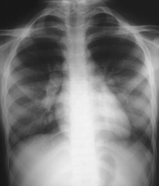
Chest radiograph showing bilateral hilar lymphadenopathy in a 10-year-old girl with sarcoidosis. |
 | View Full Size Image | |
Media type: X-RAY
|
| Media file 2:
Chest radiograph showing patchy diffuse pulmonary infiltrates involving both lung fields in a 12-year-old girl at onset of her sarcoidosis (left). A repeated study 6 months later showing almost complete resolution of the infiltrates (right). |
 | View Full Size Image | |
Media type: X-RAY
|
- Baculard A, Blanc N, Boule M, et al. Pulmonary sarcoidosis in children: a follow-up study. Eur Respir J. Apr 2001;17(4):628-35. [Medline]. [Full Text].
- Baumann RJ, Robertson WC. Neurosarcoid presents differently in children than in adults. Pediatrics. Dec 2003;112(6 Pt 1):e480-6. [Medline]. [Full Text].
- Bresnitz EA, Strom BL. Epidemiology of sarcoidosis. Epidemiol Rev. 1983;5:124-56. [Medline].
- Burton CM, Pressler T, Milman N. Pulmonary sarcoidosis in a child with cystic fibrosis. Pediatr Pulmonol. May 2005;39(5):473-7. [Medline].
- Dimitriades C, Shetty AK, Vehaskari M, et al. Membranous nephropathy associated with childhood sarcoidosis. Pediatr Nephrol. Jun 1999;13(5):444-7. [Medline].
- Fink CW, Cimaz R. Early onset sarcoidosis: not a benign disease. J Rheumatol. Jan 1997;24(1):174-7. [Medline].
- Gedalia A, Molina JF, Ellis GS Jr, et al. Low-dose methotrexate therapy for childhood sarcoidosis. J Pediatr. Jan 1997;130(1):25-9. [Medline].
- Gedalia A, Shetty AK, Ward K, et al. Abdominal aortic aneurysm associated with childhood sarcoidosis. J Rheumatol. Apr 1996;23(4):757-9. [Medline].
- Gedalia A, Shetty AK, Ward K. The diagnostic value of MRI in children with sarcoidosis presenting with fever of unknown origin. J Pedatr Orthop. 1997;17:460-462.
- Hafner R, Vogel P. Sarcoidosis of early onset. A challenge for the pediatric rheumatologist. Clin Exp Rheumatol. Nov-Dec 1993;11(6):685-91. [Medline].
- Hetherington S. Sarcoidosis in young children. Am J Dis Child. Jan 1982;136(1):13-5. [Medline].
- Heyer CM, Mueller KM, Seiffert P, et al. Pulmonary sarcoidosis in a 14-year-old boy diagnosed by low-dose CT-guided transthoracic lung biopsy. Pediatr Pulmonol. Mar 2006;41(3):269-74. [Medline].
- Heyer CM, Lemburg SP, Kagel T, et al. Evaluation of chronic infectious interstitial pulmonary disease in children by low-dose CT-guided transthoracic lung biopsy. Eur Radiol. Jul 2005;15(7):1289-95. [Medline].
- Hoffman AL, Milman N, Byg KE. Childhood sarcoidosis in Denmark 1979-1994: incidence, clinical features and laboratory results at presentation in 48 children. Acta Pediatr Scand. 2004;93:30-36.
- Hoover DL, Khan JA, Giangiacomo J. Pediatric ocular sarcoidosis. Surv Ophthalmol. Jan-Feb 1986;30(4):215-28. [Medline].
- James DG, Kendig EL Jr. Childhood sarcoidosis. Sarcoidosis. Mar 1988;5(1):57-9. [Medline].
- James DG, Neville E, Siltzbach LE. A worldwide review of sarcoidosis. Ann N Y Acad Sci. 1976;278:321-34. [Medline].
- Johns CJ, Michele TM. The clinical management of sarcoidosis. A 50-year experience at the Johns Hopkins Hospital. Medicine (Baltimore). Mar 1999;78(2):65-111. [Medline].
- Kendig EL, Brummer DL. The prognosis of sarcoidosis in children. Chest. Sep 1976;70(03):351-3. [Medline].
- Kendig EL Jr. The clinical picture of sarcoidosis in children. Pediatrics. Sep 1974;54(3):289-92. [Medline].
- Kunkel SL, Lukacs NW, Strieter RM, Chensue SW. Th1 and Th2 responses regulate experimental lung granuloma development. Sarcoidosis Vasc Diffuse Lung Dis. Sep 1996;13(2):120-8. [Medline].
- Lower EE, Baughman RP. The use of low dose methotrexate in refractory sarcoidosis. Am J Med Sci. Mar 1990;299(3):153-7. [Medline].
- Lower EE, Baughman RP. Prolonged use of methotrexate for sarcoidosis. Arch Intern Med. Apr 24 1995;155(8):846-51. [Medline].
- Mallory SB, Paller AS, Ginsburg BC, et al. Sarcoidosis in children: differentiation from juvenile rheumatoid arthritis. Pediatr Dermatol. Dec 1987;4(4):313-9. [Medline].
- Marcille R, McCarthy M, Barton JW, et al. Long-term outcome of pediatric sarcoidosis with emphasis on pulmonary status. Chest. Nov 1992;102(5):1444-9. [Medline].
- Newman LS, Rose CS, Maier LA. Sarcoidosis. N Engl J Med. Apr 24 1997;336(17):1224-34. [Medline].
- North AF Jr, Fink CW, Gibson WM, et al. Sarcoid arthritis in children. Am J Med. Apr 1970;48(4):449-55. [Medline].
- Pattishall EN, Strope GL, Spinola SM, Denny FW. Childhood sarcoidosis. J Pediatr. Feb 1986;108(2):169-77. [Medline].
- Pattishall EN, Kendig EL Jr. Sarcoidosis in children. Pediatr Pulmonol. Sep 1996;22(3):195-203. [Medline].
- Pattishall EN, Strope GL, Denny FW. Pulmonary function in children with sarcoidosis. Am Rev Respir Dis. Jan 1986;133(1):94-6. [Medline].
- Pinkston P, Bitterman PB, Crystal RG. Spontaneous release of interleukin-2 by lung T lymphocytes in active pulmonary sarcoidosis. N Engl J Med. Apr 7 1983;308(14):793-800. [Medline].
- Schurmann M, Bein G, Kirsten D, et al. HLA-DQB1 and HLA-DPB1 genotypes in familial sarcoidosis. Respir Med. Apr 1998;92(4):649-52. [Medline].
- Sharma OP. Vitamin D, calcium, and sarcoidosis. Chest. Feb 1996;109(2):535-9. [Medline].
- Sheffield EA. Pathology of sarcoidosis. Clin Chest Med. Dec 1997;18(4):741-54. [Medline].
- Shetty AK, Gedalia A. Sarcoidosis in children. Curr Probl Pediatr. May-Jun 2000;30(5):149-76. [Medline].
- Shetty AK, Gedalia A. Sarcoidosis: a pediatric perspective. Clin Pediatr (Phila). Dec 1998;37(12):707-17. [Medline].
- Shetty AK, Correa H, Galen W, Gedalia A. Pathological case of the month. Childhood sarcoidosis. Arch Pediatr Adolesc Med. Sep 1997;151(9):955-6. [Medline].
- Shetty AK, Zganjar BE, Ellis GS Jr, et al. Low-dose methotrexate in the treatment of severe juvenile rheumatoid arthritis and sarcoid iritis. J Pediatr Ophthalmol Strabismus. May-Jun 1999;36(3):125-8. [Medline].
- Singal A, Thami GP, Goraya JS. Scar sarcoidosis in childhood: case report and review of the literature. Clin Exp Dermatol. May 2005;30(3):244-6. [Medline].
- Weinberg S, Bennett H, Weinstock I. Central nervous system manifestations of sarcoidosis in children. Case report and review. Clin Pediatr (Phila). Jul 1983;22(7):477-81. [Medline].
- du Bois RM. Corticosteroids in sarcoidosis: friend or foe?. Eur Respir J. Jul 1994;7(7):1203-9. [Medline].
< |