Practice Essentials
Dyshidrotic eczema, also termed pompholyx, is a type of eczema (dermatitis). This skin condition is characterized by intensely itchy blisters that develop on the edges of the fingers, toes, palms, and soles of the feet. Dyshidrotic eczema may be acute, recurrent, or chronic, and it affects teenagers and adults. The clinical course of dyshidrotic eczema can range from self-limited to chronic, severe, or debilitating. The skin condition's unresponsiveness to treatment can be frustrating for the patient and physician.
Signs and symptoms of dyshidrotic eczema (pompholyx)
Signs and symptoms of dyshidrotic eczema (pompholyx) are as follows:
-
Symmetrical crops of clear vesicles and/or bullae (blisters)
-
Intensely purpuric (itchy)
-
Typically present on the palms and soles, as well as the lateral aspects of fingers and toes
-
Deep-seated vesicles with a tapiocalike appearance
-
May become large, form bullae, and become confluent
-
In chronic disease, fingernails may reveal dystrophic changes
-
Vesicles typically resolve without rupturing, followed by desquamation
Diagnosis of dyshidrotic eczema (pompholyx)
Diagnosis of dyshidrotic eczema (pompholyx) is as follows:
-
Typically a clinical diagnosis
-
Bacterial culture and sensitivity can rule out secondary infection
-
Patch testing to exclude allergic contact dermatitis
-
Recalcitrant cases warrant systemic evaluation
-
KOH wet mount to exclude dermatophyte infection
-
Punch biopsy for direct immunofluorescence to exclude bullous pemphigoid
Treatment of dyshidrotic eczema (pompholyx)
Treatments for dyshidrotic eczema (pompholyx) are as follows:
-
First-line treatment includes high-strength topical steroids and cold compresses; systemic steroids also used
-
Treatment for bullae (blisters): Compresses with Burow solution or 1:10.000 solution of potassium permanganate; drain large bullae with sterile syringe and leave roof intact; prescribe systemic antibiotics covering Staphylococcus aureus and group A streptococci
-
UVA or UVA-1 alone or with oral or topical psoralen
-
Topical calcineurin inhibitors
-
OnabotulinumtoxinA injections
-
For severe refractory pompholyx, azathioprine, methotrexate, mycophenolate mofetil, cyclosporine, or etanercept
-
Nickel chelators (eg, disulfiram) occasionally used in nickel-sensitive patients
-
Alitretinoin (9- cis retinoic acid)
-
Dietary avoidance of nickel and cobalt for nickel- and cobalt-sensitive patients
Background
Dyshidrotic eczema is a type of eczema (dermatitis) that is characterized by a pruritic vesicular eruption (bullae, or blisters) on the fingers, palms, and soles; typically these intensely itchy blisters develop on the edges of the fingers, toes, palms, and soles of the feet. This skin condition affects teenagers and adults and may be acute, recurrent, or chronic. A more appropriate term for this vesicular eruption is pompholyx, which means bubble. The clinical course of dyshidrotic eczema can range from self-limited to chronic, severe, or debilitating. The skin condition's unresponsiveness to treatment can be frustrating for the patient and physician (see the images below).
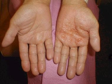 Dyshidrotic eczema (pompholyx). Tense vesicles and bullae on the palm. Courtesy of Norman Minars, MD, University of Miami, Department of Dermatology & Cutaneous Surgery.
Some believe the terms pompholyx and dyshidrosis are obsolete and favor a new term, such as "acute and recurrent vesicular hand dermatitis." The etiology of dyshidrotic eczema is unresolved and is believed to be multifactorial. Dyshidrotic eczema is considered to be a reaction pattern caused by various endogenous conditions and exogenous factors.
See All About Allergies: Be Ready for Spring, a Critical Images slideshow, to help identify a variety of allergens and symptoms.
Etiology
The hypothesis of sweat gland dysfunction has been refuted because vesicles have not been shown to be associated with sweat ducts. A 2009 case report provided clear histopathologic evidence that sweat glands do not play a role in dyshidrosis. [1] However, hyperhidrosis is an aggravating factor in 40% of patients with dyshidrotic eczema. Improvement in pruritus, erythema, vesicles, and hand dermatitis with fewer or no signs of relapse has been obtained after onabotulinumtoxinA injection. [2]
Dyshidrotic eczema may be associated with atopy and familial atopy. Of patients with dyshidrosis, 50% have atopic dermatitis.
Exogenous factors (eg, contact dermatitis to nickel, balsam, cobalt; sensitivity to ingested metals; dermatophyte infection; bacterial infection) may trigger episodes. These antigens may act as haptens with a specific affinity for palmoplantar proteins of the stratum lucidum of the epidermis. The binding of these haptens to tissue receptor sites may initiate pompholyx.
Evidence shows that the ingestion of metal ions such as cobalt can induce type I and type IV hypersensitivity reactions. In addition, they can also act as atypical haptens, activating T lymphocytes through human leukocyte antigen–independent pathways, causing systemic allergic dermatitis in the form of dyshidrotic eczema. [3, 4]
Emotional stress [5] and environmental factors (eg, seasonal changes, hot or cold temperatures, humidity) reportedly exacerbate dyshidrosis.
Dyshidrosislike eczematous eruptions with the use of intravenous immunoglobulin (IVIG) infusions have been reported. [6] A 2011 search of the literature identified pompholyx as one of the most important cutaneous adverse effects of IVIG, being present in 62.5% of the patients reported, with 75% of those patients developing the lesions after just one IVIG treatment. [7] The eruption tends to be mild and to wane over time. It usually responds very well to topical steroids, [8, 9, 10] but it may become recurrent and more aggressive after repeated doses of IVIG.
In some patients, a distant fungal infection can cause palmar pompholyx as an id reaction. In one study, one third of pompholyx occurrences on the palms resolved after treatment for tinea pedis. The factors believed to be associated with dyshidrotic eczema are discussed in more detail below.
Genetic factors
Monozygotic twins have been affected simultaneously by dyshidrotic eczema. The pompholyx gene has been mapped to band 18q22.1-18q22.3 in the autosomal dominant form of familial pompholyx. [11]
Mutations on the filaggrin gene leading to loss of filaggrin, a structural protein of the stratum corneum involved in the barrier function of the skin, causes dyskeratinization, increased transepidermal water loss, and an increase in the transepidermal antigen transfer. Combined, these features have been associated with the development of ichthyosis and atopic dermatitis, and they may be involved in the development of irritant and allergic contact dermatitis, which are well-known skin conditions associated with dyshidrotic eczema. Chronic hand dermatitis, including dyshidrotic eczema, has also been associated with defects in the skin barrier, and, in a few cases, it has been also associated with mutations in the filaggrin gene; however, these have not reached statistical significance. [12]
Aquaporins have been shown to be expressed in patients with atopic dermatitis and may also be related to exacerbation and chronicity of pompholyx. Aquaporins are channel proteins located on cell membranes that increase their permeability, in particular aquaglyceroporins. Aquaglyceroporins can transport water and glycerol. Aquaporin-3 and aquaporin-10 are normally expressed in the basal layer of the epidermis, and immunohistochemical staining had demonstrated their presence in all epidermal layers in patients with pompholyx. These channels may participate in the increase of transepidermal water loss seen in atopic dermatitis and possibly in pompholyx. Owing to osmotic gradients, among other factors, the direction of water and glycerol through aquaporins is from the skin to the environment, possibly contributing with skin dehydration, even immediately after hand washing. Hypothetically, topical and/or systemic inhibition of the expression of aquaporins in the epidermis could contribute with the preservation of water and glycerol, decreasing the frequency and severity of pompholyx exacerbations. [13]
Atopy
As many as 50% of patients with dyshidrotic eczema have reportedly had personal or familial atopic diathesis (eczema, asthma, hay fever, allergic sinusitis). The serum immunoglobulin E (IgE) level frequently is increased, even in patients who do not report a personal or familial history of atopy. Occasionally, dyshidrotic eczema is the first manifestation of an atopic diathesis.
Nickel sensitivity
This may be a significant factor in dyshidrotic eczema. Nickel sensitivity was reportedly low in some studies of dyshidrosis patients, but significantly elevated in other studies. Increased nickel excretion in the urine has been reported during exacerbations of pompholyx. Ingested metals have been found to provoke exacerbations of pompholyx in some patients.
Low-nickel diets have reportedly decreased the frequency and severity of pompholyx flares. A high palmoplantar perspiration rate has been suggested to result in a local concentration of metal salts that may provoke the vesicular reaction. Contact allergy has been documented in 30% of patients with dyshidrotic eczema.
Cobalt sensitivity
The oral ingestion of cobalt manifests systemic allergic dermatitis as dyshidrotic eczema less frequently than does the oral ingestion of nickel. Much more common is the simultaneous occurrence of nickel and cobalt allergy seen in 25% of nickel-sensitive patients developing pompholyx. In these cases, the eczema is usually more severe. When suspected as the cause of the dyshidrotic eczema, high oral ingestion of cobalt should be taken in consideration, regardless of the patch test results. [3]
A point-based, low-cobalt diet has been proposed to help patients limit cobalt ingestion and to keep the serum level below the threshold for developing flares, which is approximately less than 12 mcg/d. This diet has demonstrated higher compliance than an avoidance diet list. In addition, this diet reduces the amount of nickel consumed. [3]
Exposure to sensitizing chemicals or metals
Dyshidrotic eczema outbreaks are sometimes associated with exposure to sensitizing chemicals or metals (eg, chromium, cobalt, carba mix, fragrance mix, diaminodiphenylmethane, dichromates, benzoisothiazolones, paraphenylenediamine, perfumes, fragrances, balsam of Peru, Primula plant).
Id reaction
Controversy surrounds the possible existence of an id reaction, which is considered to be a distant dermatophyte infection (tinea pedis, kerion of scalp) triggering a palmar pompholyx reaction (also termed pompholyx dermatophytid).
Fungal infection
Pompholyx occasionally resolves when a tinea pedis infection is treated, then relapses when the fungal infection recurs, supporting the existence of this reaction pattern. Of patients who have a vesicular reaction to intradermal trichophytin testing, less than one third have experienced a resolution of pompholyx after treatment with antifungal agents.
Emotional stress
This is a possible factor in dyshidrotic eczema. Many patients report recurrences of pompholyx during stressful periods. Improvement of dyshidrotic eczema using biofeedback techniques for stress reduction supports this hypothesis.
Other factors
Isolated reports describe other possible causative factors, such as aspirin ingestion, oral contraceptives, cigarette smoking, and implanted metals, among others. A 3-year prospective study of the causes of dyshidrotic eczema (pompholyx) in 120 patients found causes of pompholyx related to contact exposure (67.5%), including to cosmetic products (31.7%) and metals (16.7%); interdigital-plantar intertrigo (10%); and internal factors (6.7%), with an additional 15% of patients having undiagnosed (idiopathic) causes probably related to atopic factors. [14]
Contact allergy was found in 89 (74.2%) of the 120 patients. The most frequent allergens were nickel, shower gel, chromium, fragrance, shampoo, and balsam of Peru. Less frequent allergens were lanolin, cobalt, thiuram, lauryl sulfate, fresh tobacco, p -phenylenediamine (PPD), formaldehyde, parabens, and octyl gallate. In 97 of 193 positive patch test results, correlation existed between the application of the agent and pompholyx recurrence. The relevance of the analysis was confirmed in 81 (67.5%) of the 120 patients. In summary, the most frequent causes of pompholyx related to contact with substances were hygiene product intolerance (46.7%), metal allergy (25%), and others (28.3%).
Intertrigo occurred in 19 (15.8%) of the 120 patients. Of those individuals, 80% presented with dermatophytosis and 20% presented with candidiasis. After 3 weeks of antifungal therapy, 13 of 19 patients remained asymptomatic of pompholyx.
With regard to internal causes, 30 patients presented with a positive patch test result for metals, but only 2 presented with exacerbations of the lesions after a challenge test.
Of 58 patients with a history of smoking tobacco, 5 presented with a positive reaction on a tobacco patch test, and 2 of those were considered relevant. Drug allergy was determined to be the causative agent in 3 patients (amoxicillin in 2 and intravenous immunoglobulin in 1). Food-related pompholyx was detected in 4 patients, and, after a challenge test, reactivation occurred in 3 of these patients (2 for paprika and 1 for orange juice).
Ultraviolet A light
In a case series, 5 patients with prior diagnosis of pompholyx developed lesions morphologically and histologically consistent with a vesicular dermatitis after provocation with long-wavelength ultraviolet A (UVA) light. Further workup ruled out contact dermatitis, polymorphic light eruption, and heat as the culprit, confirming that the reaction was due to true photosensitivity rather than to photoaggravation. [15]
Pompholyx caused by UVA exposure may possibly be considered a variation of seasonal (summer) pompholyx. In the United States, dyshidrotic eczema is more commonly seen in warmer climates and during the spring and summer months. A study in Turkey also revealed a higher prevalence of dyshidrotic eczema in the summer months. [16]
In a case series, three patients with histories of frequent pompholyx exacerbations, mostly during summer (ie, photoaggavated pompholyx), were subjected to photoprovocation testing, with positive development of pompholyx lesions in two of them. Patients reported lesions after exposure to solar-simulated ultraviolet radiation and broadband UVA. They were treated with photoprotective measures in addition to the standard treatment for pompholyx, resulting in a decrease in the frequency and severity of the exacerbations. These authors suggested that the skin condition may be underdiagnosed and recommended recognition and early detection to institute sun protection as soon as possible and to avoid starting phototherapy or photochemotherapy in this particular subset of pompholyx patients. [17]
Of interest, UVB phototherapy and photochemotherapy are well-known, efficient treatments for pompholyx.
Epidemiology
Occurrence in the United States
Dyshidrotic eczema occurs in 5-20% of patients with hand eczema and more commonly develops in warmer climates and during spring and summer months (seasonal or summer pompholyx).
International occurrence
Dyshidrotic eczema accounted for 1% of initial consultations in a 1-year Swedish study. In a study of 107,206 Swedish individuals, 51 (0.05%) were diagnosed with dyshidrosis. Of all hand dermatitis cases in that population, 3% had dyshidrosis. [18]
In a retrospective study reviewing records of 714 Portuguese patients during a 6-year period, Magina et al found dyshidrotic eczema to be the third most common type of hand dermatitis (20.3%). [19]
Sex- and age-related demographics
The male-to-female ratio for dyshidrotic eczema has variably been reported as 1:1 and 1:2. Dyshidrotic eczema affects individuals aged 4-76 years; the mean age is 38 years. The peak incidence of the skin condition occurs in patients aged 20-40 years. After middle age, the frequency of dyshidrotic eczema episodes tends to decrease.
Prognosis of Dyshidrotic Eczema
Dyshidrotic eczema follows a chronic, intermittent course, with fewer episodes occurring after middle age. Some mildly affected patients experience spontaneous resolution within 2-3 weeks. (See Treatment and Medication.)
Patient Education
Inform individuals with this skin condition about the difficulty of achieving successful treatment.
Inform patients that the typical first-line treatments for the blisters of dyshidrotic eczema include high-strength topical steroids and cold compresses.
Additionally, stress reduction, including possibly consultation with a mental health professional, has been helpful for some patients. This may include biofeedback therapy.
Bed rest may be necessary if patients develop large blisters on the feet. Work activities or activities of daily living may be hampered by blisters on the hands.
Instruct nickel- or cobalt-sensitive dyshidrotic eczema patients to avoid contact with these allergens, which may include avoiding certain activities, foods, and beverages.
A hand care regimen, including moisturizers and emollients may be helpful.
For patient education information, see the Skin Conditions and Beauty Center, as well as Eczema (Atopic Dermatitis). (See Treatment and Medication.)
-
Dyshidrotic eczema (pompholyx). Tense vesicles and bullae on the palm. Courtesy of Norman Minars, MD, University of Miami, Department of Dermatology & Cutaneous Surgery.
-
Dyshidrotic eczema (pompholyx). Close-up view of tense vesicles and bullae of the palm. Courtesy of Norman Minars, MD, University of Miami, Department of Dermatology & Cutaneous Surgery.
-
Dyshidrotic eczema (pompholyx). Discrete yellow pustules on the sole of the foot. Courtesy of Norman Minars, MD, University of Miami, Department of Dermatology & Cutaneous Surgery.
-
Dyshidrotic eczema (pompholyx). Multiple tense vesicles on the palm.
-
Dyshidrotic eczema (pompholyx). Small tense vesicles on the fingers.
-
Dyshidrotic eczema (pompholyx). Small, discrete, coalesced vesicles on the dorsal hand.
-
Dyshidrotic eczema (pompholyx). Small, discrete, coalesced vesicles on the fingers.
-
Dyshidrotic eczema (pompholyx). Palms and soles of a patient with a dyshidrosis flare. The patient unroofed a large bulla on the right sole.
-
Dyshidrotic eczema (pompholyx). Small discrete vesicles of the lateral fingers.