Practice Essentials
Osteosarcoma is the third most common cancer in adolescence–only lymphomas and brain tumors occur more frequently. The lesion is characterized by the production of osteoid and is thought to arise from a primitive mesenchymal bone-forming cell.
The mainstay of therapy is surgical removal of the lesion. Using modern techniques, limb-sparing procedures can often preserve function of the affected limb. Systemic treatment using chemotherapy is used to treat micrometastatic disease, which is present but not detectable in most patients at diagnosis.
No preventive measures for childhood cancers are known.
The following image is a plain radiograph typical of osteosarcoma at the time of diagnosis.
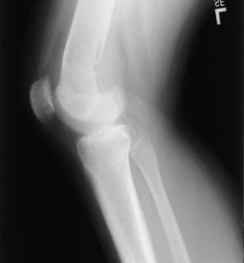 Lateral plain radiograph of the knee reveals an osteosarcoma of the distal femur. The lesion is mainly posterior, with disruption and elevation of the periosteum (Codman triangle), and extends beyond the bone into the soft tissue.
Pathophysiology
Osteosarcoma can occur in any bone. It most commonly occurs in the long bones of the extremities near metaphyseal growth plates. The most common sites include the femur (42%), with 75% of those tumor arising in the distal femur; tibia (19%), with 80% of tumors in the proximal tibia; and humerus (10%), with 90% of tumors in the proximal humerus. [1] Other locations of note include the skull or jaw (8%) and pelvis (8%).
Any sarcoma that arises from bone is technically called an osteogenic sarcoma. Therefore, this term includes fibrosarcoma, chondrosarcoma, and osteosarcoma, all named for their morphologic characteristics. The focus of this article is osteosarcoma. Numerous variants of osteosarcoma are known and include conventional types (ie, osteoblastic, chondroblastic, fibroblastic types) and telangiectatic, multifocal, parosteal, and periosteal types.
Etiology
The exact cause of osteosarcoma is unknown. However, numerous risk factors are known.
-
Rapid bone growth appears to predispose patients to osteosarcoma, as suggested by the increased incidence during the adolescent growth spurt, [2] the high incidence among large dogs (eg, Great Danes, St Bernards, German shepherds), and the typical location of osteosarcomas near the metaphyseal growth plate of long bones.
-
Exposure to radiation is the only known environmental risk factor.
-
There appears to be a cluster of tumor suppressor genes on chromosome 3. [3]
-
A genetic predisposition may be present. [4, 5]
Retinoblastoma, especially the combination of a constitutional mutation of the RB gene (germline retinoblastoma) with radiation therapy, is associated with a particularly high risk of osteosarcoma development. Of note, the genetic locus retinoblastoma at band 13q14 has also been implicated in the pathogenesis of sporadic osteosarcoma.
Bone dysplasias, including Paget disease, fibrous dysplasia, enchondromatosis, and hereditary multiple exostoses, increase the risk for osteosarcoma.
Li-Fraumeni syndrome (germline TP53 mutation) is a predisposing factor for osteosarcoma.
Rothmund-Thomson syndrome (ie, autosomal recessive association of congenital bone defects, hair and skin dysplasias, hypogonadism, cataracts) is associated with an increased risk of osteosarcoma.
Epidemiology
United States data
The incidence is 400 cases per year (4.8 cases per million persons < 20 y). [6]
Race-, sex-, and age-related demographics
The incidence is slightly higher in African Americans than in Caucasians (data from the National Cancer Institute [NCI] Surveillance, Epidemiology, and End Results [SEER] Study Pediatric Monograph, 1975-1995). [1] In African Americans, the annual incidence is 5.2 cases per million population younger than 20 years. In Caucasians, the annual incidence is 4.6 cases per million population younger than 20 years.
The incidence is slightly higher in male individuals than in female individuals. In male individuals, the incidence is 5.2 cases per million population per year. In female individuals, the incidence is 4.5 cases per million population per year. The male to female ratio is 1.4:1. [7]
The incidence of osteosarcoma increases steadily with age; a relatively dramatic increase in adolescence corresponds with the growth spurt. Osteosarcoma is rarely diagnosed in patients younger than 5 years (about 1% of cases). [8]
In children aged 5-9 years, the annual incidence is 2.6 cases for African Americans and 2.1 cases for Caucasians per million population. In children aged 10-14 years, the annual incidence is 8.3 cases for African Americans and 7 cases for Caucasians per million population. In adolescents aged 15-19 years, the annual incidence is 8.9 cases for African Americans and 8.2 cases for Caucasians per million population.
Patients whose disease is diagnosed during their growth spurt are taller than average, although patients identified in adulthood have average height.
Prognosis
Patients with the periosteal type of osteosarcoma have a more favorable outcome. In an analysis of 119 patients, the overall survival was 83% at 10 years. [9]
The prognosis for patients with conventional high-grade osteosarcoma primarily depends on whether metastases are detectable at diagnosis. Patients who present with metastases or with multifocal disease have a poor prognosis, with long-term survival rates of less than 25%.
For patients with initially localized disease, the prognosis depends mainly on 2 variables: resectability and the response to chemotherapy. Those who have completely resectable disease and those whose tumors have an excellent histologic response to neoadjuvant chemotherapy have the best likelihood for a cure.
Before the 1970s, the 5-year survival rate of patients with nonmetastatic osteosarcoma was less than 20%, even with aggressive surgery (mostly amputations).
The fact that most relapses occurred at metastatic sites (primarily the lung) attests to the fact that most patients have undetectable metastatic disease at diagnosis (ie, micrometastatic disease).
With the introduction of postoperative (adjuvant) chemotherapy, survival rates began to improve. According to data from the NCI SEER program, the 5-year survival rate from 1975-1984 was 49% and from 1985-1994 was 63%. [1] For the latter period, female patients fared slightly better than male patients (5-year survival rates of 70% vs 59%). In a small dataset of patients younger than 5 years, outcome appeared to be similar to that of older patients. [8]
Results of the most recent cooperative group trial conducted by the Children's Oncology Group suggest that the addition of ifosfamide to standard 3 drug regimen was not helpful, but that the addition of the immune-enhancing drug muramyl tripeptide increased 6-year overall survival from 70% to 78% for localized disease. [10, 11] The use of MTP-PE requires further investigation before becoming standard therapy.
Surgical resection of recurrent disease can achieve cure in about 25% of patients. [12]
In a cohort study of 733 long-term (>5 y) survivors of osteosarcoma, Nagarajan et al reported overall survival of 88.6% at 20 years. Of interest in this group was the incidence of second malignancy (5.4%), those who reported at least 1 chronic medical condition (86.9%), and those who reported activity limitations (29.1%). The cohort includes a larger number of patients with amputations than would be seen in recently treated patients. [13]
Improving the survival rate and functional outcome and minimizing the short-term and long-term adverse effects remain goals of clinical trials for osteosarcoma.
The major challenge is curing patients with unresectable metastatic disease. Strategies currently under consideration include dose intensification (eg, anthracycline dose-escalation facilitated by dexrazoxane cardioprotection), immune modulators, monoclonal antibodies targeting tumor-cell antigens (eg, Her2/neu), and antiangiogenic agents that target components of the tumor vascular supply. High-dose administration of the bone-seeking radioisotope samarium is also under investigation (with autologous stem-cell support) for safety and efficacy in metastatic or nonresectable osteosarcoma limited to bone.
Finally, the role of the emerging field of oncolytic viruses for the treatment of osteosarcoma is currently being explored (ClinicalTrials.gov, NCT00503295 and NCT00931931).
Morbidity/mortality
The overall 5-year survival rate for patients whose condition was diagnosed between 1974 and 1994 was 63% (59% for male patients, 70% for female patients).
Complications
The following complications have been reported:
-
Cardiomyopathy is primarily a result of anthracycline (doxorubicin) use. Patients should receive routine follow-up echocardiography studies after they complete therapy and periodically long-term
-
Reports have included vascular inflammation, dyslipidemia, and early atherogenesis. [14]
-
Secondary malignant neoplasms may arise as a result of chemotherapy, particularly with alkylating agents.
-
Infertility is a nearly universal effect of the high-dose alkylating agents used to treat osteosarcoma.
-
Hearing loss is common due to cisplatin and should be monitored carefully during treatment. [15]
Patient Education
Chemotherapy
Parents and patients (if appropriate) must undergo formal education about chemotherapy to learn about the adverse effects of their medications. They must know what is expected to happen as a result of the therapy, and they should encouraged to call with any questions.
Central venous catheters
When patients have central venous catheters that exit the skin (eg, Hickman or Broviac catheters), the patient or the parents must learn to properly care for the line. This care usually involves daily heparin flushes.
Patients must also know their limitations (eg, restriction from swimming).
Patients with subcutaneous catheters (eg, Mediport catheters) do not need to perform daily-care routines, but they should learn to apply a topical anesthetic (eg, lidocaine-prilocaine [EMLA] cream) at least 1 hour before an anticipated needle stick.
-
Lateral plain radiograph of the knee reveals an osteosarcoma of the distal femur. The lesion is mainly posterior, with disruption and elevation of the periosteum (Codman triangle), and extends beyond the bone into the soft tissue.
-
Anteroposterior plain radiograph of the same with distal femoral osteosarcoma as shown in image above. The osteolytic lesion is apparent on the right side of the image.
-
MRI of the same distal femoral osteosarcoma as in images shown under the plain radiography section above; the uninvolved side is shown for comparison.
-
Close-up MRI of the same distal femoral osteosarcoma as shown in image above, and in images shown under the plain radiography section above.