Background
Eales disease is an idiopathic obliterative vasculopathy that usually involves the peripheral retina of young adults. In 1880, Henry Eales first described it in healthy young men with abnormal retinal veins and recurrent vitreal hemorrhages.
Clinical findings in Eales disease are characterized by avascular areas in the retina periphery, followed posteriorly by microaneurysms, dilatation of capillary channels, tortuosity of neighboring vessels, and spontaneous chorioretinal scars. It is a diagnosis of exclusion, as many other retinal disorders can mimic Eales disease, especially conditions of retinal inflammation or neovascularization. Note the clinical image below.
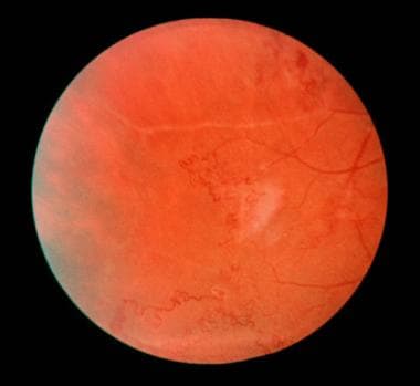 Eales disease. Fundus photo of the peripheral retina, revealing vascular tortuosity and peripheral retinal neovascularization.
Pathophysiology
Eales disease is believed to be a primary, noninflammatory disorder of the walls of peripheral retinal vessels, namely the shunt vessels. This often leads to vascular occlusions, peripheral neovascularization, and vitreous hemorrhage. The microvascular abnormalities are seen at the junction of perfused and nonperfused zones of the retina. Associations with tuberculosis and multiple sclerosis have been suggested, but the pathophysiology of Eales disease is unclear. Studies relating the association of Eales disease with sensitivity to tuberculin protein suggests that this disease may be associated with immunologic phenomena after Mycobacterium tuberculosis inoculation. [1, 2, 3]
Epidemiology
Frequency
United States
Eales disease is uncommon, with most reports based upon a series of cases.
International
Eales disease is most commonly seen in India and portions of the Middle East.
Mortality/Morbidity
No known mortality is associated with Eales disease. Visual compromise is seen in patients with recurrent vitreous hemorrhage; however, the visual acuity resolves to better than 20/200 in greater than 70% of patients. If retinal nonperfusion extends into the macula, the visual acuity usually is worse than 20/400.
Race
No racial predilection is known in Eales disease; however, the disease is more prevalent in India and portions of the Middle East.
Sex
Young adult males have been reported to have an increased prevalence of Eales disease; however, a study of 55 patients by Gieser and Murphy found that men and women are affected equally. [4]
Age
Peak age of onset for Eales disease is 20-35 years, with a reported range of 13-63 years.
Prognosis
More than 90% of patients with Eales disease can be brought to a morphological standstill with unchanging visual acuity.
Gieser and Murphy reported that 67% of patients had a final visual acuity in the better eye of 20/40 or better, 24% ranged from 20/50 to 20/200, and 9% were worse than 20/250. [4]
In another study from India, 72% of eyes that underwent vitrectomy for complications of Eales disease maintained a vision of 20/200 or better over 5 years of follow-up care. [5]
Patient Education
Patients with Eales disease should be educated to report visual symptoms of floaters or decreased vision to their ophthalmologist as soon as possible, in order to implement effective treatment and to prevent the need for vitrectomy surgery for vitreous hemorrhage or retinal detachment.
-
Eales disease. Fundus photo of the peripheral retina, revealing vascular tortuosity and peripheral retinal neovascularization.
-
Eales disease. Fluorescein angiogram of late leakage from peripheral retinal neovascularization.


