Background
Preseptal cellulitis (periorbital cellulitis) is a common infection of the eyelid and periorbital soft tissues that is characterized by acute eyelid erythema and edema. Preseptal cellulitis may be caused by bacteria, viruses, fungi, or helminths. Bacterial infections may result from local spread of an adjacent sinusitis or dacryocystitis, from external ocular infection, or from trauma to the eyelids.
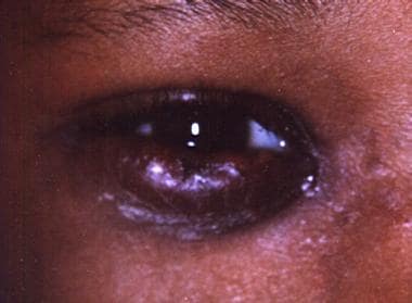 Preseptal cellulitis. This image shows an 8-year-old patient who presented with unilateral eyelid swelling and erythema.
Preseptal cellulitis tends to be a less severe disease than orbital cellulitis (postseptal cellulitis), which can have a similar initial presentation. Preseptal cellulitis differs from orbital cellulitis in that it is confined to the soft tissues that are anterior to the orbital septum.
Preseptal and orbital cellulitis exist on an anatomic continuum. Preseptal cellulitis can spread posterior to the septum, progressing to form subperiosteal and orbital abscesses. Infection in the orbit can spread posteriorly and cause cavernous sinus thrombosis or meningitis.
Orbital cellulitis has a higher morbidity, requires aggressive treatment, and may require surgical intervention, whereas preseptal cellulitis usually is managed medically. Delineation of the exact location of inflammation is necessary for proper diagnosis and treatment. Pain or limited eye movement, chemosis, afferent pupil, or resistance to retropulsion indicates orbital extension of the infection. However, preseptal cellulitis is much more common than orbital cellulitis, and both types of cellulitis are more common in children than in adults. [1]
Orbital septum
Periorbital inflammation is classified by location and severity. One of the major anatomic landmarks in determining the location of disease is the orbital septum. The orbital septum is a thin membrane that originates from the orbital periosteum and inserts into the anterior surfaces of the tarsal plates of the eyelids. The septum separates the superficial eyelid from the deeper orbital structures, and it forms a barrier that prevents infection in the eyelid from extending into the orbit.
Patient education
Patients should be instructed that loss of vision or pain with eye movements is an indication that the infection has spread to the orbit and may necessitate surgical intervention. Increased edema, redness, or localized pain also are warning signs.
Etiology
Direct inoculation and spread from adjacent tissues can cause preseptal cellulitis. Upper respiratory tract infections, especially paranasal sinusitis, commonly precede orbital cellulitis and some cases of preseptal cellulitis. In 2 large case series, nearly two thirds of cases of cellulitis were associated with upper respiratory tract infection. One half of these cases were from sinusitis. It also is relevant to note that some cases have been associated with dental abscess as a risk factor as this also may present with a risk for adjacent spread. [2]
The most common organisms are Staphylococcus aureus, Staphylococcus epidermidis,Streptococcus species, and anaerobes, reflecting the organisms that commonly cause upper respiratory infections and external eyelid infections. Blood and skin culture results tend to be negative.
Prior to the introduction of the Haemophilus influenzae type b (Hib) polysaccharide vaccine in 1985, H influenzae was the most common organism isolated in blood cultures. [3, 4, 5, 6] One study prior to the introduction of the vaccine noted that blood culture results were more likely to be positive (42%) if the patient had an upper respiratory infection and that subcutaneous aspirates were more likely to be positive (44%) if the patient had eyelid trauma or external ocular infection.
Since the vaccine came into widespread use, the rate of Haemophilus -positive blood cultures has dropped; studies have reported that the rate of any positive blood culture now is less than 4%.
A study specifically looking at periorbital and orbital cellulitis since the advent of the vaccine likewise found that the rates of Hib-related cellulitis dropped, from 11.7% to 3.5%. Total cases per year from all pathogens also declined, suggesting that H influenzae may have played a facilitative role in the pathogenesis of cellulitis.
Periorbital cellulitis also has been reported with anthrax and smallpox. [7, 8, 9]
Risk factors
Antecedent events in preseptal cellulitis may include the following recent eyelid lesions:
-
Bug bites [10]
-
Lesions caused by a recent surgical procedure near the eyelids [11]
-
Lesions caused by oral procedures
-
Dacryocystitis
An upper respiratory tract infection, especially sinusitis, [10] may be present concurrently with preseptal cellulitis or may recently have occurred. Many systemic diseases have been reported with concurrent preseptal cellulitis, including the following:
-
Varicella
-
Adenovirus
-
Herpes simplex virus
-
Asthma
-
Nasal polyposis
-
Neutropenia [12]
Epidemiology
According to the National Center for Disease Statistics in 1995, approximately 5000 inpatients in the United States had a primary discharge diagnosis of deep inflammation of the eyelid, as specified in the International Classification of Diseases, 9th revision (ICD-9).
Preseptal cellulitis primarily is a pediatric disease, with approximately 80% of patients being younger than 10 years and most patients being younger than 5 years. Patients with preseptal cellulitis tend to be younger than patients with orbital cellulitis. [1]
Prognosis
If preseptal cellulitis is identified and treated promptly, the prognosis for complete recovery without complication is excellent.
Morbidity occurs from the spread of pathogens to the orbit, which can threaten vision and result in central nervous system (CNS) spread. Untreated orbital cellulitis can lead to the development of an orbital abscess or can spread posteriorly to cause cavernous sinus thrombosis. Systemic spread of bacteria may lead to meningitis and sepsis.
In a study of pediatric patients with intracranial infection, high-risk features included the following [13] :
-
Ocular trauma
-
Age older than 7 years
-
Subperiosteal abscess
-
Headache [14] and fever persisting despite the use of intravenous (IV) antibiotics
Patients who are immunocompromised or diabetic have a higher likelihood of developing fungal infections, which can rapidly become fatal. Aggressive management, including imaging studies of the brain and early IV therapy, along with a high index of suspicion, is indicated for these patients. Otolaryngologist consultation also should be ordered if fungal infection is suspected.
Further, infection with β-hemolytic Streptococcus could lead to negrotizing fasciitis, though this is a rare complication.
-
This image shows a 10-year-old child who presented with fever, acute unilateral eyelid erythema, and limited extraocular motions. The presentation is suspicious for orbital cellulitis.
-
Preseptal cellulitis. This image shows an 8-year-old patient who presented with unilateral eyelid swelling and erythema.
