Continually Updated Clinical Reference
 
 
  All Sources     eMedicine     Medscape     Drug Reference     MEDLINE
 
eMedicine - Optic Neuropathy, Anterior Ischemic : Article by

Quick Find
Authors & Editors
Introduction
Clinical
Differentials
Workup
Treatment
Medication
Follow-up
Miscellaneous
Multimedia
References

Related Articles
Central Retinal Vein Occlusion

Ocular Hypotony

Papilledema

Pseudopapilledema


Patient Education
Click here for patient education.


Author: Brian R Younge, MD, Professor of Ophthalmology, Mayo Clinic School of Medicine

Brian R Younge is a member of the following medical societies: American Medical Association, American Ophthalmological Society, and North American Neuro-Ophthalmology Society

Editors: Edsel Ing, MD, FRCSC, Assistant Professor, Department of Ophthalmology & Vision Sciences, University of Toronto, Sunnybrook and Women's Health Sciences Center, Toronto East General Hospital; Simon K Law, MD, PharmD, Assistant Professor of Ophthalmology, Jules Stein Eye Institute; Chief of Section of Ophthalmology Surgical Services, Department of Veterans Affairs Healthcare Center, West Los Angeles; Lance L Brown, OD, MD, Ophthalmologist, Affiliated With Freeman Hospital and St John's Hospital, Regional Eye Center, Joplin, Missouri; Hampton Roy Sr, MD, Associate Clinical Professor, Department of Ophthalmology, University of Arkansas for Medical Sciences

Author and Editor Disclosure

Synonyms and related keywords: anterior ischemic optic neuropathy, AION, nonarteritic anterior ischemic optic neuropathy, NAION, arteritic anterior ischemic optic neuropathy, ischemic optic neuropathy, ION, giant cell arteritis, optic atrophy, optic nerve, optic disc

Background

Field defects typical of ischemic optic neuropathy were probably first described by Knapp in 1875. Miller and Smith first used the term ischemic optic neuropathy in 1966, and Hayreh later added the term anterior. In 1924, Uhthoff first described severe visual loss, with field defects and swollen optic discs.1

Anterior ischemic optic neuropathy (AION) is the most common cause of acute optic neuropathy in older age groups. It can be nonarteritic (nonarteritic anterior ischemic optic neuropathy [NAION]) or arteritic, the latter being associated with giant cell arteritis. It is characterized by visual loss associated with optic disc swelling of a pallid nature, sometimes with flame hemorrhages on the swollen disc or nearby neuroretinal layer, and sometimes with nearby cotton-wool exudates. Visual loss is usually sudden, or over a few days at most, and it is usually permanent, with some recovery possibly occurring within the first weeks or months. Optic atrophy of varying degrees ensues within the next few weeks, and it is usually generalized but may be sectorial (NAION).

Although the pathophysiology differs in the arteritic form of AION, ischemia is the end result, as the results on vision are the same. According to Rucker, temporal arteritis probably was first described very early by Ali Ibn Isa (AD 940-1010).2 In modern times (1890), Hutchinson described a disease of this nature, and, in 1932, Horton and colleagues at the Mayo Clinic used the term temporal arteritis.3, 4 To better describe the histologic features, Gilmour suggested the term giant cell arteritis in 1941. Treatment with steroids was started at the Mayo Clinic in 1949.
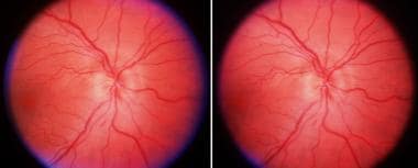
Pathophysiology

AION is thought to be an ischemic process affecting the posterior circulation of the globe, principally vessels (ie, short posterior ciliary arteries) supplying the optic nerve at its exit from the eye. Only glial cells support the optic disc at this site, and it is the only site in which swelling can occur. More posterior ischemia results in a similar condition, without visible swelling, and is termed posterior ischemic optic neuropathy. Early observations of optic disc photographs suggested that patients with small discs having smaller or nonexistent cups have an anatomical predisposition for NAION. As an ischemic episode evolves, the swelling compromises circulation, with a spiral of ischemia resulting in further neuronal damage. The ischemic spiral is less implicated in the arteritic type of AION, in which the entire ophthalmic arterial circulation to the eye and orbit may be compromised.

Frequency

United States

Patients with both arteritic and nonarteritic forms of AION are usually older than 50 years, with females predominating in the arteritic group. The incidence of the nonarteritic type is 2.3-10.3 per 100,000 in the United States, and, for the arteritic type, it is 0.36 per 100,000. In the arteritic group, incidence, like that of giant cell arteritis, increases almost exponentially with advanced age. The literature seems to support the notion that whites are affected more commonly than blacks in the nonarteritic group, and people of Scandinavian or European ancestry are the most commonly affected ethnic group in the arteritic type.

International

In the nonarteritic group, incidence is higher in whites and uncommon in other races.

The countries with the highest incidence of arteritic AION are the Scandinavian countries (ie, Norway, Denmark, Sweden), followed by Germany. The arteritic form is not as well recognized in non-whites. Recent genetic evidence may help to explain this incidence.

Mortality/Morbidity

NAION is not commonly associated with life-threatening conditions, although the presence of other vascular conditions is frequent (eg, hypertension, 46.9%; diabetes, 23.9%; myocardial infarction, 11%). The role of smoking in this disease is unclear. By contrast, the arteritic form of AION is associated with a more morbid condition, giant cell (cranial) arteritis. This condition affects many organs of the body, and the incidence of death associated with it is higher than that of the general population. Late-onset abdominal aortic aneurysms contribute to the morbidity and mortality of giant cell arteritis.

Bilateral visual loss is more common in the arteritic form of the disease, especially if treatment is delayed, and approximates 50% in some earlier series. By contrast, bilateral visual loss may be seen in 12-19% of NAION, and it usually occurs sequentially instead of simultaneously.

Progressive visual loss in the contralateral eye can occur in either form of AION, despite steroids, anticoagulation, or hyperbaric oxygen. Progressive visual loss is less common and usually stabilizes in a few days.

Race

NAION is most common in whites (95%); it is less common in African Americans (2%), Asians (3%), and Hispanics (1%). The arteritic form of the disease is predominantly described among whites of European descent, particularly Scandinavian and German.

It was a past misconception that African American patients did not succumb to giant cell arteritis. However, numerous documented cases of giant cell arteritis in blacks exist; giant cell arteritis in African American patients is not uncommon.

Sex

Females dominate the incidence in both forms of AION, but only slightly in the nonarteritic form (1.2:1), as compared to the arteritic type (2:1).

Age

Both disorders are found in older age groups. In the nonarteritic group, age ranges from the late 40s and older. The arteritic group almost always is older than 50 years, with an exponential increase with advanced age (90% of patients are >60 y). Rare cases of AION occur before 40 years, and the differentiation from optic neuritis associated with demyelinating disease is important in this crossover age group.


History

  • Visual loss is painless in at least 90% of patients with NAION. The vision loss is noticed upon awakening, perhaps due to nocturnal hypotension.
  • Patients with arteritic AION often have symptoms other than visual loss, such as malaise, headache, scalp tenderness and tender temporal arteries, jaw pain on mastication (jaw claudication), generalized muscle aches, and swelling.
  • The earlier manifestations of the disease include malaise, weight loss, fever, vague abdominal or GI pains, and anorexia.
  • Late manifestations, often years later, include a much higher incidence of abdominal aortic aneurysm.

Physical

  • Nonarteritic anterior ischemic optic neuropathy
    • NAION has typical findings of visual loss and field loss in an otherwise asymptomatic individual.
    • A small cup disc ratio is usually noted. Initially, the optic disc is swollen and pale, often in a generalized or diffuse manner.
    • Sectorial disc edema, especially of the superior disc, is classic.
    • Visual loss with NAION usually is not as severe as with arteritic AION, but vision loss as severe as no light perception has been described.
  • Arteritic anterior ischemic optic neuropathy
    • In patients with arteritic AION, the disc is classically described as chalky white, pale, and swollen.
    • Ischemia in multiple vascular territories is not uncommon (eg, central retinal artery occlusion, choroidal infarction, anterior segment ischemia, extraocular muscle ischemia causing diplopia).
    • The temporal arteries may be quite prominent, ropey, and tender.
    • Oral, tongue, or even scalp ulcers rarely may be seen.

Causes

AION is an ischemic disease, but the cause is yet to be found definitively. In the nonarteritic form, atherosclerosis is assumed to be the basis, with its effect on the circulation of the optic nerve head. The posterior ciliary arteries feed the optic nerve head, and, despite variable results in animal primate models with ligation of the posterior ciliary arteries, their susceptibility to atherosclerosis and arteriosclerosis in a widespread manner seems to be the underlying cause. In the arteritic form, the basis for the ischemia is identical in pattern, with a giant cell arteritis involving most of the orbital vessels, including the central retinal artery, and the posterior ciliary arteries. Involvement of the branch retinal arterioles is rare presumably because of the lack of internal elastic lamina.

  • Elucidating the genetic predisposition to giant cell arteritis has yet to be completed, but it has promise. Incidence in families of Scandinavian origin is high, and it may be possible to genetically determine those persons who are predisposed to this disorder. Human leukocyte antigen (HLA) haplotypes also may provide some interesting relationships, as there are very rare instances of giant cell arteritis in patients with true rheumatoid arthritis. The proximity of the gene locus in these 2 diseases seems to preclude the expression of both diseases in the same individual.
  • According to Miller's edition of Walsh and Hoyt's Clinical Neuro-ophthalmology, the causes and associated conditions of AION are as follows5:
    • Vasculitides
      • Giant cell arteritis
      • Polyarteritis nodosum
      • Systemic lupus
      • Buerger disease
      • Allergic vasculitis
      • Postviral vasculitis
      • Postimmunization
      • Syphilis
      • Radiation necrosis
    • Systemic vasculopathies
      • Hypertension
      • Atherosclerosis
      • Diabetes mellitus
      • Migraine
      • Takayasu disease
      • Carotid occlusive disease
    • Hematologic
      • Polycythemia vera
      • Sickle cell disease (trait)
      • Acute hypotension (shock)
      • Glucose-6-phosphate dehydrogenase deficiency (G-6-PD)
    • Ocular
      • Postcataract (possibly)
      • Low-tension glaucoma


Central Retinal Vein Occlusion
Ocular Hypotony
Papilledema
Pseudopapilledema

Other Problems to be Considered

Diabetic papillitis


Lab Studies

  • The most important test in any patient with AION is the erythrocyte sedimentation rate (ESR). In patients with arteritic AION, the ESR is usually elevated, although 10% of patients may have a normal ESR. In NAION, the ESR is more likely to be normal, assuming no comorbid condition is present. The Westergren ESR is thought to be more reliable than the Wintrobe ESR.
  • A hematology group is useful. Mild anemia may be present.
  • Other blood tests, such as the C-reactive protein (CRP), have been found useful in diagnosing giant cell arteritis.

Imaging Studies

  • Ultrasound of the temporal arteries and ocular Doppler ultrasound have been described, but their use in mainstream diagnosis of arteritic AION versus NAION remains to be seen.
  • Ocular plethysmography (OPG) may be advocated. OPG is thought to be abnormal in patients with arteritic AION.
  • MRI is useful in younger individuals who may have demyelinating disease. It is not useful in older age groups, in either the arteritic or nonarteritic form of AION.
  • Computed tomography is not useful in either the arteritic or nonarteritic form of AION.
  • Fluorescein angiography has been suggested as a possible method of distinguishing arteritic AION from NAION. With arteritic AION, a markedly prolonged choroidal filling time is usually present.
  • Angiography of the cerebral circulation has been useful in giant cell arteritis, showing segmental stenosis or even occlusion of the extracranial vessels. However, this invasive study has fallen into less frequent use.

Procedures

  • Temporal artery biopsy is used to diagnose giant cell arteritis. It is especially useful in patients with any of the symptoms of giant cell arteritis or in patients with visual loss and a high ESR. A normal result of the temporal artery biopsy is often used to exclude the diagnosis of giant cell arteritis in older patients with AION.
  • Whenever possible, a biopsy specimen of at least 2-3 centimeters should be obtained to minimize the possibility of skip lesions. Bilateral temporal artery biopsy should be considered if giant cell arteritis is still suspected despite an initial negative result of the temporal artery biopsy. Delaying the second side a few weeks may improve the yield of a positive biopsy result on that second side.
  • Biopsy should generally be performed within 4 weeks of initiation of steroid treatment, although positive biopsy results can be obtained months after steroids have begun.

Histologic Findings

The idiopathic form of ischemic optic neuropathy has no characteristic pathology other than obliterative occlusion of the cilioretinal arteries and ischemic necrosis of the optic nerve head in variable degree.

Giant cell arteritis has a characteristic inflammatory infiltrate that has a granulomatous appearance, sometimes with giant cells. Complete occlusion of the ophthalmic artery within the orbit may result in ischemic changes of the globe in its entirety. The use of frozen section for temporal artery biopsy is very useful in determining arteritis, and it may establish the diagnosis with a single temporal artery biopsy. Rarely, if the initial temporal artery biopsy result is negative, the contralateral biopsy result may be positive due to minimal involvement or skip areas. Inflammatory infiltrate in the adventitia often is considered to be sufficient evidence for diagnosis, even with the elastica intact.


Medical Care

Comanagement with an internist, especially a rheumatologist, is helpful in patients with giant cell arteritis. Control of blood pressure and diabetes, often comorbid conditions, is helpful in the general sense, but it is of little use in the recovery of visual loss.

  • In giant cell arteritis, the steroid regimen is as follows:
    • The initial dose is 40-60 mg/d of prednisone, depending on the size of the patient and the severity of the disease. If starting at 40 mg/d, hold for 2-4 weeks; then, reduce as below. If starting at 60 mg/d, reduce by 10 mg every 2 weeks to 40 mg, followed by 5-mg reductions every 1-2 weeks to 20 mg/d, and then 2.5 mg every 1-2 weeks. Below 10 mg/d, reduce 1 mg per month. The reduction schedule depends of the course on the patient.
    • Obtain ESR and/or CRP at monthly intervals to monitor the course of the patient. Brief interviews at monthly intervals are helpful. If recurrences develop, the reduction schedule needs to be delayed, and, sometimes, small increments need to be given again for flare-ups. Avoid large increments for flare-ups if possible.
  • Some authors have advocated larger doses, even intravenous doses of 1 gram daily for several days, followed by the standard treatment as above. Support for this is currently lacking, but, in an ongoing study at the Mayo Clinic, a double-masked study is underway to determine if intravenous doses accelerate the recovery and shorten the need for months of long-term steroids.
  • At a later stage in the steroid management, it is sometimes useful to add antimetabolites, such as methotrexate or cyclosporin, to reduce the dosage of steroids, particularly if adverse effects are becoming a problem. Careful monitoring of liver function and blood counts is essential and is best left to the rheumatologist.
  • Steroid treatment for the nonarteritic type of AION has its advocates, but data do not support its use. In those cases where the diagnosis is in question, a short-term trial is warranted. Once temporal arteritis has been ruled out, there is little point in continuing, as the long-term complications of steroids are considerable.

Surgical Care

Optic nerve fenestration was advocated for AION until the completion of the Ischemic Optic Neuropathy Decompression Trial (IONDT). This study conclusively showed no effect of the surgery. Advocates for decompression in the patient with progressive AION still exist, but, to date, no evidence is available to establish the effectiveness of this treatment.

Temporal artery biopsy is warranted for diagnosis in those cases in which arteritis may be the etiology.

Consultations

  • Consultation with a rheumatologist is advisable if any indication of giant cell arteritis is present.
  • Consultation with other specialists on a case-by-case basis may be required. Giant cell arteritis is a systemic disease and can affect multiple organ systems.
  • Numerous complications of steroid use require medical monitoring with the help of a primary care physician or an internist.


Oral steroids are of little or no use in NAION and are usually of little benefit in established arteritic AION.

It is critical that systemic steroids are initiated early in the case of giant cell arteritis, especially if one eye is involved. Usually, the treatment prevents the second eye from becoming involved, but, sometimes, vision is lost despite steroids.

Drug Category: Corticosteroids

Have anti-inflammatory properties and cause profound and varied metabolic effects. In addition, these agents modify the body's immune response to diverse stimuli. In those cases that overlap with the optic neuritis (inflammatory, demyelinating) group (ie, patients in their fourth to fifth decade), a trial of steroids (intravenous) is useful.

Drug NameMethylprednisolone (Solu-Medrol, Depo-Medrol)
DescriptionDecreases inflammation by suppressing migration of polymorphonuclear leukocytes and reversing increased capillary permeability.
Adult Dose1 g IV qd for 3 d, followed by 100 mg of prednisone qd for 10 d
Pediatric DoseNot established
ContraindicationsDocumented hypersensitivity; viral, fungal, or tubercular skin infections
InteractionsCoadministration with digoxin may increase digitalis toxicity secondary to hypokalemia; estrogens may increase levels of methylprednisolone; phenobarbital, phenytoin, and rifampin may decrease levels of methylprednisolone (adjust dose); monitor patients for hypokalemia when taking medication concurrently with diuretics
PregnancyC - Fetal risk revealed in studies in animals but not established or not studied in humans; may use if benefits outweigh risk to fetus
PrecautionsHyperglycemia, edema, osteonecrosis, peptic ulcer disease, hypokalemia, osteoporosis, euphoria, psychosis, growth suppression, myopathy, and infections are possible complications of glucocorticoid use


Further Outpatient Care

  • Patients with AION need to be observed for several reasons, as follows:
    • They may develop visual loss in the other eye.
    • Underlying medical conditions (eg, hypertension, diabetes) that need ongoing care may be present.
    • In patients with giant cell arteritis, a long-term plan of steroids and other medications to control the arteritis is needed.

In/Out Patient Meds

  • Systemic steroids, ranging from 100 mg of prednisone daily to lower doses that are tolerated more easily on a long-term basis, may be indicated. Alternate day steroid therapy is not recommended, especially in the acute treatment of arteritic AION. Titration of dosage with clinical symptoms and ESR seems to be the best regimen. Treatment may be continued for a year or more, and it may be augmented by other antimetabolites, such as Imuran or methotrexate. Ultimately, a low dose of 2.5-5 mg daily, which is very close to physiologic secretion by the adrenals, is desirable.

Transfer

  • Transfer to a rheumatologist is desirable for care of those patients with arteritic forms of AION. Long-term adverse effects of steroids are common and problematic; these adverse effects require careful control.

Deterrence/Prevention

  • No medications of known value are available in the prevention of AION in the other eye. Currently, most practitioners advise use of an aspirin daily in patients who can tolerate this medication. Its long-term efficacy is not proven.
  • The use of levodopa in NAION remains to be seen.

Complications

  • Other than visual loss in the second eye, which may occur simultaneously with that in the first eye, few ocular complications accompany AION. Ocular palsies in the arteritic form of the disease and ischemia of the entire globe have been reported. Rarely, scalp necrosis can occur.
  • Steroids have well-known and significant adverse effects. Such adverse effects are beyond the scope of this article, and an internist or a rheumatologist best manages them.
  • Occasional complications of temporal artery biopsy include hemorrhage or wound infection. Rare complications of temporal artery biopsy include facial nerve palsy, scalp necrosis, and cerebrovascular accident (if the superficial temporal artery supplies a critical collateral to the internal carotid circulation).

Prognosis

  • Prognosis for visual recovery generally is poor. However, in the IONDT study, it was found that more recovery of vision and visual field occurred than was expected. Part of this finding may be explained by adaptation, but the measured visual acuity and parameters of the fields did seem to improve substantially in many cases. If there is any good news about the nonarteritic form of ischemic optic neuropathy, it is that a second attack has never been documented in an eye that has already suffered one attack. Thus, the vision that the patient has, even if both eyes have been affected, will remain stable. However, a second attack in the same eye has been found on occasion with the arteritic form of ischemic optic neuropathy associated with giant cell arteritis.

Patient Education

  • AION is a very frustrating disease, to both patients and physicians, because little can be performed to treat it. Investigation of large vessel diseases, scanning of the brain, and treatment modalities have proven fruitless. Once visual loss has occurred, little can be performed to restore it.
  • Awareness of the entity of giant cell arteritis is important to both physicians and patients, as the intervention of steroids may prevent loss of vision in the other eye, as well as prevention of considerable comorbidity in other organ systems.
  • General health measures (eg, control of blood pressure, obesity, and diabetes; not smoking) are important, but bear little result in recovery of vision that is already lost.


Medical/Legal Pitfalls

  • AION has been found in association with many conditions. NAION also has been reported after several surgical procedures (eg, coronary artery bypass grafts, heart valve procedures and replacements, lumbar surgery) and after diagnostic and interventional arteriography procedures. The linkage between such procedures is most likely atherosclerosis and, perhaps, embolism in predisposed individuals.

Special Concerns

  • AION has been associated with drugs. In particular, amiodarone, which commonly is used to control arrhythmias of a very lethal nature, has been reported with AION, but it has not been definitively linked with a cause-and-effect relationship. Reports continue to accumulate, but a masked study has never been undertaken to determine a statistically significant role of the etiology of this drug with AION.
  • Several more references have been appended from recent articles in the literature for those readers who might be interested.


Media file 1:  Anterior ischemic optic neuropathy. Swollen pale disc that can be seen in stereo by converging the eyes and fusing the central image.
Click to see larger pictureClick to see detailView Full Size Image
Media type:  Photo

Media file 2:  Anterior ischemic optic neuropathy, late stage. Optic atrophy has supervened, and the atrophic pale disc with a more pronounced cup can be seen in stereo.
Click to see larger pictureClick to see detailView Full Size Image
Media type:  Photo


  1. Uhtoff W. Zu den entzundlichen sehnerven: Affectionen bei arteriosklerose. Ber Dtsch Ophthalmol Gesampte. 1924;44:196-198.
  2. Ali Ibn Isa. Memorandum Book of a Tenth-Century Oculist. (Translated by CA Wood). Chicago: Northwestern University; 1936.
  3. Hutchinson J. Diseases of the arteries. Arch Surg (London). 1890;1:323.
  4. Horton BT, Magath TB, Brown GE. An undescribed form of arteritis of the temporal vessels. Proc Staff Meet Mayo Clinic. 1932;7:700.
  5. Miller NR. Anterior ischemic optic neuropathy. In: Walsh and Hoyt's Clinical Neuro-Ophthalmology. Vol 1. 1982:212-226.
  6. Bielory L, Ogunkoya A, Frohman LP. Temporal arteritis in blacks. Am J Med. Jun 1989;86(6 Pt 1):707-8. [Medline].
  7. Collignon-Robe NJ, Feke GT, Rizzo JF 3rd. Optic nerve head circulation in nonarteritic anterior ischemic optic neuropathy and optic neuritis. Ophthalmology. Sep 2004;111(9):1663-72. [Medline].
  8. Costello F, Zimmerman MB, Podhajsky PA. Role of thrombocytosis in diagnosis of giant cell arteritis and differentiation of arteritic from non-arteritic anterior ischemic optic neuropathy. Eur J Ophthalmol. May-Jun 2004;14(3):245-57. [Medline].
  9. Crawley B, Scherer R, Langenberg P, Dickersin K. Participation in the Ischemic Optic Neuropathy Decompression Trial: sex, race, and age. Ophthalmic Epidemiol. Sep 1997;4(3):157-73. [Medline].
  10. Foroozan R, Varon J. Bilateral anterior ischemic optic neuropathy after liposuction. J Neuroophthalmol. Sep 2004;24(3):211-3. [Medline].
  11. Glueck CJ, Wang P, Bell H, Rangaraj V, Goldenberg N. Nonarteritic anterior ischemic optic neuropathy: associations with homozygosity for the C677T methylenetetrahydrofolate reductase mutation. J Lab Clin Med. Mar 2004;143(3):184-92. [Medline].
  12. Hattenhauer MG, Leavitt JA, Hodge DO, Grill R, Gray DT. Incidence of nonarteritic anterior ischemic optic neuropathy. Am J Ophthalmol. Jan 1997;123(1):103-7. [Medline].
  13. Ischemic Optic Neuropathy Decompression Trial. Characteristics of patients with nonarteritic anterior ischemic optic neuropathy eligible for the Ischemic Optic Neuropathy Decompression Trial. Arch Ophthalmol. Nov 1996;114(11):1366-74. [Medline].
  14. Johns LN, Arnold AC. Incidence of nonarteritic anterior ischemic optic neuritis (population based study). J Neuroophthalmol. 1994;14:38-49.
  15. Kuprjanowicz L, Goslawski W, Karczewicz D, Szych Z. [Evaluation of retinal nerve fiber thickness with scanning laser polarimetry in patients with anterior ischemic optic neuropathy]. Klin Oczna. 2004;106(3 Suppl):440-2. [Medline].
  16. Love DC, Rapkin J, Lesser GR, Shmookler BM, Kolsky MP, Jackson B, et al. Temporal arteritis in blacks. Ann Intern Med. Sep 1986;105(3):387-9. [Medline].
  17. Munteanu M, Lehaci C. [Acute anterior ischemic optic neuropathy in association with optic nerve drusen]. Oftalmologia. 2004;48(3):16-9. [Medline].
  18. Purvin V, King R, Kawasaki A, Yee R. Anterior ischemic optic neuropathy in eyes with optic disc drusen. Arch Ophthalmol. Jan 2004;122(1):48-53. [Medline].
  19. Salomon O, Rosenberg N, Steinberg DM, Huna-Baron R, Moisseiev J, Dardik R, et al. Nonarteritic anterior ischemic optic neuropathy is associated with a specific platelet polymorphism located on the glycoprotein Ibalpha gene. Ophthalmology. Jan 2004;111(1):184-8. [Medline].
  20. The Ischemic Optic Neuropathy Decompression Trial Research Group. Optic nerve decompression surgery for nonarteritic anterior ischemic optic neuropathy (NAION) is not effective and may be harmful. The Ischemic Optic Neuropathy Decompression Trial Research Group. JAMA. Feb 22 1995;273(8):625-32. [Medline].

Optic Neuropathy, Anterior Ischemic excerpt

Article Last Updated: Nov 26, 2007