Overview
Evoked potentials (EPs), or evoked responses, measure the electrophysiologic responses of the nervous system to a variety of stimuli. In theory, almost any sensory modality can be tested; however, in clinical practice, only a few are used on a routine basis. The EPs most frequently encountered are the following:
-
Visual evoked potentials (VEPs; these include both flash and checkerboard types)
-
Short-latency somatosensory evoked potentials (SEPs)
-
Short-latency brainstem auditory evoked potentials (BAEPs)
Late evoked responses are generally used for studying higher cortical functions (eg, P300 in Alzheimer disease). Their clinical usefulness is limited by the experimental paradigm, and they are not used routinely or widely in general clinical neurology. Nevertheless, late evoked responses show promise and may make more inroads into clinical settings in the near future. Some centers have developed testing paradigms for olfactory and gustatory evoked responses as well.
Clinical use of EPs has changed over time. Progressive advances in imaging technology, especially in magnetic resonance imaging (MRI), have reduced the use of EP testing in clinical practice. The basic difference between the 2 studies is as follows:
-
MRI largely remains an imaging, structural, or anatomic test and therefore gives more accurate information about structural problems
-
EP testing assesses functionality and thus supplies information about the physiology of a certain anatomic pathway, providing much less spatial or localizing information than MRI does
In some circumstances, MRI and EP studies may be complementary. However, most clinical questions are answered better by MRI of the pertinent neurologic structures. SEPs have shown promise in predicting outcome after postanoxic coma.
Visual Evoked Potential
The visual evoked potential (VEP) tests the function of the visual pathway from the retina to the occipital cortex. It measures the conduction of the visual pathways from the optic nerve, optic chiasm, and optic radiations to the occipital cortex. It is important to keep in mind that although the axons from the nasal half of the retina decussate at the optic chiasm, the temporal axons do not. Therefore, retrochiasmatic lesions may not be detected by full-field checkerboard stimulation.
The usual waveform is an initial negative peak (N1 or N75), followed by a large positive peak (P1 or P100), followed by another negative peak (N2 or N145). Maximum value for P100 is 115 msec in patients younger than 60 years; after this age, it rises to 120 msec in women and 125 msec in men. Even though published norms are available in the medical literature, each individual laboratory should have its own norms to control for laboratory-to-laboratory variations in technique.
The W morphology, in the author’s experience, is most often an individual variation, though decreasing the stimulation frequency from the ubiquitous 2 Hz to 1 Hz usually converts the W shape into a conventional P100 peak. Check size and alternation rate are factors in this; the responses can be manipulated to a W or a conventional P100 response by changing these parameters. Large checks tend to produce VEPs similar to those produced by flash stimulation.
VEPs are most useful for testing optic nerve function and less useful for assessing postchiasmatic disorders. In patients with retrochiasmatic lesions, MRI is a more useful test. Partial-field studies may be useful for retrochiasmatic lesions; however, they are not performed routinely in clinical settings. It is also important to note that the macula projects to the occipital pole, whereas the rest of the retina projects to the mesial calcarine cortex.
Although the VEP is very useful for detecting an anterior visual conduction disturbance, it is not specific with regard to etiology. A tumor compressing the optic nerve, an ischemic disturbance, or a demyelinating disease may cause delay in the P100; only additional clinical history and, often, MRI are needed to uncover the etiology.
Physiologic basis
The generator site for VEPs is believed to be the peristriate and striate occipital cortex. Prolongation of P100 latency is the most common abnormality and usually represents an optic nerve dysfunction. VEP is clearly more sensitive than physical examination in detecting optic neuritis.
Ikeda et al investigated current source generators (dipoles) of human VEP to pattern-onset stimuli. [1] A visual stimulus (a checkerboard pattern) was presented for 250 msec in each of the 8 quadrants. Central and peripheral parts of each of the 4 quadrant fields were evaluated. The VEPs, consisting of initial positive-late negative waves, were recorded mainly on the occipital region contralateral to stimulated visual fields. The initial positive waves of VEP were divided into the following 2 components:
-
Early component with an approximate peak latency of 70-90 msec
-
Late component with an approximate peak latency of 100-120 msec
The results from these analyses of VEP indicated topographic localization of the dipoles around the calcarine fissure. [1] This was comparable to the retinotopy of the human occipital lobe based on clinicopathologic studies.
In a multicenter study, Brigell et al described the pattern VEP using standardized techniques, concluding that the peak latency of pattern-reversal VEP is a sensitive measure of conduction delay in the optic nerve caused by demyelination. [2] To establish whether pattern-reversal VEP could be standardized for use as a measure in multicenter therapeutic trials for optic neuropathy or multiple sclerosis (MS), the investigators evaluated stimulus and recording variables at 4 centers.
Overall, pattern-reversal VEPs were recorded from 64 healthy subjects and 15 patients with resolved optic neuritis; the results showed equivalent latency and amplitude data from all centers, indicating that the VEP test can be standardized satisfactorily for multicenter clinical trials. [2] Furthermore, the authors concluded that the N70 and P100 peak latencies and N70-P100 interocular amplitude difference were sensitive measures of resolved optic neuritis.
Abboud et al, in a study that used flash VEP to assess left-right asymmetry in the potential amplitude on the scalp in brain-damaged patients who had sustained a stroke, determined that the VEP amplitude was smaller over the ischemic hemisphere than over the intact hemisphere. [3] This finding indicates that the left-right asymmetry in scalp VEPs of patients after brain damage may be a result of changes in the conductivity of the volume conductor, attributable to the ischemic region between the source and the electrodes.
Ipata et al, in a study designed to assess interhemispheric visual transfer of information in humans, obtained estimates of interhemispheric transfer time that ranged from 5.77 to 12.54 msec, depending on the type of component and the location of the electrode sites. [4] More anterior locations yielded shorter values, and overall transfer time tended to be 7 msec shorter for the N70 component than for the P100 component.
Factors influencing visual evoked potentials
The usual VEPs are evoked by checkerboard stimulation. Because the cells of the visual cortex are maximally sensitive to movement at the edges, a pattern-shift method is used, with a frequency of 1-2 Hz. The size of the checks affects the amplitude of the waveform and the latency of the P100.
In addition, pupillary size, gender, and age all affect the VEP. Deterioration of visual acuity, up to 20/200, does not alter the response significantly; large checks may be required. In some studies, women have slightly shorter P100 latencies. Sedation and anesthesia abolish the VEP. Some subjects, by fixating beyond the plane of stimulation, may alter or suppress P100 altogether.
Certain drugs (eg, carbamazepine) prolong VEPs. In a study of the effects of carbamazepine and sodium valproate monotherapy on VEPs in 18 epileptic children, Yuksel et al found that carbamazepine slows down central impulse conduction. [5] . Pattern-reversal VEPs were determined before administration of the antiepileptic drugs and after 1 year of therapy; the VEP amplitude showed no consistent changes after 1 year of therapy, but VEP P100 latencies were significantly prolonged after 1 year of carbamazepine therapy.
According to Trip et al, atrophy of the optic nerve was correlated with decreased VEP amplitude. [6]
Sannita et al evaluated the correlation between amplitude and latencies of the pattern-reversal VEP and serum glucose level in healthy volunteers. Pattern VEP and serum glucose levels were obtained at 2-hour intervals during an 8-hour experimental session. At serum glucose concentrations within the physiological range of variability (55-103 mg/dL), the P100 latency increased with increasing serum glucose level, with a 6.9% estimated latency difference between lower and higher glucose concentrations. [7]
Technical aspects
A checkerboard pattern (or, less often, a flash) is used as stimulation. Responses are collected over Oz, O1, and O2 and with hemifield studies at T5 and T6 with the standard electroencephalographic (EEG) electrode placement. Monocular stimulation is used to avoid masking of a unilateral conduction abnormality. Sedation should not be used, and note should be taken of medications that the patient is taking regularly.
Testing circumstances should be standardized, including a seating distance of 70-100 cm from the monitor screen, which gives a check size of approximately 30 seconds of visual angle. The vision should be corrected to the extent possible in case of a visual problem. Pupil size and any abnormality should be noted. The P100 waveform is at its maximum in the midoccipital area. Stimulus rates of 1-2 Hz are recommended, and the filter setting should be in 1- to 200-Hz bandwidth (the outside limits are 0.2 and 300 Hz).
The recommended recording time window (ie, the sweep length) is 250 msec; 50-200 responses are to be averaged. A minimum of 2 trials should be given. The responses are averaged, and the P100 positive polarity waveform that appears in the posterior head region is analyzed. The mean latency is about 100 msec. Normative data should be assembled on a laboratory-by-laboratory basis.
Pitfalls
A check size of 27 seconds of visual angle may result in normal P100 latency in a patient with cortical blindness; smaller checks (ie, ≤ 20 seconds of visual angle) should be used to demonstrate the abnormality. If cortical blindness is suspected, large checks should not be used.
In conditions such as retinal disease or refractory errors, the amplitude may be smaller and, at very small check sizes, the latency may increase. For this reason, proper refraction is of great importance.
Because the VEP measures the pathway from the retina to area 17, a normal P100 does not exclude lesions of the visual pathway beyond area 17. Consequently, the VEP may be normal in patients with the diagnosis of cortical blindness. Note that in such cases, the VEP is still useful, in that it rules out disease up to area 17 in patients with a normal response.
The usefulness of VEP is limited in malingering and hysterical visual loss. It is useful when a normal VEP is recorded, but abnormal responses are of limited diagnostic value in such cases. Baumgartner et al reported that as many as 5 of 15 healthy subjects were able to suppress their pattern VEPs. [8]
Clinical utility
With an abnormal VEP, the differential diagnostic considerations include the following:
-
Optic neuropathy
-
Optic neuritis
-
Ocular hypertension
-
Diabetes - Szabela et al found abnormal VEP in 22% of type 2 diabetics [11]
-
Toxic amblyopia
-
Leber hereditary optic neuropathy
-
Aluminum neurotoxicity [12]
-
Manganese intoxication [13]
-
Retrobulbar neuritis
-
Ischemic optic neuropathy
-
MS [14]
-
Tumors compressing the optic nerve - Optic nerve gliomas, meningiomas, craniopharyngiomas, giant aneurysms, and pituitary tumors
Factors contributing to the clinical usefulness of VEP testing include the following:
-
It is more sensitive than MRI or physical examination for prechiasmatic lesions
-
It is an objective and reproducible test for optic nerve function
-
The abnormality observed persists over long periods
-
It is inexpensive as compared with MRI
-
Under certain circumstances, it may be helpful for positively establishing optic nerve function in patients with a subjective complaint of visual loss; a normal VEP virtually excludes an optic nerve or anterior chiasmatic lesion
In general, the VEP is preferable in cases of optic nerve and anterior chiasmatic lesions, whereas MRI is clearly superior in retrochiasmatic disease. Note that the VEP is nonspecific with regard to the underlying etiology and pathology.
Optic neuritis and neuropathy
The VEP characteristically shows an increase in P100 latency of the involved side. The use of steroids in this condition has been controversial.
Trauzettel-Klosinski et al observed that oral prednisone initially had a positive effect on VEP latencies in acute optic neuritis. [15] In this study, 48 patients with acute optic neuritis were treated with either oral methylprednisolone (100 mg/day initially with dosage reduction every 3 days; n=15 [treatment group]) or oral thiamine (100 mg/day; n=33 [control group]), 36 of them in a double-blind procedure. Oral methylprednisolone yielded a faster improvement in VEP latency in the initial phase but had no benefit after 12 weeks or 12 months.
Elvin et al used Doppler ultrasonography, MRI, and VEP measurements to study abnormal optic nerve function. [16] VEP assessments were performed in 16 patients. Patients with impairment of visual acuity and a prolonged VEP initially had a more swollen nerve and increased flow resistance in the affected optic nerve. Statistically significant side-to-side differences were found in the optic nerve diameter and in the resistance to flow in the central retinal artery between the affected and unaffected eyes.
Atilla et al found that VEP amplitude decrease was more significant in patients with ischemic optic neuropathy, whereas latency prolongation was more significant in those with optic neuritis. [17]
Yukagawa et al found delayed P100 latencies in 7 of 46 eyes in patients with uveitis due to human T-lymphotropic virus type 1 (HTLV-1). [18]
Multiple sclerosis
The McDonald criteria have incorporated VEPs into the diagnosis of multiple sclerosis. In patients in whom an insidious neurologic progression has occurred, VEPs are recommended when MRI documents at least 4, but no more than 8, T2 lesions consistent with MS. [19]
Adrenoleukodystrophy
Adrenoleukodystrophy is an X-linked metabolic disorder with very-long-chain fatty acid (VLCFA) accumulation and multifocal nervous system demyelination, often with early involvement of visual pathways. Kaplan et al found that pattern-reversal VEPs were abnormal in 17% of men with adrenoleukodystrophy; no evidence indicated that reduction of VLCFA levels improved or retarded visual pathway demyelination. [20]
Idiopathic intracranial hypertension
Kesler et al used VEP studies to assess 20 patients with chronic idiopathic intracranial hypertension and found that 55% of the patients had prolonged VEPs. [21] These latencies tended to correlate best with visual field deficits, but other clinical findings were less congruent. The prolongation of the latencies suggested demyelination as the pathogenic process occurring in these individuals.
Classic and common migraine
Shibata et al recorded pattern-reversal VEP to transient checkerboard stimulus in 19 patients with migraine with visual aura (ie, classic migraine), 14 patients with migraine without aura (ie, common migraine) in the interictal period, and 43 healthy subjects. [22] Latencies and amplitudes of pattern-reversal VEPs in each group were analyzed.
P100 amplitude was significantly higher in patients with classic migraine than in healthy subjects, whereas latencies of pattern-reversal VEPs did not differ significantly. [22] No significant differences were noted in latency between the common migraine group and healthy subjects or in latencies and amplitudes of pattern-reversal VEP between the classic migraine and common migraine groups.
Zgorzalewicz found prolongation of P100 and N145 latencies and reduction in amplitude in migraine patients in 1 hemisphere. [23]
These results suggest that patients with classic migraine may have hyperexcitability in the visual pathway during interictal periods and that the increased amplitude of pattern-reversal VEPs after attacks may be due to cortical spreading depression.
Brainstem Auditory Evoked Potentials
The brainstem auditory evoked potential (BAEP), or brainstem auditory evoked response (BAER), measures the functioning of the auditory nerve and auditory pathways in the brainstem (see the image below).
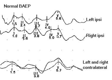 Normal brainstem auditory evoked potentials.
The short-latency BAEP generally is used for clinical purposes. The test can be performed with the patient under either sedation or general anesthesia. Standard broadband monaural click stimulation is used on the ear tested while a masking noise 30-40 dB lower in intensity is used on the contralateral ear. The intensity of the click should be 65-70 dB above the click perception threshold, and clicks should be repeated at a rate of about 10 Hz.
BAEPs are useful in estimating or aiding in the assessment of hearing loss. The most commonly used method for this is evoked response audiometry. The frequency of stimulation is 50-70 Hz, and at least 3 different intensities should be used. Wave V latency shifts are used to estimate the amount of hearing loss.
In children, especially those younger than 2 years, the BAEP can be used to screen for those in whom auditory amplification might facilitate achievement of more normal speech and language development. However, some children with a normal BAEP have abnormal hearing; Kileny showed middle latency abnormalities in some of these cases. [24] Nevertheless, the role of BAEP is to identify patients who could benefit from a hearing aid. Obviously, with a normal BAEP, a hearing aid would not be useful for correcting the hearing loss.
Physiologic basis
BAEPs predominantly activate the pathways in the brainstem that are ipsilateral to the side of click stimulation. In particular, lesions in the middle to upper pons tend to lead to ipsilateral BAEP abnormalities. The structures involved in the generation of BAEPs may be concerned more with sound localization than with hearing itself.
Whether nuclei, tracts, or both generate the peak latencies is not known. Currently, the generators are postulated to be as follows:
-
Wave I - Action potential of cranial nerve (CN) VIII
-
Wave II - Cochlear nucleus (and CN VIII)
-
Wave III - Ipsilateral superior olivary nucleus
-
Wave IV - Nucleus or axons of lateral lemniscus
-
Wave V - Inferior colliculus
Factors influencing brainstem auditory evoked potentials
Factors influencing peak latencies of BAEPs include the following:
-
Patient age
-
Patient sex
-
Auditory acuity stimulus repetition rate
-
Stimulus intensity
-
Signal polarity
Rarefaction (ie, movement of the earphone diaphragm away from the eardrum) produces an increase in wave I amplitude. In severe hearing loss, all waveforms may be delayed, wave I may be absent with waves II through V delayed, or all waveforms may be absent. Note that in patients with hearing loss, BAEPs still can be obtained to assess central conduction time by increasing stimulation intensity.
Kern et al studied the effects of insulin-induced hypoglycemia on the auditory brainstem response (ABR) in humans. [25] ABRs were examined in 30 healthy men during euglycemia and after 20 minutes and 50 minutes of steady-state hypoglycemia of 2.6 mmol/L induced with insulin. Hypoglycemia increased interpeak latencies III-V and I-V, whereas changes in the latency of wave I were not significant.
BAEPs are very resistant to alteration by anything other than structural pathology in the brainstem auditory tracts. They are not significantly affected by barbiturate doses sufficient to render the electroencephalogram (EEG) “flat” (ie, isoelectric) or by general anesthesia (though Garcia-Larrea et al reported BAEP loss with combined lidocaine and thiopental infusion [26] ).
Disorders of the peripheral vestibular system do not affect BAEP. Thus, 21 patients who had labyrinthine diseases (ie, Ménière disease, labyrinthitis, vestibular neuronitis) had no BAEP interwave latency abnormalities in terms of the limits employed for clinical neurologic purposes.
Technical aspects
An electrode is placed on each ear lobe and at Cz. The first 10 msec are averaged; 2000-4000 responses may be averaged. At least 2 separate trials should be performed. The recording montage is at least (and usually) a 2-channel montage: channel 1 is from the ipsilateral ear to the vertex, and channel 2 is from the contralateral ear to the vertex. Because of relative vertex positivity, the waveforms are recorded as upward deflections. The normal response is a series of waveforms within a 10-msec time window.
Clinically, the first 5 waves are used, with more significance placed on waves I, III, and V. Peak and interpeak latencies are measured, side-to-side differences are calculated, and wave I-V ratios may be used. Audiometry is very helpful and should be done within a reasonable interval after the BAEP study. This helps delineate any hearing loss that might influence the test results. Hearing loss in the 2000- to 4000-Hz frequency range is especially important in that it may delay the BAEP.
Recording a neonatal BAEP is technically different from recording an adult BAEP. The skin of a newborn is very sensitive; accordingly, special nonallergenic tape should be used to fix the electrode, and collodion or other irritant chemicals are to be avoided. To avoid collapse of the earlobe and obstruction of the auditory canal in premature babies, the earphone should be held slightly above the ear.
The earphone is best held by hand, and the recording preferably should be performed with the neonate asleep. This helps reduce those high-frequency EEG components that might interfere with BAEP recording. Because of the slower response, the sweep should be set at 15-20 msec and the low-frequency cutoff filter at 20-30 Hz.
Clinical utility
The most common uses of the BAEP are in multiple sclerosis (MS) and in acoustic neuroma. It is a useful screening test, though it has some limitations; magnetic resonance imaging (MRI) may be preferable when a small lesion is under consideration.
Increased I-III interpeak latency indicates a lesion from CN VIII to the superior olivary nucleus, whereas increased III-V interpeak latency suggests a lesion from the superior olivary nucleus to the inferior colliculus ipsilateral to the ear stimulated. Intraoperative monitoring during cerebellopontine angle tumor surgery may be useful in helping the surgeon preserve as much function as possible.
Cerebellopontine angle lesions (acoustic neuromas)
The BAEP may be abnormal when audiometry fails to disclose a lesion. The characteristic findings are increased I-V and increased I-III interpeak latencies ipsilateral to the lesion. Meningiomas and other cerebellopontine angle tumors may not produce any abnormalities until they are large enough to be externally compressive.
Demyelinating disease
An abnormal response may be seen with higher frequency in symptomatic patients with demyelinating disease. Sometimes, however, a positive test may be recorded in the absence of clinical brainstem symptoms.
Migraine
Zgorzalewicz found significant prolongation of waves III and IV in children and adolescents with migraine, a finding that supports the idea that the brainstem contributes to the pathologic mechanism of migraine. [27]
Multiple sclerosis
BAEP evaluation should be considered if the clinical symptoms implicate a lesion outside the brainstem. In this case, an abnormal BAEP would further support the diagnosis of MS. If, however, the clinical sign (eg, diplopia) points to the brainstem, a BAEP abnormality is merely confirmatory. In various studies, about 20% of the patients tested for a second lesion have an abnormal BAEP, and about half of these go on to develop MS in the next 1-3 years.
Purves et al reported that pattern-shift visual evoked potentials (VEPs) were abnormal in 45% of patients without brainstem signs, somatosensory evoked potentials (SEPs) were abnormal in 35%, and BAEPs were abnormal in 14%. [28] When the 3 modalities were considered together, 97% of patients with definite MS, 86% of patients with probable MS, and 63% of patients with possible MS had abnormal findings on at least 1 of these tests. [28] Similar findings were reported by Ferrer et al. [29]
Kjaer reported a 38% rate of abnormal BAEPs in MS patients with silent lesions, whereas 50% of these patients had an abnormal VEP and only 13% an abnormal SEP. [30] Kjaer also reported 22 patients with only spinal symptoms, 55% of whom showed an abnormal BAEP. [31]
Chiappa found that the BAEP was positive in 21% of clinically unsuspected cases of MS. [32] Most authors have concluded that of the 3 tests, BAEP yields the smallest percentage of patients; however, it still adds to the detection rate because it is abnormal in a different subset of patients.
Brainstem tumor
Bilateral prolongation of latencies and interpeak latencies may be seen. Gordon et al evaluated the efficacy of ABR as a screening test for small acoustic neuromas by assessing the diagnostic sensitivity of BAEP in these tumors. [33] Patients with surgically proven acoustic neuromas underwent preoperative BAEP tests within 2 months of surgery. A result was considered abnormal if the interaural wave I-V latency difference exceeded 0.2 msec, the absolute wave V latency was abnormally prolonged, or waveform morphology was abnormal or absent.
Of the 105 patients, 92 (87.6%) had an abnormal BAEP result, and 13 (12.4%) had completely normal waveforms and wave latencies. [33] Of the 18 patients who had tumors larger than 2 cm, 12 had tumors 2.5 cm or larger and 6 had tumors between 2.1 and 2.4 cm; all 18 had abnormal BAEPs. Of the 29 patients with tumors between 1.6 and 2 cm, 25 (86%) had abnormal BAEPs. Of the 45 patients with tumors between 1 and 1.5 cm, 40 (89%) had an abnormal response. Of the 13 with tumors 9 mm or smaller, only 9 (69%) had an abnormal ABR finding.
These data show that BAEP sensitivity decreases with decreasing tumor size. [33] Therefore, MRI scanning is the preferred study in this setting because the accuracy for detection of tumors smaller than 1 cm through BAEP testing is 70%. Nevertheless, BAEP studies are useful in patients who have implanted medical devices (eg, pacemakers) that prevent MRI scanning.
Meningomyelocele
Taylor et al studied BAEP and VEP in 47 infants with meningomyelocele in an effort to determine whether evoked potentials EPs reflect early neurologic status and whether BAEPs and VEPs have prognostic value for neurologic outcome. [34] The infants, aged 1 day to 3 months, were tested while still in hospital after the meningomyelocele repair.
Normal BAEPs were found in 41% of the patients and normal VEPs in 62%. BAEPs were abnormal in 9 infants studied who had symptomatic Arnold-Chiari malformation; VEPs were abnormal in only 55% of symptomatic infants. VEPs did not appear to be sensitive enough to have prognostic value in these infants. Nevertheless, BAEPs were consistently abnormal in patients with symptomatic Arnold-Chiari malformation and showed a positive predictive value of 88% and an accuracy of 84% in predicting central neurologic sequelae.
Brainstem stroke
The response to BAEP studies is variable in cases of brainstem stroke; some lesions cause abnormal latencies, and some do not (eg, negative BAEP in lateral medullary syndrome).
Respiratory insufficiency after encephalitis
Schwarz et al showed prolonged interpeak latencies (I-III, I-V, III-V, IV-V) and delayed absolute latencies of waves II, III, V, and I, at least on 1 side, in the BAEP. [35] The auditory pathways are near the respiratory control centers in the brainstem; therefore, the electrophysiologic abnormalities of wave III and the IV-V complex may be a reflection of the disturbed central control of ventilation.
Outcome prediction in coma
BAEP can be done while the patient is sedated. It can be used as a prognostic indicator. Survival is unlikely in the absence of BAEP. The brain-dead patient invariably has abnormal BAEPs—either the absence of all waveforms or the presence of wave I and the absence of all subsequent waveforms.
BAEP and SEP studies were performed within 72 hours of admission in 127 children with severe head injury to predict the outcome of posttraumatic coma (brain death or survival). On first assessment, 50 comatose children had normal BAEP and SEP; 78% survived and 22% deteriorated and died. Of the 45 who had abnormal findings; 69% improved and survived, whereas 31% deteriorated and died. All 32 children who did not have recordable BAEP and SEP died. These data suggest that BAER is useful for predicting neurologic outcome in this setting.
Outcome prediction in perinatal asphyxia
In a study of 78 children with perinatal asphyxia who were screened with BAEPs, only 37 of the 78 developed neurodevelopmental deficits, consisting of static encephalopathy or developmental delay. [36] BAEP abnormalities were seen in 40.5% of these patients and demonstrated a specificity of 87.8%. With these findings, BAEPs may help predict adverse neurodevelopmental outcomes after perinatal asphyxia.
Childhood speech disorders
Maassen et al found that children with speech and language deficit showed abnormal auditory evoked potentials in 95% of the study group. [37]
Dementia
P300 long-latency auditory EPs have been considered for using in evaluation for Alzheimer disease. In Alzheimer disease, the posterior scalp components are selectively lost. This test was also found to be strongly correlated with hypometabolism on positron emission tomography in this disease population. Although the test shows promise, its findings are considered nonspecific and nondiagnostic and are not limited to Alzheimer dementia. [38]
Somatosensory Evoked Potentials
The first evoked potential (EP) measurement is credited to Richard Caton of Liverpool, England, in 1913, but he could not record his results because he had no camera. About 34 years later, the first human scalp recording was accomplished. Major improvements included introduction of the first signal averager (by Dawson in 1954) and of the first modern averager, in 1958.
The actual somatosensory EP (SEP) is considered to be the result of summated effects of action potentials and synaptic potentials in a volume conductor. The short-latency SEP (SLSEP) is considered to be generated from volleys traversing the large-fiber sensory system (ie, the posterior columns and medial lemnisci). Studies have shown that simply changing the size and shape of the volume conductor can create voltage differences at the surface.
Physiologic basis
Large-diameter group Ia fibers and group II cutaneous afferents are primarily responsible for SEPs. Because of the ease of delivery and quantification, electrical stimulation is typically used, though other types of sensory stimuli have been tried with success. When a mixed nerve is stimulated, Ia muscle afferents are activated. In the spinal cord, the dorsal columns are mainly responsible for conduction of the activity that generates the SEP. In the brain, the lemniscal and thalamocortical pathways are involved. Extralemniscal pathways also may play a role.
Drews et al, in a study investigating the contribution of group I muscle afferent activation to the production of H reflexes and SEP in humans, found that in most subjects, H reflex was correlated with SEP size. [39] If 2 identical stimuli were applied to the posterior tibial nerve with an interval of 1 sec, the second H reflex was 30% smaller than the first one. The corresponding SEPs were reduced only slightly. Postactivation depression presumably results from intrinsic properties of synapses of group I muscle afferents.
In this study, electrical stimuli were applied to the tibial nerve in the popliteal fossa to study how the information is transferred from group I muscle afferents to motor neurons and to the somatosensory cortex. [39] For control purposes, identical stimuli were applied to the skin. The SEP evoked by skin stimulation alone had a peak latency that was 5 msec longer than the SEP to transcutaneous nerve stimulation. The threshold intensity to evoke an H reflex was at least twice as high as the threshold for an SEP.
The generators of median nerve SEP are as follows:
-
Erb point – Brachial plexus
-
N11, N13 – Dorsal column, nucleus cuneatus
-
P14 – Medial lemniscus
-
N18 – Subcortical
-
N20 – Primary sensory cortex
-
P22 – Primary motor cortex
Buchner et al researched the origin of P16 of the median nerve SEP. [40] After median nerve stimulation, several monophasic peaks were recorded at the scalp in the 15- to 18-msec time range. Source analysis using 3 different methods modeled a source near the center of the head with an orientation toward the activated hemisphere and a peak activity at 16 msec after the stimulus. Magnetic recordings detected no signals in this time range, which confirmed that the source had a subcortical location.
From dipole localization, the exact origin of the P16 source could not be assigned to either the subthalamic level or the thalamocortical radiation, because of the limited spatial resolution at the center of the spherical head model. An estimate of the conduction velocity of the medial lemniscus suggested a subthalamic origin. [40] The P16 source was preserved in 2 patients with a lesion of the thalamocortical radiation and the ventral thalamus. Further evidence for a subthalamic location was derived from the physical mechanisms generating far-field potentials.
The generators of tibial SEP are as follows:
-
N22 – Dorsal gray and root entry zone at lumbosacral spine
-
N29 – Nucleus gracilis
-
P31 – Brainstem
-
N34 – Brainstem
-
P37 – Primary sensory cortex
Factors influencing somatosensory evoked potentials
Age, height, and limb length
Vaney et al, in a study examining the role of physical parameters (including height, age, and upper limb length) in relation to the median nerve SEP, concluded that SEP studies might be affected by such parameters. [41] The authors recorded SEPs after median nerve stimulation and identified the following 3 major positive and negative peaks:
-
P1 (16 msec), N1 (20 msec)
-
P2 (28 msec), N2 (33 msec)
-
P3 (43 msec), N3 (50 msec)
N1 and P1 were correlated significantly with height and limb length.
Sleep stage
Noguchi et al, in a study that evaluated the changes of frontal and parietal SEP in the awake state and compared them with SEPs in different stages of sleep in 10 healthy adult subjects, found that frontal and parietal SEP components were affected differently as sleep stages progressed. [42] The amplitudes of frontal components were increased in sleep, whereas the amplitudes of parietal components were decreased.
The most discordant changes occurred in stages III/IV. The amplitudes for the frontal N18-P22-N30 complex and the parietal N20-P26-N32 complex increased from stage II to stages III/IV, whereas those for frontal N30-P40 and parietal N32-P40 decreased. [42] P14 and frontal N18 latencies did not change significantly. The further latencies showed progressive prolongation from the awake state to slow-wave sleep.
The SEP waveforms and latencies in rapid eye movement (REM) sleep were similar to those in the awake state. [42] Amplitudes for frontal peaks remained slightly higher and amplitudes for parietal peaks slightly lower. Apparently both excitatory and inhibitory influences may mediate these sleep stage–related changes.
Note that significant neuropathy may be a complicating factor in acquiring SEP, and the development of cortical potentials may be irregular, delayed, dispersed, and of poor amplitude. Generally, some of these difficulties can be overcome by increasing the number of samples collected.
Water immersion
Water immersion therapy is used for cardiovascular, respiratory, and orthopedic conditions and also appears to benefit some neurologic patients, although little is known about how it affects neural activity. Sato et al examined the effect of water immersion on median nerve SLSEPs and found that immersion significantly reduced SLSEP components known to originate in several cortical areas. This attenuation suggests that water immersion influences the cortical processing of somatosensory inputs. [43]
Technical aspects
Stimulation techniques
Mixed nerve stimulation need not be supramaximal; a small twitch is sufficient. The duration of the stimulus is typically 200-300 msec, though some prefer longer stimuli. The repetition rate is usually 3 Hz, though some patients tolerate only 1-2 Hz. There is no change in SEP until the 15-Hz stimulation frequency and no need for random stimuli (thought they may be needed to trigger off the electrocardiogram [ECG]).
Cutaneous nerve stimulation is used with the trigeminal and lateral femoral cutaneous nerves; dermatomal studies may be used to study segmental innervation disturbances. Dermatomal SEPs may be technically difficult and therefore are not used routinely. Current MRI technology has largely replaced dermatomal SEP studies in suspected radiculopathies and spinal cord lesions. In expert hands, however, the latter may have value in selected cases.
Recording and filtering
Surface or needle electrodes may be used. Cephalic bipolar or referential recording montages are appropriate. The general setting for filtering is 10-2500 Hz.
Upper-extremity channels are as follows:
-
Channel 1 - C3/C4 referenced to Fz
-
Channel 2 - Second cervical spinous process to Erb point
-
Channel 3 - Fz to contralateral cortex
Lower-extremity channels are as follows:
-
Channel 1 - L1, L3 to hip
-
Channel 2 - Cz- Fpz
Normal upper-limb (median nerve) SLSEP
For this test, at least 3 channels are needed, but 4 channels are generally used, as follows:
-
Erb point to Fz
-
Nuchal midline C2 spine electrode to Fz
-
Contralateral somatosensory area scalp electrode to Fz
-
Extra channel - Erb point to contralateral scalp; polarity is arranged so that active electrodes, Erb point, cervical, and cortical result in an upward deflection, representing negativity, with Fz used as the reference electrode for the upper-extremity EP; this electrode location on the scalp is not inactive and may attain electronegativity at 19-20 msec after stimulation that is equal or nearly equal to that at the contralateral cortical site and abolishes N19 in Cc-Fz derivation
N/P13 deflection
Most human clinicopathologic correlations suggest that the N/P13 waveform is generated in the lower medulla, probably in the dorsal column nuclei. The negativity recorded in the Fz-Cc derivation (N19) is the difference in negativity between the 2 electrode sites and thus is a “derived” waveform. N19 generally is believed to originate in the primary sensory cortex; however, good human clinicopathologic data are available to suggest that much of N19 is generated in the thalamus.
Chiappa et al and Goldie et al published data on a series of patients with instructive lesions, indicating that much of the negativity after 15 msec was generated in the thalamus. [44, 32] Regli and Despland studied 50 patients who had acute infarctions and found N19 to be preserved in small lesions confined to the postcentral gyrus but absent in large lesions involving the underlying white matter and thalamus. [45] Epileptogenic lesions of the sensory cortex often produce augmentation of P22 but not of N19.
These data suggest that the negative deflection appearing 16-19 msec after stimulation of the median nerve at the wrist is probably generated in the thalamus. The widespread negative activity seen at 25-30 msec with lower-limb stimulation (ie, the posterior tibial nerve at the ankle) is also believed to be generated in the thalamus. The subsequent positive activity (N/P37) is probably generated in the primary sensory cortex.
When lower-limb stimulation is employed, absence of the cauda equina potential (LP) suggests the presence of a lesion at or below that level. Technical considerations (eg, muscle artifact) also may obscure the LP potential.
Clinical interpretation is based on the time interval between wave peaks. Registering a good Erb point or LP potential is important for allowing measurement of central conduction times. Side-to-side comparisons of latencies can be useful in clinical diagnosis. Like BAEPs, SEPs are fairly resistant to changes by widespread influences other than structural pathology in somatosensory tracts. SLSEPs are not significantly altered by barbiturate doses sufficient to render the electroencephalogram (EEG) isoelectric or by general anesthesia.
Yiannikas et al found that both SEPs and electromyography (EMG) were of limited clinical utility in patients with cervical spondylosis who presented with symptoms but without neurologic signs of root compression. [46] In patients with clinical signs of neurologic deficit, EMG and SLSEPs may be helpful or confirmatory.
In patients with clinical evidence of myelopathy, performing SLSEPs from both upper and lower extremities may be informative and may reveal or disprove a conduction block. [46] Patients with syringomyelia often have abnormal central conduction time after upper-limb stimulation. In cervical spondylosis, an abnormal latency difference may be noted between brachial plexus (EP) and lower medullary (N/P13) components after upper limb stimulation.
Le Pera et al documented selective abnormality of the N13 spinal SEP to dermatomal stimulation in patients with cervical monoradiculopathy. [47] Scalp SEPs to dermatomal stimulation have proved to be only partially useful in the diagnosis of monoradiculopathy, mostly in cases without motor impairment.
The aim of the study was to test the sensitivity of the spinal N13 potential in uncovering lesions of single cervical roots. [47] The investigators studied 5 patients suffering from cervical monoradiculopathy by using a technique that allowed specific recording of the genuine N13, which probably is generated by dorsal horn cells.
None of the patients showed signs of muscle impairment, and needle EMG findings were always normal. [47] In 4 patients, the N13 SEP was absent after stimulation of the dermatome corresponding to the damaged root, whereas both the lemniscal P14 and the cortical N20 components were normal. SEP recorded after stimulation of upper-limb nerves showed no abnormality in any of these patients.
Apparently, loss of N13 potential after dermatomal stimulation could be due to deafferentation of dorsal horn neurons; the N13 is particularly sensitive to initial root compression. A montage allowing recording of the genuine N13 SEP, therefore, can improve the sensitivity of dermatomal SEP recording in patients with suspected cervical monoradiculopathies.
Clinical utility
Multiple sclerosis
The SEP is positive in a number of patients with multiple sclerosis (MS). Abnormalities may include prolonged latencies or lack of development of the SEP. Lower-extremity studies are more often abnormal because of the longer pathway. Upper-limb SLSEPs are abnormal in about 40-60% of MS patients; lower-limb SLSEPs have an abnormality rate of about 70%, presumably because of the greater length of white matter involved. American Academy of Neurology (AAN) guidelines suggest that SEPs may be useful for diagnosing clinically silent MS lesions. [48]
Eisen et al reported abnormal results in trigeminal nerve stimulation in 41% of patients with MS. [49] Abnormalities of trigeminal SLSEPs have also been reported in patients who had MS, Wallenberg syndrome, cerebellopontine angle tumors, trigeminal neurinomas, or meningeal sarcoidosis with facial paralysis.
Approximately one third of SLSEP abnormalities in MS are unilateral. One fifth of the bilateral abnormalities are asymmetric. Some patients with MS show loss of N/P13 with preservation and normal latency of N19; this pattern is difficult to explain if SLSEP generator sources are assumed to be linked in series.
Comparisons of SEP, visual evoked potential (VEP), and brainstem auditory evoked potential (BAEP) have found that VEP testing and SEP testing are about equally sensitive for revealing clinically unsuspected lesions and that BAEP is one half to one third less sensitive. The 3 tests should be viewed as complementary.
Lumbosacral disk disease
Sitzoglou et al identified clear dermatomal SEP abnormalities that correlated with radiculopathy in as many as 83% of the cases studied. [50] Electromyography (EMG) yielded positive results in about 63% of the same subjects. Thus, dermatomal SEPs may complement routine electrophysiologic testing of patients with radiculopathy and may provide a sensitive noninvasive technique for defining the level of disk prolapse.
This study included 24 patients with unilateral radiculopathy, all of whom had clinical signs and symptoms of disk prolapse and positive findings on neuroradiologic testing. [50] The latency and the amplitude of the first positive SEP waveform were measured, and both peripheral nerve conduction studies and EMG were performed.
Tsonidis et al reported on the diagnostic value of dermatomal SEPs by correlating the neurophysiologic data with clinical, neuroradiologic, and operative findings in 12 patients with surgically treated lumbar disk protrusion. [51] The retrospective study disclosed correlation of the SEPs after dermatomal stimulation and surgical findings in 83% of cases.
In this study, the diagnostic workup included a history, a neurologic examination, routine lumbar spine films, and computed tomography (CT) and magnetic resonance imaging (MRI) of the lumbar spine, in addition to neurophysiologic investigations, especially conduction velocity studies, and standard SEPs and dermatomal SEPs. [51]
Cervical syringomyelia
Patients with cervical cord syrinxes may show abnormalities in median nerve SEPs, indicating a lesion in the upper cervical cord, with relative sparing of lower-limb SEPs. Wagner et al monitored median nerve SEPs intraoperatively in 28 patients with cervical or cervicothoracic syringomyelia. [52] Analysis was focused on SEP components: N13 (spinal cord), P14 (brain stem), and N20 (cortex).
N13 was absent in about 87% of patients because of a combined effect of syringomyelia and general anesthesia, and it did not recover. [52] P14 showed a significant intraoperative latency increase in 2 patients; this was irreversible in 1 patient who had a postoperative worsening of sensory function. N20 showed no significant alterations. Pure motor deficits after surgery were not predicted by SEP monitoring.
Thus, intraoperative P14 recordings helped to identify, and thereby prevent, injury to the dorsal columns, whereas N13 recording did not contribute to the intraoperative monitoring of spinal cord function in syringomyelia. [52]
Spondylosis
Berthier et al evaluated the effects of spondylosis on SEP to ascertain its potential role in the preoperative assessment of cervical myelopathy in patients with MRI abnormalities and clinical findings of either segmental spinal cord or dorsal column dysfunction; using median and tibial nerve SEP, they found no clear correlation between the severity of MRI abnormalities and that of clinical presentation or SEP abnormalities. [53]
In this study, some patients with MRI evidence of cervical cord impingement or intramedullary T2 hyperintensity showed normal SEPs, whereas 8 of 13 patients without evidence of cord narrowing or T2 signal abnormality showed abnormal SEPs. [53] This discrepancy suggests that MRI and SEPs may evaluate different aspects of the disease process.
On the basis of this study, the SEP does not appear to be a good measure of anatomic deficit; however, spinal cord dysfunction detected by SEP may be present in patients with an unremarkable MRI image. SEP recording may therefore be useful in the preoperative assessment of symptomatic patients without MRI evidence of cervical cord compression.
Lyczak et al reported abnormal SEPs in 56% of patients with cervical myelopathy. [54] The tibial nerve SEPs were used, and abnormal central conduction times were observed. A significant reduction of the abnormalities occurred after the operation. Abnormal SEPs before the procedure correlated with the severity of myelopathy, and improvement in SEPs after the procedure correlated strongly with clinical improvement.
L5/S1 radiculopathy
Dumitru and Dreyfuss applied strict criteria to define a group of 20 patients with a unilateral/unilevel L5/S1 radiculopathy, concluding that the clinical utility of both segmental and dermatomal SEPs is dubious in patients with known unilateral/unilevel L5 and S1 nerve root compromise. [55]
Castello et al studied the effects of lumbar nerve root decompression on SEP and demonstrated a significant improvement in postoperative SEP latency in patients with lateral recess stenosis. [56]
Intraoperative SEP
The basis of intraoperative SEP as a representative index of motor function is based on the fact that the vascular compromise that may cause motor dysfunction or loss also affects the lateral corticospinal tract and the dorsal spinocerebellar tract. In general terms, the regions of both motor and sensory pathways (lateral corticospinal tract and dorsolateral spinal cord and the alpha motor neurons) are served by the same vascular supply.
Intraoperative SEP poses a special challenge and requires close cooperation with the operating room staff, as well as special equipment and careful attention to limit electrical noise. Careful checking of the ground is important, as is good shielding. The list of interfering factors is long and includes electrocautery equipment, nerve stimulators, and electric drills. Anesthetic agents and level of anesthesia also are interfering factors.
The following considerations should be kept in mind:
-
Access to the patient is important, but the recording should not interfere with the operative procedure; a baseline recording should be done before the operation to establish the patient’s own normal value under less stressful circumstances
-
The operative plan and what the surgeon expects from the monitoring must be discussed in advance; communication with the entire staff is important
-
A note should be made of any preexisting diseases (eg, diabetes) that might interfere with successful SEP testing
Stimulus pulse duration was evaluated by Luk et al, who found a duration of 0.3 msec to be the recommended choice for tibial SEPs during intraoperative monitoring. [57] Stimulus duration affected amplitude but not latency.
SEP monitoring during lumbosacral spinal stenosis is a well-known procedure. Weiss found that even in routine surgical procedures, monitoring the SEP along with EMG helps the surgical team to avoid neurogenic complications. [58]
Recording motor evoked potential (MEP) and SEP during thoracoabdominal aortic surgery to assess ischemia of the spinal cord has been valued by a number of authors as a means of lowering the risk of postoperative neurologic injury. [59] Polo et al found SEP and MEP to be useful in scoliosis surgery for assessing hypotension-related anoxic cord injury. [60] Weigang et al found SEP monitoring to be useful in thoracoabdominal aortic endovascular stent grafting for prevention of spinal cord anoxia. [61]
Arrington et al used similar methods in pelvic fractures and acetabular surgery and found that combined SEP and MEP recording prevented damage to the sciatic nerve. [62] Mills et al found that monitoring radial nerve SEP was helpful in humeral nailing procedures. Schwartz et al reported that monitoring ulnar SEP was predictive of brachial plexus injury during surgery for correction of scoliosis. [63] However, Deutsch et al found that the false-negative rate was 9% for SEP in anterior spinal surgical approaches. [64]
Hyun et al reviewed 85 cases of intraoperative monitoring using a combination of SEP and MEP and found that the combination of SEP and MEP had a higher sensitivity for detecting postoperative motor abnormalities than either modality alone did. [65]
In this study, no postoperative neurologic abnormalities were seen when SEP and MEP parameters remained stable. [65] In 20 of the 85 cases, the MEP was abnormal, with or without SEP changes. In 7 of these cases, the MEP recovered, and there were no resulting neurologic deficits. The remaining 13 patients did not have recovery of the MEP and had transient or permanent neurologic abnormalities. Four patients had SEP changes with no MEP abnormalities that did not correlate to postoperative motor dysfunction.
Baba et al reported the results of spinal cord EP monitoring for cervical and thoracic compressive myelopathy, finding that the preoperative EPs were not clearly helpful in predicting outcome but that early postoperative recovery of the EPs correlated with clinical improvement. [66]
In this study, epidural spinal cord EPs were recorded in 95 patients undergoing surgery for cervical or thoracic compressive myelopathy. [66] Abnormal spinal cord EPs correlated significantly with the severity of spinal cord compromise and symptoms (eg, myelopathy). All of the thoracic myelopathy cases and 91% of the cervical myelopathy cases exhibited abnormal EPs.
Davis et al reported the use of ulnar nerve SEPs to detect and prevent position-related neuropathy in the first pediatric patient in the world to undergo robotic-assisted thyroidectomy; they concluded that upper-extremity SEPs should be routinely performed during this procedure. [67] Patient positioning for this approach, as well as retraction during exposure, has the potential to result in postoperative brachial plexopathy similar to what is seen in other types of surgery.
Acute transverse myelitis
Few studies have evaluated the role of EP changes in acute transverse myelitis. Misra et al assessed 10 patients who had lower-limb and upper-limb weakness by using detailed clinical, MRI, and neurophysiologic evaluation in conjunction with median and tibial SEPs, upper- and lower-limb MEPs, and concentric needle EMG; they found that MRI and MEPs were useful in assessing clinical outcome but that SEP played only a limited role. [68]
Tuberculous myelopathy
Misra et al investigated the value of SEP and MEP changes in patients with Pott paraplegia and found that MEP and SEP correlated with respective motor and sensory impairments, as well as with outcome. [69]
Intracranial neoplasm
Rowed et al used SEP to identify the somatosensory cortex in an effort to help remove intracranial neoplasms and spare eloquent cortex. [70] SEPs were recorded in response to contralateral median nerve stimulation from the cortical surface. Polarity reversal of SLSEP waves was used to identify the position of the central sulcus in 46 consecutive craniotomies for removal of metastases, gliomas, or meningiomas located in, near, or overlying sensorimotor cortex.
SEPs were recorded successfully in 43 of 46 cases (94%), with demonstration of polarity reversal in 42 of 43 cases (98%). SEP localization led to modification of 14 of 42 procedures (33%), most frequently because of either displacement or involvement of sensorimotor cortex by tumor. Six patients (14%) developed new neurologic deficits, but none of these were attributable to incorrect identification of sensorimotor cortex. Routine use of this technique should be considered in all procedures for lesions located near the central sulcus. [70]
Diabetic polyneuropathy
SEP is used in the context of diabetes mainly for purposes of confirmation, though it can also be used in selected cases when the central conduction time is needed. In general, the SEP is prolonged in patients with clinically significant diabetic neuropathy. Although SEP may be confirmatory, it is not used often, since routine nerve conduction studies readily yield the diagnosis in diabetic neuropathy.
Palma et al studied SEPs in individuals with non–insulin-dependent diabetes who had different degrees of neuropathy. [71] The wrist–Erb point conduction velocity is decreased and the Erb point–N13 interpeak latencies are increased in patients with diabetes. The N11-N13, N13-N20, and N13-P22 interpeak latencies are within the normal range. The wrist–Erb point conduction velocity was proportional to the degree of neuropathy. The degrees of neuropathy have no influence on the EP-N13 interpeak latency.
Neurologic conditions
Giant SEPs have been reported in cortical reflex myoclonus. Kofler et al described enlarged cortical responses in 14 patients with progressive supranuclear palsy (PSP), attributing this finding to cortical hyperexcitability. [72] Given that frontal lobe dementia is frequently present in patients with PSP, striatofrontal deafferentation and intracortical disinhibition may explain the increase in the size of the SEP.
Ferri et al observed large-amplitude middle latency SEPs in children with benign epilepsy of childhood with centrotemporal spikes. The mechanism of this is not known. An age-related decrease in amplitude and a lack of SEP after age 12 years were noted. The findings may be interpreted as maturational changes.
Rinsho described a 66-year-old woman with corticobasal degeneration, cortical reflex myoclonus with related cortical spike, aphasia, clumsiness, and dystonia with rigidity. The right side was affected, and median nerve stimulation elicited a giant SEP over the left scalp. He also described a 58-year-old woman with reflex myoclonus cortical myoclonic tremor and a giant cortical SEP. In a study of 2 patients with corticobasal degeneration, 1 showed a giant SEP and 1 did not.
Striano et al studied a family with cortical tremor, myoclonus, and epilepsy and found giant SEP potentials and enhanced long latency reflex I; genetic study revealed linkage on chromosome 2p. [73]
Valeriani studied cortical myoclonus and concluded that the initial giant SEP corresponded to physiologic potentials evoked in healthy subjects, whereas the late giant SEP could be explained by the hyperpolarization that follows the postsynaptic excitation of the early components.
Tsuda et al described Lafora body myoclonus with giant SEP; positron-emission tomography revealed no increase in glucose metabolism in the somatosensory cortex. [74]
Ugawa reported that the dipole responsible for the giant SEP is localized in the sensory cortex. Some patients showed the localization for the dipole in the superior frontal gyrus in the paracentral lobule.
In a study by Schmitt et al, increased amplitude of SEP was noted in all cases of ceroid lipofuscinosis.
Myoclonus and giant SEP have been described in herpes simplex encephalitis. Triggs described giant SEP with anterior spinal artery syndrome. The hypothesis has been advanced that this is due to lack of inhibition of the anterolateral inhibitory influences on the dorsal column medial lemniscal system.
Saitoh described giant SEP in the syndrome of mitochondrial myopathy, encephalopathy, lactic acidosis, and strokelike episodes (ie, MELAS syndrome). Lu reported dyssynergia cerebellaris myoclonica in 3 brothers in whom alcohol decreased both the myoclonus and the giant SEP amplitude.
Vitamin B-12 deficiency
Both upper- and lower-limb SEPs have been found to be abnormal in patients with vitamin B-12 deficiency, often showing either no components or only the peripheral EP peak. Puri et al reported that serum vitamin B-12 level correlated well with the latencies of P37 and sural SNAP. [75] On treatment, normalization of P100, MRI signal, and N20 and partial recovery of P37 latencies were seen at 6 months, 9 months, and 1 year, respectively. [75]
Hyperthyroidism
Takahashi and Fujitani studied median SEPs in 14 patients and found that the N19-P23 amplitude was significantly higher in the patients than in the healthy control subjects. [76]
Hypothermia
Guerit et al studied the use of the SEP to determine the optimal degree of hypothermia during circulatory arrest and found that the delay of SEP reappearance after restoration of blood flow correlated significantly with cardiac arrest duration. [77] They concluded that neurophysiologic monitoring of brainstem activity, as provided by SEPs, enables determination of the optimal temperature for hypothermic circulatory arrest.
The investigators sequentially recorded subcortical (P14) and cortical (N20) SEPs in 32 patients undergoing deep hypothermic circulatory arrest. [77] Under normal hemodynamic conditions, hypothermia initially produced N20 disappearance at a mean nasopharyngeal temperature of 20.4 ± 2.6° C (range, 14.5-26.1°C) and P14 disappearance at a mean of 16.9 ± 2.0°C (range, 12.4-20.2°C). On rewarming, P14 reappeared at mean temperatures of 19.3 ± 4.0°C (range, 13.5-29.2°C) and N20 at a mean of 21.1 ± 4.1°C (range, 14.3-29.6°C). [77]
Neurologic recovery was uneventful in 23 patients; 5 patients presented with neurologic sequelae (minor or transient in 4; no recovery from anesthesia and death after 48 hours in 1), and 4 patients died during the operation. [77] Of the 24 surviving patients in whom P14 disappeared when the hypothermia was deep enough to cause cardiac arrest (duration, 17-94 min), 23 had a normal neurologic outcome. By contrast, all surviving patients in whom cortical SEPs disappeared at higher temperatures presented neurological sequelae.
Carotid endarterectomy monitoring
Duffy et al compared the results of cerebral oximetry and SEP monitoring in carotid endarterectomy. [78] A 50% decrease in SEP amplitude and a 10% decrease in regional oxygen saturation (rSO2) were considered clinically significant. Compared with SEP, rSO2 had a sensitivity of 50% and a specificity of 96%. [78] Clinical experience with this evolving technology is ongoing. Its role in neurovascular procedures has yet to be established.
Fiori and Parenti used 2-channel electroencephalography (EEG) and SEP monitoring for selective shunting during 255 carotid endarterectomies for severe carotid stenosis; they found that computerized EEG is an easily interpretable method of monitoring and reveals rapidly developing cerebral ischemia but that severe SEP changes can occur in spite of a normal EEG pattern when cerebral ischemia has a slow onset. SEP monitoring is a slower recording method of but can yield a finer distinction of less severe cerebral ischemia.
Myotonic dystrophy
Patients with myotonic dystrophy have a prolonged interpeak latency between the Erb point and N/P13, indicating a sensory system involvement in this disorder.
Epidural sensory block
The existence of differential sensory block during epidural analgesia has been confirmed by some authors and disputed by others. Zaric et al evaluated epidural sensory block by thermal stimulation, laser stimulation, and recording of the SEP and found that recording of SEP did not demonstrate significant difference between responses from the sites with the most superficial sensory block and those from the sites with the most intense sensory block.
In this study, the zone of anesthesia was smaller than the zone of any other investigated variable. [79] The cranial spread of analgesia and motor block was lower than that of laser-assessed block. Partial block of laser perception and thermal perception lasted longer than analgesia and motor block. No consistent segmental or temporal differences were found between the thermal test and laser methods.
During epidural block, prolongation of latencies and reduction in amplitudes of SEP produced at the most cranial analgesic dermatome did not differ significantly from those produced at the anesthetic dermatome. [79] No differential block of small nerve fibers was found during epidural analgesia by thermal test and argon laser stimulation.
Postanoxic coma
Zandbergen et al found SLSEP and neuron-specific enolase (NSE) to be correlated with unfavorable outcomes. [80] Patients who were unconscious at 72 hours and had an abnormal SLSEP had poor outcomes. Even after 24 hours, the predictive value of the SLSEP was very strong.
Cardiac arrest
Wijdicks et al reviewed several studies evaluating the use of SEPs and prognosis after cardiac arrest and found that the absence of N20 on bilateral SEPs of the median nerve was associated with negative outcome in terms of patient recovery. [81] The false-positive rate for this finding was 0.7%. The presence of an N20 response did not correlate with a positive or negative outcome. Accordingly, the authors concluded that SEPs can predict poor neurologic outcomes accurately in this population when evaluated between days 1 and 3 after cardiac arrest. [81]
Stroke
Tzvetanov et al attempted to answer the predictive value of median nerve SEP in the early phase of stroke; the results were mixed. [82]
Maturational changes with children
Myelination occurs earlier in the peripheral nerves than in the central pathways. Consequently, changes are noted in SEPs seen as children mature. These changes are seen in the latency and morphology of waveforms. As with peripheral nerve conduction studies, the conduction velocities are decreased and do not reach adult values until age 3-4 years for peroneal nerve stimulation.
With respect to the SEP waveforms, the mean Erb point latency will not change with maturation. Central conduction times are increased in infants and decrease as individuals mature. The morphology is comparable to that of cortical waveforms, which are broader in the young and decrease with age.
Finally, it is important to understand when SEPs can be reliably recorded in infants. The tibial SEP is absent in patients younger than 31 weeks’ gestational age but is present in 50% of newborns. The median nerve SEP is reported to be present in 66-85% of term infants. Therefore, if the SEP is abnormal, serial SEPs would be warranted because of the variable presence of the response in this group. [83]
Current Clinical Role of Evoked Potentials
During the last decade, the rapid advancement in magnetic resonance imaging (MRI) technology has diminished the utilization of electrophysiologic testing. To an extent, this is justified by the high yield and generally good correlation of MRI findings with the underlying pathology. In addition, MRI is often better able to visualize the pathologic anatomy underlying a disease state.
Nevertheless, there are some situations in which either the disturbance is not readily visible by an imaging modality or MRI is neither feasible nor cost-effective. For such eventualities, evoked potential (EP) studies—including visual evoked potential (VEP), brainstem auditory evoked potential (BAEP), and somatosensory evoked potential (SEP)—are uniquely suited. Their current clinical role may be briefly summarized as follows.
In a wide variety of primary and secondary visual system diseases, VEP testing provides a sensitive extension of the clinical examination. MRI is a highly accurate localizing modality, whereas VEP is useful primarily in assessing optic nerve function in the anterior (prechiasmatic) portion (it is lateralizing but not localizing to the lesion). In patients with insidious onset and appropriate MRI findings for multiple sclerosis (MS), VEPs are recommended to help confirm the diagnosis.
BAEP is useful in acoustic neuroma, but in the past few years its yield in small lesions has been surpassed by that of MRI, and in most cases MRI is clearly superior. However, MRI may be inapplicable in some patients (eg, the increasingly large percentage of elderly persons who are equipped with pacemakers), whereas BAEP studies can be done in patients with a variety of implanted devices. BAEP provides good anatomic definition.
SEP, though limited with respect to spatial localization, is a good functional tool; its primary use is to determine compromised central nervous system (CNS) conduction. It may help confirm symptoms when few physical findings are noted. It may also reveal asymptomatic lesions, thereby facilitating the workup of suspected MS. SEP testing and MRI may serve as complementary modalities. SEP is of limited value in spinal disorders. Degenerative disk disease, spinal stenosis, and compressive lesions show poor physioanatomic correlation.
SEP may confirm or reject the presence of a suspected conduction block. BAEPs and short-latency SEPs (SLSEPs) may be able to establish an anatomic region where the conduction disturbance or block occurs. They provide a sensitive tool for assessment of brainstem auditory and somatosensory tracts and nearby structures. Abnormalities demonstrated by these tests are etiologically nonspecific and must be integrated carefully into the clinical situation by a physician familiar with the clinical uses and limitations of these tests.
In patients who have experienced a cardiac arrest, MRI of the brain frequently is unremarkable for pathology and therefore is less helpful in predicting neurologic outcomes from the injury. In these situations, SEP studies may be useful for helping predict negative outcomes.
With advances in technology and increases in the frequency with which certain implants and medical devices are used in patients, the number of people who are unable to undergo MRI scanning is growing. For these patients, EP studies may be a suitable avenue for diagnosis. Finally, intraoperative EPs may provide timely information in the operating room that has the potential to improve neurosurgical or neurovascular surgical outcomes.
Questions & Answers
Overview
What are evoked potentials (EPs)?
What are the differences in the clinical use between MRI and evoked potentials (EPs)?
What are visual evoked potential (VEP) tests?
What is the physiologic basis of visual evoked potential (VEP)?
Which factors influence results of visual evoked potentials (VEPs)?
What are the technical aspects of visual evoked potentials (VEPs) for clinical use?
What are pitfalls in the use of visual evoked potentials (VEPs) for clinical use?
Which disorders may be identified with visual evoked potentials (VEPs)?
Which factors contribute to the clinical usefulness of visual evoked potential (VEP) testing?
What is the role of visual evoked potential (VEP) testing in the workup of multiple sclerosis?
What is the role of visual evoked potential (VEP) testing in the workup of adrenoleukodystrophy?
What is the role of visual evoked potential (VEP) testing in the workup of migraine?
What are brainstem auditory evoked potential (BAEP) tests?
What is the physiologic basis of auditory evoked potentials (BAEPs)?
Which factors contribute to the clinical usefulness of brainstem auditory evoked potentials (BAEPs)?
What are technical aspects of brainstem auditory evoked potential (BAEP) testing?
What are most common uses of the brainstem auditory evoked potential (BAEP) testing?
What is the role of brainstem auditory evoked potential (BAEP) testing in the workup of migraine?
What is the role of brainstem auditory evoked potential (BAEP) testing in the workup of dementia?
What are somatosensory evoked potential (SEP) tests?
What is the physiologic basis of somatosensory evoked potentials (SEPs)?
What are the generators of median nerve somatosensory evoked potential (SEP)?
What are generators of tibial somatosensory evoked potential (SEP)?
How does sleep stage affect the results of somatosensory evoked potentials (SEPs) testing?
How does water immersion affect the results of somatosensory evoked potential (SEP) testing?
Which stimulation techniques are used in somatosensory evoked potential (SEP) testing?
How are somatosensory evoked potentials (SEPs) recorded and filtered?
What is the role of (N/P13) deflection in somatosensory evoked potential (SEP) testing?
What is the role of somatosensory evoked potentials (SEPs) in the workup of multiple sclerosis?
What is the role of somatosensory evoked potentials (SEPs) in the workup of cervical syringomyelia?
What is the role of somatosensory evoked potentials (SEPs) in the workup of spondylosis?
What is the role of somatosensory evoked potentials (SEPs) in the workup of L5/S1 radiculopathy?
What are intraoperative somatosensory evoked potentials (SEPs)?
What is the role of somatosensory evoked potentials (SEPs) in the workup of tuberculous myelopathy?
What is the role of somatosensory evoked potentials (SEPs) in the workup of intracranial neoplasm?
What is the role of somatosensory evoked potentials (SEPs) in the workup of diabetic polyneuropathy?
What is the role of somatosensory evoked potentials (SEPs) in the workup of neurologic conditions?
What is the role of somatosensory evoked potentials (SEPs) in the workup of vitamin B-12 deficiency?
What is the role of somatosensory evoked potentials (SEPs) in the workup of hyperthyroidism?
What is the role of somatosensory evoked potentials (SEPs) in the workup of hypothermia?
What is the role of somatosensory evoked potentials (SEPs) in carotid endarterectomy monitoring?
What is the role of somatosensory evoked potentials (SEPs) in the workup of myotonic dystrophy?
What is the role of somatosensory evoked potentials (SEPs) in the workup of epidural sensory block?
What is the clinical role of evoked potentials?
-
Normal brainstem auditory evoked potentials.