Practice Essentials
Fibromuscular dysplasia (FMD) is an angiopathy that affects medium-sized arteries predominantly in young women of childbearing age. Among patients with identified FMD, renal involvement occurs in 60-75%, cerebrovascular involvement in 25-30%, visceral involvement in 9%, and arteries of the limbs in about 5%. [1, 2] FMD occurs in most other medium-to-large arteries as well, including the coronary arteries, [3] the pulmonary arteries, [4] and the aorta. [5] See the image below.
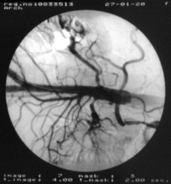 Angiogram of the descending aorta demonstrates the stenoses of FMD in the renal arteries bilaterally.
Signs and symptoms
Most patients with craniocervical FMD are asymptomatic. No particular symptoms are pathognomonic for FMD. Any history compatible with a stroke in younger individuals may indicate underlying FMD. The family history should include information about relatives who have had vascular events at a young age.
In symptomatic patients, manifestations of FMD may include the following:
-
Nonspecific problems, such as headache, light-headedness, vertigo, and tinnitus
-
Neck pain or carotidynia
-
History of transient or permanent neurologic deficits of the face or extremities (eg, weakness or numbness)
-
Visual changes or speech difficulties
-
Symptoms compatible with a sentinel bleed (eg, a sudden explosive headache followed later by neck stiffness): May indicate an aneurysm that may be associated with FMD
-
Symptoms suggestive of noncraniocervical FMD, such as hypertension (renal involvement), abdominal pains or a history of ischemic bowel (mesenteric or visceral artery involvement), or intermittent leg claudication (extremity artery involvement)
Physical examination should include the following:
-
Thorough neurologic examination
-
Neurovascular examination, including auscultation for carotid and vertebral artery bruits
-
General physical examination that includes a search for signs of renal, visceral, and extremity artery involvement
See Clinical Presentation for more detail.
Diagnosis
Routine laboratory investigations are usually nonproductive but may show renal impairment.
Imaging considerations include the following:
-
A history of stroke or transient ischemic attack (TIA) in a young individual or a subarachnoid hemorrhage in a person of any age should prompt cerebrovascular imaging; such imaging is also indicated for any individual known to have FMD
-
Conventional angiography is standard for detecting FMD and its associated vascular lesions
-
Conventional cerebrovascular ultrasonography is unlikely to depict the carotid lesions of FMD
-
The sensitivity and specificity of computed tomography (angiography (CTA), time-of-flight (TOF) magnetic resonance angiography (MRA), or contrast-enhanced MRA (CE MRA) in this setting remain to be established
-
Conventional CT and MRI may be useful in finding ischemic strokes caused by arterial dissection or the FMD lesions themselves, as well as for detecting subarachnoid hemorrhage
Pathologically, FMD is classified into 3 main types, as follows [10, 11, 12] :
-
Intimal fibroplasia (< 10% of all cases of renal FMD)
-
Medial fibroplasia: Further classified into 3 subtypes: medial dysplasia (80% of renal cases and most carotid cases), perimedial fibroplasia (10-15% of all renal cases), and medial hyperplasia (1-2% of all renal cases)
-
Adventitial fibroplasia (< 1% of all renal cases)
Because these frequency figures are largely based on findings from renal studies, they may not reflect the distribution of FMD types in carotid disease.
See Workup for more detail.
Management
Partly because of the unknown etiology of FMD, no curative therapy exists. Fortunately, FMD is often benign when asymptomatic, and medical treatment is not indicated. In symptomatic cases, management depends on the presentation, as follows:
-
Presentation with hypertension: The patient should be evaluated by a nephrologist and possibly considered for vascular intervention
-
Presentation as a TIA or ischemic stroke: If the patient presents within 3 hours of onset, consider intravenous tissue plasminogen activator (tPA); intra-arterial mechanical embolectomy and intra-arterial pharmacologic fibrinolysis may be considered to extend the acute treatment window to 6 hours; if tPA treatment is employed, avoid anticoagulants and antiplatelet agents for at least 24 hours
-
The diagnosis of FMD should be considered in any young individual presenting with a stroke or subarachnoid hemorrhage. Fortunately, cerebral angiography is the investigation of choice to detect not only FMD but also arterial dissection, vasculitis, and aneurysms, which are other major etiologies of stroke in this population. Thus, cerebral angiography should be performed if another cause for the stroke is not clear. The treatment options are influenced by the findings on angiography
-
Presentation with only FMD identified on angiography: Administer antiplatelet agents
-
Presentation with arterial dissection and FMD identified on cerebral angiography: Initially, focus on the dissection; consider anticoagulation after cerebral hemorrhage has been ruled out
-
Presentation with subarachnoid hemorrhage: Focus acute treatment on preventing rebleeding, arterial vasospasm, and further ischemic cerebral injury; close aneurysms; subsequently, manage conservatively unless further history is consistent with thromboembolic phenomena
Considerations for surgical treatment include the following:
-
Surgical vascular reconstruction of renal FMD has met with good success, [13] but the role of surgery in carotid and vertebrobasilar FMD is not well understood
-
With respect to stroke prophylaxis, the lesions in FMD are not amenable to endarterectomy; thus, surgical management is used as a last resort
-
A few cases with vascular graft placements and surgical bypass of FMD lesions have been reported
-
Aneurysms that may coexist with FMD should be managed in a similar manner to those that are not associated with FMD
-
Interventional radiologic management of FMD lesions may be suitable for some patients, especially those who are not good surgical candidates
See Treatment and Medication for more detail.
Background
Fibromuscular dysplasia (FMD) was first observed in 1938 by Leadbetter and Burkland in a 5-year-old boy, and described as a disease of the renal arteries. Involvement of the craniocervical arteries was recognized in 1946 by Palubinskas and Ripley.
FMD is an angiopathy that affects medium-sized arteries predominantly in young women of childbearing age. FMD most commonly affects the renal arteries and can cause refractory renovascular hypertension. Of patients with identified FMD, renal involvement occurs in 60-75%, cerebrovascular involvement occurs in 25-30%, visceral involvement occurs in 9%, and arteries of the limbs are affected in about 5%. [1, 2] Case reports have shown FMD in most other medium-to-large arteries as well, including the coronary arteries [3] , the pulmonary arteries [4] , and the aorta [5] . In 26% of patients, disease is found in more than one arterial region [6] .
In patients with identified cephalic FMD, 95% have internal carotid artery involvement and 12-43% have vertebral artery involvement. Although FMD can affect arteries of any size [7] , involvement of smaller ones, including intracranial vessels, is rare. Although an early autopsy series of 819 consecutive patients found the prevalence of FMD in the internal carotid arteries to be 1% [8] , a larger, more recent autopsy series of 20,244 patients recently identified the overall prevalence of FMD of the internal carotid arteries to be only 0.02% [9] . From a neurologic perspective, FMD is an important cause of stroke in young adults.
Pathophysiology
The etiology of FMD is not known, although the histopathologic findings have been described in detail (see Histologic Findings).
Although the etiology of FMD is unknown, several other associated vascular pathologies have been identified. In 1982, Mettinger and Ericson [14] scrutinized 4000 consecutively performed cerebral angiographies and found 37 that were consistent with FMD. Of these, 19 patients had aneurysms. In 1988, Cloft et al performed a meta-analysis including 498 FMD patients as well as examined 117 of their own patients and found a combined prevalence of aneurysms to be 7.3%. [15]
In 1975, Stanley et al found that 8 of their 17 cerebrovascular FMD cases had intracranial aneurysms, and they proposed a classification system that includes a "medial fibroplasias with aneurysms" subtype. [11] The beadlike dilatations observed within FMD lesions share gross and histologic characteristics of aneurysms. The casual link between FMD and aneurysms is less clear but is possibly related to an underlying connective tissue problem that results in loss of arterial wall strength. This wall weakness may allow for vessel dilation (aneurysm formation and beading in FMD) as well as injury, which then causes compensatory fibroplasia. Besides aneurysms, many case series and reports have identified FMD in patients presenting with arterial dissection. [16, 17]
FMD is a predisposing factor in 15% of spontaneous cervical carotid dissections. Dissections in FMD are more commonly multiple than in patients without an identified underlying arteriopathy.
FMD lesions likely predispose the artery to dissection through weakening of the arterial wall. Although the multiple manifestations of a structural arteriopathy in FMD hint of a genetic cause, such as collagen or elastin mutation, epidemiologic data suggesting familial transmission are generally weak.
The increased incidence of FMD in women as compared with men suggests a possible hormonal or genetic influence. Some authors have proposed the sex difference to be related to immune system functioning, but overt inflammation, as is observed in most classic autoimmune diseases, is histologically lacking.
Many reports exist of familial occurrences of FMD, mostly in siblings. Some studies have even suggested that familial occurrence is relatively common. For example, Rushton in 1980 suggested familial occurrences in relatives of 12 out of 20 identified probands. [18] However, histologic proof was established in only the index cases, and vascular events such as early strokes and hypertension were used to identify the other affected family members. Most large series have reported that the great preponderance of FMD cases are sporadic. Bilateral renal FMD has been noted in a pair of identical twins. [19]
In case reports, FMD has been associated with mutations in collagen [20] , with cutis laxa [21] , and with alpha1-antitrypsin deficiency [22] . Associative links to neurofibromatosis, Alport syndrome, and pheochromocytoma have also been suggested. [2]
Epidemiology
Frequency
United States
Although early autopsy and radiologic series suggested that FMD involving the craniocervical arteries occurs at a frequency of approximately 1%, a more recent large series looking at FMD in the carotid arteries only suggests a lower frequency, on the order of 0.02%. [9]
International
The frequency is unknown.
Mortality/Morbidity
FMD generally follows a benign course and is frequently an incidental finding. However, cranial involvement bears worse prognosis because of the occurrence of dissection and strokes and the coexistence of saccular aneurysms. Specific mortality and morbidity data are lacking.
Regarding the risk of recurrent carotid artery dissection, de Bray et al prospectively reviewed 103 consecutive patients with carotid artery dissection with follow-up for an average of 4 years. Of those, 5 had recurrent dissections and 4 of the 5 patients with recurrent dissections were diagnosed with FMD. If considering the presentation of recurrent dissection of the carotid artery, FMD was associated in 80% of their series. [23]
Race
Whites are considered to be more commonly affected than blacks, although specific statistics on racial predilection are not available.
Sex
FMD occurs more frequently in women, at a ratio of approximately 3:1 to 4:1.
Age
FMD most commonly presents in young to middle-aged adults. One angiographic series found a mean age of 48 years with a range of 24-70 years. [14] Cases have even been described in the pediatric population, including infantile-onset cases. [24]
-
Digital subtraction angiogram of the right internal carotid artery demonstrates an irregular extracranial portion that is consistent with FMD.
-
Conventional angiogram of the left carotid artery demonstrates a 1.5-cm, long, smooth, severe stenosis of the extracranial internal carotid artery. Note that the artery is not completely occluded and a thin continuous string of contrast is present along the length of the stenosis. This smooth tubular stenosis is suggestive of the intimal fibroplasia form of FMD but can be observed with any of the subtypes.
-
Cerebral angiogram of the left carotid artery territory demonstrates a long, irregular stenosis with a string-of-beads appearance along the entire extracranial length of the internal carotid artery (ICA). This is consistent with the most common medial dysplasia form of fibromuscular dysplasia. Also note similar involvement of the first 3 cm of the external carotid artery (ECA). Such extensive ICA involvement, as well as ECA involvement, is atypical. Note sparing of the carotid bulb.
-
Lateral view of a right carotid angiogram demonstrates multiple stenoses of FMD of the internal carotid artery. The string of beads appearance is suggestive of the medial dysplasia form of FMD.
-
Anteroposterior view of a right carotid angiogram demonstrates FMD of the extracranial portion of the right internal carotid artery.
-
Angiogram of the descending aorta demonstrates the stenoses of FMD in the renal arteries bilaterally.
-
Angiogram of the right vertebral artery demonstrating irregular stenoses of fibromuscular dysplasia at the level of C2-3.
-
Illustration of the operative approach of graduated dilatation of the internal carotid artery (ICA). The common carotid and external carotid arteries are cross-clamped, and the superior thyroid artery is clipped while the ICA is isolated, opened, and dilated with progressively larger dilators. This technique has been shown to be successful in the management of medically refractive FMD stenoses.
-
Illustration depicts the intraluminal appearance of graduated dilatation of the stenoses of FMD. The dilator is passed into the vessel and opens the bandlike narrowings.
-
Illustration depicts the locations of FMD lesions, which differentiate regions with typical and atypical angiographic appearances of this disease.
-
Digital subtraction angiography of the left internal carotid artery distribution demonstrates a large 1.5-cm-diameter aneurysm of the right anterior communicating artery. Aneurysms may be associated with systemic vasculopathies such as FMD.
-
Small infarct in woman with fibromuscular dysplasia from dissected vertebral artery. An incidental aneurysm, or ovoid diverticula, is noted in the supraclinoid left internal carotid artery.
-
Small infarct in woman with fibromuscular dysplasia from dissected vertebral artery. An incidental aneurysm, or ovoid diverticula, is noted in the supraclinoid left internal carotid artery. Dissected vertebral artery.
-
Small infarct in woman with fibromuscular dysplasia from dissected vertebral artery. An incidental aneurysm, or ovoid diverticula, is noted in the supraclinoid left internal carotid artery. Internal carotid angiogram.


