Continually Updated Clinical Reference
 
 
  All Sources     eMedicine     Medscape     Drug Reference     MEDLINE
 
eMedicine - Limb-Girdle Muscular Dystrophy : Article by

Quick Find
Authors & Editors
Introduction
Clinical
Differentials
Workup
Treatment
Follow-up
Multimedia
References

Related Articles
Congenital Muscular Dystrophy

Congenital Myopathies

Dermatomyositis/Polymyositis

Emery-Dreifuss Muscular Dystrophy

Endocrine Myopathies

Facioscapulohumeral Dystrophy

Inclusion Body Myositis

Metabolic Myopathies

Spinal Muscular Atrophy

Thyroid Disease


Patient Education
Click here for patient education.


Author: Glenn Lopate, MD, Associate Professor, Department of Neurology, Division of Neuromuscular Diseases, Washington University School of Medicine; Chief of Neurology, St Louis ConnectCare, Consulting Staff, Barnes Jewish Hospital

Glenn Lopate is a member of the following medical societies: American Academy of Neurology, American Association of Neuromuscular and Electrodiagnostic Medicine, and Phi Beta Kappa

Editors: Raj D Sheth, MD, Professor, Departments of Neurology and Pediatrics, Director of Comprehensive Epilepsy Program, Department of Neurology, University of Wisconsin at Madison; Francisco Talavera, PharmD, PhD, Senior Pharmacy Editor, eMedicine; Agapito S Lorenzo, MD, Laboratory Director, Associate Professor, Departments of Neurology, Creighton University and University of Nebraska Medical Center; Matthew J Baker, MD, Consulting Staff, Collier Neurologic Specialists, Naples Community Hospital; Nicholas Y Lorenzo, MD, Chief Editor, eMedicine Neurology; Consulting Staff, Neurology Specialists and Consultants

Author and Editor Disclosure

Synonyms and related keywords: limb-girdle muscular dystrophy, sarcoglycanopathy, alpha-dystroglycanopathy, LGMD, LGMD1, LGMD2, LGMD2C, LGMD2D, LGMD2E, LGMD2F, myofibrillar myopathy, desmin-storage myopathy,  LGMD2A, calpainopathy, LGMD2B, dysferlinopathy, telethoninopathy, TRIM32-related dystrophy, LGMD2J, titinopathy, myotilinopathy, laminopathy, caveolinopathy, desminopathy, alpha-β-crystallinopathy, myotilinopathy, filamin C myopathy, selenoprotein N myopathy, laminopathy, LGMD2J, titin protein

Background

Walton and Nattrass first proposed limb-girdle muscular dystrophy (LGMD) as a nosological entity in 1954.1 Their definition included the following characteristics:

  • Expression in either male or female sex
  • Onset usually in the late first or second decade of life (but also middle age)
  • Usually autosomal recessive and less frequently autosomal dominant
  • Involvement of shoulder or pelvic-girdle muscles with variable rates of progression
  • Severe disability within 20-30 years
  • Muscular pseudohypertrophy and/or contractures uncommon

This classification allowed for a wide range of phenotypic variability. Improved diagnostic methods have demonstrated that a large group of neuromuscular disorders, including some that were not truly LGMD, were included in this definition.

LGMD classification has been revolutionized with the advent of molecular genetics. A recent classification scheme, proposed during a workshop in 1995, is based on clinical and molecular characteristics. It divides cases into autosomal dominant (LGMD1) and autosomal recessive (LGMD2) syndromes. This list continues to expand, and, as of this writing, specific mutations are known for 3 autosomal dominant LGMDs and 12 autosomal recessive LGMDs.  

Although not truly limb-girdle syndromes, diseases classified as myofibrillar myopathies share several phenotypic characteristics with the LGMDs. They are usually adult-onset diseases with slowly progressive weakness involving proximal (and distal) muscles. Many patients have respiratory failure, cardiomyopathy, and neuropathy.

Cardinal morphologic features on muscle biopsy are vacuolated muscle fibers and inclusions that were initially given different names in the 1970s. In 1980, desmin was noted to accumulate in the inclusions, and the name desmin storage myopathy was coined. However, in the mid 1990s, other proteins were also found to accumulate in the abnormal muscle fibers, and molecular genetic studies revealed several chromosomal loci. Since then, the relatively generic term myofibrillar myopathy has been adopted. These diseases are discussed here in part because mutations in 2 genes can present with either an LGMD or a myofibrillar myopathy phenotype.

Pathophysiology

Limb-girdle muscular dystrophy (LGMD) protein defects occur in several pathways involved in the biologic function of muscle and can be divided into groups based on cellular localization. These include proteins associated with the sarcolemma (see Media file 1), proteins associated with the contractile apparatus (see Media file 2), and various enzymes involved in muscle function. However, although the primary defect in many LGMDs is known, the precise mechanism leading to the dystrophic phenotype has not always been elucidated. Specific protein function and abnormalities are discussed below with each LGMD.

The cause of myofibrillar myopathies is unknown but is discussed below for each of the different mutations that have been associated with the disease. Of interest, several mutations that result in myofibrillar myopathy are in genes that code for Z-disk proteins.

Frequency

International

  • Autosomal recessive LGMDs are more common than the autosomal dominant forms of the disease, which probably account for about 10% of all LGMDs. The overall frequency of all LGMD syndromes has been estimated to be 5-70 per 1 million population in several countries.
  • Different populations often have different frequencies of the various LGMDs.
  • A study involving 6 centers in the United States examined muscle biopsy results and performed Western blotting in 266 patients with a referral diagnosis of LGMD. Of these, 18% had dysferlinopathy, 15% had sarcoglycanopathy, 15% had alpha-dystroglycanopathy, 12% had calpainopathy, and 1.5% had caveolinopathy.2
  • LGMD2A may be the most common LGMD in some populations, accounting for about 25% of all cases.
  • LGMD2B may be the most common form in other populations, accounting for approximately 20% of all cases.
  • As a group, the sarcoglycanopathies (LGMD2C-2F) account for about another 20-25% of all cases, but a high percentage of severe cases. As with other LGMDs, different sarcoglycanopathies are overrepresented or underrepresented in different populations, with some populations having representative cases of all 4 sarcoglycanopathies and with other populations having only 1 mutation type, which is probably related to founder effects and population inbreeding (consanguinity). LGMD2C is common in Tunisia; LGMD2D is common in Europe, the United States, and Brazil; and LGMD2E and LGMD2F are common in Brazil. Overall, LGMD2D (alpha-sarcoglycanopathy) is twice as common as LGMD2C (gamma-sarcoglycanopathy) and 2E (beta-sarcoglycanopathy), and LGMD2F (delta-sarcoglycanopathy) is the least common.
  • LGMD2I due to a mutation in the fukutin-related protein gene (FKRP) is also relatively common. Recent studies show that patients with this mutation account for 6-38% of cases of LGMD, with a particularly high prevalence in parts of Northern Europe.
  • The remaining autosomal recessive LGMDs are rare and are often observed in selected populations (eg, Hutterite population of Manitoba for LGMD2H).
  • Myofibrillar myopathies are rare disorders. Their prevalence and incidence are unknown. The largest series was from the Mayo Clinic and consisted of 63 patients with myofibrillar myopathy who were examined between 1977 and 2003.

Mortality/Morbidity

  • Morbidity and mortality rates vary. With early onset, the course is generally rapid.
  • Patients can become wheelchair bound in their early teens and die from respiratory complications in their late teens.
  • Patients with slowly progressive LGMD may be able to ambulate for more than 30 years after disease onset, and they may not become wheelchair bound until much later than their teens. Some patients with confirmed mutations have had nearly normal strength.

Race

LGMD is reported in races and countries throughout the world.

Sex

Autosomal dominant and autosomal recessive forms of LGMD affect both sexes equally.

Age

  • The age of onset varies among the different mutations.
  • Age of onset can vary among families with the same mutation. Reported age of onset of LGMDs is between 1 and 50 years, although some patients may be asymptomatic.  Myofibrillar myopathies can present in the first decade of life up until the 60s or 70s.  


History

Autosomal recessive limb-girdle muscular dystrophy (LGMD): All patients have a history of progressive, proximal muscle weakness. Described below are the major distinguishing characteristics. The Brazilian population has been particularly well studied, and much of the population data comes from this group.

  • LGMD2A (calpainopathy)
    • LGMD2A may be the most common autosomal recessive LGMD, accounting for about 30% of all cases in the Brazilian and other populations. In some areas, including the Basque region of Spain (where a founder mutation is identified), LGMD2A accounts for almost 80% of all cases of LGMD.
    • About two thirds of patients present at 8-15 years of age, with a range of 2-40 years.
    • The most typical presentation is of weakness due to scapular-humeral-pelvic weakness that may be similar to the presentation of facioscapulohumeral dystrophy. LGMD2I may also have a similar phenotype.
    • Hip-girdle weakness is most prominent in the gluteus maximus and hip adductors. Along with abdominal weakness, this leads to a wide-based, lordotic gait.
    • Atrophy is often prominent.
    • Progression tends to be slow, and wheelchair use begins 11-28 years after the onset of symptoms.
    • The clinical course varies widely among and within families.
    • Atypical presentations include a severe Duchenne-like course, exercise-induced stiffness and myalgia before the onset of weakness, early and clinically significant contractures (especially of the ankles, elbow, and neck) similar to those of Emery-Dreifuss muscular dystrophy.
    • Presentation with asymptomatic hyperCKemia has been reported in about 10% of cases.
    • Facial and cardiac involvement have not been reported.
  • LGMD2B (dysferlinopathy)
    • LGMD2B is also a common cause of autosomal recessive LGMD, accounting for about 20% of cases in the Brazilian population. However, in some populations (eg, Cajun, Arcadian groups), it accounts for about 40% of cases.
    • Two main phenotypes can occur: a limb-girdle phenotype and a distal-myopathy phenotype (Miyoshi myopathy).
    • With either phenotype, presentation usually occurs at 15-25 years, but it can be as early as 12 years.
    • In the limb-girdle presentation, pelvic and femoral muscles are affected first, with the proximal portions of the arms becoming weak later. Gastrocnemius weakness and wasting may be prominent. The patient's gait is unique, with a waddling component combined with inability to raise his or her heels off the ground.
    • With Miyoshi myopathy, the presentation includes gastrocnemius weakness and difficulty with toe walking. The forearm muscles are weak and atrophic, with sparing of intrinsic hand muscles.
    • Late in the course of disease, the 2 modes of presentation usually become indistinguishable.
    • Either phenotype can occur in the same family.
    • The disease slowly progresses, and patients are usually confined to a wheelchair 10-30 years after the onset of weakness.
    • Rare cases present with distal anterior leg weakness and foot drop or with asymptomatic hyperCKemia.
  • Sarcoglycanopathies (LGMD2C-2F)
    • In general, sarcoglycanopathies tend to cause a severe Duchenne-like phenotype, but mild Becker-like phenotypes have been described. Overall, these diseases account for about 20-25% of all LGMDs, but they are overrepresented among severe cases. LGMD2D (alpha-sarcoglycan [adhalin]) accounts for 40% of the sarcoglycanopathies, LGMD2C and 2E (gamma-sarcoglycan and beta-sarcoglycan) each account for about 23% and LGMD2F (delta-sarcoglycan) accounts for 14% of cases in the Brazilian population.
    • Onset is usually at ages 6-8 years, but onset at or before 2 years and as late as the teens (or even adulthood) has been reported.
    • Some delay in motor milestones is not uncommon.
    • Weakness affects the hip and abdominal and shoulder musculature. Scapular winging is more common in LGMD2C-2F than in Duchenne muscular dystrophy.
    • Hypertrophy of the calf is common, and the tongue muscles may become enlarged.
    • Progression tends to be more rapid than that of other LGMDs, with loss of ambulation usually at 12-16 years but can be as early as 10 years. Patients with a late onset tend to have a slowly progressive course.
    • Intelligence is normal.
    • Cardiomyopathy is reported in about 30% of cases and is most common with LGMD2E or 2F.
    • Progressive weakness leads to restrictive lung disease and hypoventilation and the need for ventilatory assistance.
    • Death can occur as early as in the second decade of life, although some patients live into adulthood without respiratory assistance.
  • LGMD2G (telethoninopathy)
    • To date, LGMD2D has been reported only in Brazilian patients, with great phenotypic variability between and within families.
    • The age of onset is 2-15 years.
    • Weakness is predominantly proximal, but one half of patients may present with foot drop, and nearly all eventually develop distal weakness.
    • Calf hypertrophy occurs in about 50%.
    • Wheelchair confinement occurs in the third to fourth decade.
    • Mild cardiac involvement occurs in about 50%.
  • LGMD2H (tripartite motif–containing gene 32 [TRIM32]–related dystrophy) 
    • LGMD2H has been observed only in the Hutterite people of Manitoba.
    • This disease is allelic with sarcotubular myopathy (see Congenital Myopathies).
    • Most patients have a mild phenotype, with limb-girdle weakness and a waddling gait at presentation. The proximal arm muscles and the distal leg muscles are involved late.
    • The age of onset is 8-27 years; some patients are asymptomatic in their third decade.
    • Back pain and fatigue are common symptoms.
    • Progression is slow, with continued ambulation until around 50 years of age or later.
  • LGMD2I (fukutin-related proteinopathy) 
    • LGMD2I may be a fairly common cause of autosomal recessive LGMD, causing 11% of all cases in Brazil and 38% of cases in Denmark.
    • The disease is allelic with congenital muscular dystrophy with secondary laminin alpha-2 deficiency (MDC 1C). (See Congenital Muscular Dystrophy.)
    • The presentation of patients with a mutation in fukutin-related protein (FKRP) gene can vary from severe congenital muscular dystrophy to mild, late-onset LGMD.
    • The LGMD phenotype is variable. Patients can have a severe Duchenne-like presentation with delay in motor milestones, hypotonia, and severe proximal weakness. Progression to wheelchair by the teenage years and restrictive respiratory failure (even when patients are ambulant) is common. The most common presentation is likely a Becker-like onset with normal early motor milestones. An adult-onset form occurs at 11-40 years of age and is slowly progressive.
    • In a large study in Denmark, 2 groups of patients could be delineated based on genotype-phenotype correlations. Of the 38 patients studied, 27 (71%) had a homozygous mutation (826A>C) while 11 (29%) had a compound heterozygous mutation.3 
      • The patients with a homozygous mutation had a later onset (mean of 18 y) and slower progression than patients with a compound heterozygous mutation. Only 15% lost the ability to ambulate by their mid 40s. Presentation with exertional myoglobinuria, calf hypertrophy and cardiomyopathy were all more common than in patients with a compound heterozygous mutation.
      • The patients with a compound heterozygous mutation had an earlier onset (mean of 5 y) and more rapid progression. All lost the ability to ambulate by their mid 20s. Tongue hypertrophy, more severe respiratory failure, contractures, and spine abnormalities were more common than in patients with a homozygous mutation.
  • LGMD2J (titinopathy)
    • LGMD2J has been described only in Finnish patients. Some family members have a typical limb girdle phenotype with severe proximal weakness (LGMD2J) while other family members have distal dominant weakness (Finnish [tibial] muscular dystrophy).
    • The onset of the LGMD syndrome is at 10-30 years, with proximal weakness. Some patients later develop distal weakness.
    • The disease slowly progresses, and wheelchair confinement usually occurs within 20 years, but some patients are ambulant past 60 years.
    • No facial weakness or cardiac involvement is noted.
  • LGMD2K
    • LGMD2K has been described in Turkish and Italian families.
    • The disease is allelic with Walker-Warburg syndrome. (See Congenital Muscular Dystrophy.)
    • The age of onset is 1-6 years.
    • LGMD2K is characterized by severe proximal muscle weakness with slow progression. Contractures may be present.
    • Results of ophthalmologic and funduscopic examinations, including electroretinography, are normal.
    • Facial dysmorphic features and mental retardation may occur, though brain MRIs are normal.
  • LGMD2L
    • LGMD2L has recently been proposed as the name for a LGMD syndrome described in 3 patients in 2 families with a mutation in the fukutin gene.4
    • The disease is allelic with Fukuyama congenital muscular dystrophy.
    • Patients presented with hypotonia before age 1 year. Progression was moderate with proximal greater than distal weakness affecting the legs more than the arms. The affected children, now aged 7-9 years can still walk.
    • Interestingly, these children have worsening weakness during febrile illnesses, and like boys with Duchenne muscular dystrophy, their weakness improves with steroids. 
    • All children had normal intelligence and normal brain MRIs. 

Autosomal-dominant LGMD: Autosomal dominant LGMD is less common than autosomal recessive LGMD, accounting for about 10% of all cases. In general, patients with autosomal dominant LGMD have a later onset and slower course than those of autosomal recessive LGMD. Creatine kinase (CK) elevations are also not as great in autosomal dominant LGMD as in autosomal recessive LGMD. 

  • LGMD1A (myotilinopathy, also Myofibrillar myopathies)
    • Onset varies from young adulthood to the mid 70s.
    • Weakness can initially be proximal or distal but progresses to clinically significant proximal and distal weakness in all patients.
    • The progression is slow, with late loss of ambulation. 
    •  Dysarthria may be prominent.
    • Cardiomyopathy is noted in 50%.
    • Neuropathy noted in over 50% may account for distal weakness.
  • LGMD1B (laminopathy, allelic with autosomal dominant Emery-Dreifuss muscular dystrophy). 
    • Onset can be from childhood (<10 y) to the mid 30s.
    • LGMD1B results in proximal weakness with slow progression.
    • Distal limb and facial weakness may be late manifestations.
    • Cardiac disease begins by the 30s-50s and affects two thirds of patients. Atrioventricular (AV) block progresses from first degree to complete. Dilated cardiomyopathy and ventricular arrhythmias may also be present.
  • LGMD1C (caveolinopathy)
    • Onset is usually in the first or second decade, but it may manifest at a later age with a distal myopathy or elevation of CK levels.
    • Patients have mild-to-moderate proximal weakness with slow-to-moderate progression.
    • Exercise-induced cramps may be present.
    • Calf hypertrophy affects some patients.
    • Adults usually remain ambulant.
    • Patients may also present with elevated CK levels without weakness but with myalgia and cramps, distal weakness, hypertrophic cardiomyopathy, or rippling-muscle disease. The last condition is mechanical or activity-induced, electrically silent muscle contraction that moves laterally in wavelike fashion across the muscle. Myoedema, or mounding of the muscle after percussion, may be observed. Patients may also have proximal weakness, muscle hypertrophy, or myalgias.
  • LGMD1D (Online Mendelian Inheritance in Man [OMIM] %603511)
    • Two families have been described. 
    • Onset is in adulthood, with proximal weakness. 
    • Progression is slow. 
    • Dysarthria may be present.
  • LGMD1E (Dilated cardiomyopathy with conduction defect and muscular dystrophy, OMIM %602067)
    • One large family has been described. 
    • Onset is in early adulthood, with proximal weakness. 
    • Progression is slow. 
    • Cardiac arrhythmia and cardiomyopathy are noted in all patients beginning 1-2 decades after weakness and may lead to sudden death.
  • LGMD1F
    • One large family has been described.
    • Onset is from the first year of life to the mid 50s. 
    • LGMD1F causes early proximal weakness with progression to distal weakness in most patients.  
    • Patients with a young onset may have rapid progression and require use of a wheelchair by their 20s-30s. They may also have facial and respiratory weakness and/or spinal deformity.  
  • LGMD1G
    • One family has been described.
    • Onset is in the 30s-40s.
    • LGMD1G causes slowly progressive proximal weakness.

Clinical features to distinguish the main LGMDs are often most helpful early in the disease.

  • LGMD1A: Dysarthria is common.
  • LGMD1B: Cardiac complications include cardiomyopathy and arrhythmia. Contractures are common.
  • LGMD1C: Onset is usually in childhood, but patients may present with asymptomatic elevations of CK levels. Calf hypertrophy may be prominent.
  • LGMD2A: Patients have prominent atrophy of the periscapular, biceps, gluteus maximus, thigh adductors, and hamstring muscles, with sparing of the hip abductors. Contractures are common, in which case the disease needs to be differentiated from Emery-Dreifuss muscular dystrophy, Bethlem myopathy, and laminin-alpha2 deficiency.
  • LGMD2B: Patients may have early weakness and/or atrophy of the gastrocnemius (might be detected only on MRI), inability to walk on toes, waddling gait, atrophic distal biceps, and spared periscapular and deltoid muscles. Onset is in the late teens or early 20s.
  • LGMD2C-2F: Patients may have Duchenne- and/or Becker-like weakness but with additional involvement of the periscapular muscles causing scapular winging. Muscle hypertrophy is common. Mental development is normal. Cardiomyopathy may be present in some.
  • LGMD2G: Patients may have initial anterior tibial weakness causing foot drop or a typical LGMD phenotype.
  • LGMD2H: Patients may have a late onset, slow progression, no cardiac symptoms, but mild ECG changes. This form is reported in the Hutterite population.
  • LGMD2I: This form has a widely variable spectrum with prominent muscle hypertrophy and cardiomyopathy (Duchenne like). Patients may have prominent tongue hypertrophy and severe weakness and wasting of upper arms, neck flexors, and axial muscles; these features can help in distinguishing this disease from Duchenne muscular dystrophy.

Myofibrillar myopathies (MFM) 

  • Myofibrillar myopathies, also called desmin-storage myopathies (desmin was the first protein found and is the most consistent protein in the aggregates that are characteristic of these disorders) refers to a group of hereditary myopathies with homogeneous morphological features. The initial morphological change in MFM is disintegration of the Z-disk, and then of the myofibrils, followed by abnormal ectopic accumulation of multiple proteins. 
  • Age at onset varies from 7-77 years, with a mean of 54 years, except for patients with mutations in selenoprotein N who have onset at birth and the 1 described patient with a lamin A/C mutation who presented at age 3.
  • Clinically, this group of disorders is heterogeneous, with slowly progressive weakness affecting the proximal and distal muscles in most patients, but about 25% present with distal predominant weakness, and 25% present with only proximal weakness. They are included in this article because some mutations are in the same genes that cause LGMD phenotypes.
  • Rare findings include the following:
    • Facial weakness
    • Asymmetric weakness
    • Severe atrophy
    • Respiratory failure, which may be severe or at presentation
    • Contractures
    • Distal sensory deficits (neuropathy diagnosed in about 20%)
  • Cardiac disease may be present either as cardiomyopathy or arrhythmias and conduction block is present in about 50%.
  • Specific mutations
    • Desminopathy: Onset is generally in the 20s or 30s with slow progression. Patients often present with distal weakness that progresses proximally, but limb-girdle, scapuloperoneal, and distal weakness combined with proximal weakness have all been described. Inter- and intrafamilial variability exists. Those with autosomal recessive disease may have an early onset. Cardiac disease occurs in about 60% and may follow or precede myopathy, or it may be isolated. Respiratory failure may be severe and may be present at presentation. Facial and bulbar weakness may occur late. About 75% of patients eventually need assistance with ambulation.
    • Alpha-β-crystallinopathy: Onset varies from early to mid adulthood. Patients present with proximal more often than distal weakness. They may also present with respiratory failure. Patients may have neuropathy, cardiac failure, or conduction abnormality. Other mutations can cause dominant congenital posterior polar cataract.
    • Myotilinopathy: The first mutations described were in 2 patients with an LGMD phenotype (see LGMD1A). Since then, several patients have been found with a myofibrillar myopathy. Onset is usually in mid-to-late adulthood. Most patients present with distal greater than proximal weakness. Neuropathy occurs in about 50%. Cardiomyopathy affects about 50%. Dysarthria, joint contractures and myalgias are present in about 33%. One family with spheroid body myopathy, a congenital myopathy, has been found with a mutation in the myotilin gene.
    • Filamin C (gamma-filamin) myopathy: Age at onset is 37-57 years, with proximal greater than distal weakness. Respiratory failure occurs in about 50% of patients. Neuropathy affects about 40%. Cardiac disease may be present.
    • Z-band alternatively spliced PDZ motif-containing protein (ZASPopathy): Onset is at age 44-73 years, and patients most often present with distal more than proximal weakness, though proximal weakness can occur alone. Cardiac disease occurs in about 25% of patients and may be the presenting or predominant feature. Neuropathy affects approximately 45% of patients. Mutations are allelic with Markesbery distal myopathy and dilated cardiomyopathy +/- isolated noncompaction of left ventricular myocardium.   
    • Selenoprotein N myopathy: Selenoprotein N mutations were originally found in patients with congenital muscular dystrophy with rigid spine syndrome or minicore congenital myopathy. A study has shown that some patients with Mallory-body desmin-related myopathy also have a mutation in the selenoprotein N gene. Onset is at birth with hypotonia as well as axial and proximal weakness. Contractures and scoliosis are common and cardiac disease may occur. Death or the need for ventilatory support occurs before adulthood due to progressive respiratory failure.
    • Laminopathy: Besides presenting with a limb girdle phenotype (see LGMD1B), a recent case was described with a myofibrillar myopathy. The patient presented at age 3 with difficulty running and at age 5 was noted to have limb-girdle weakness.

Physical

See History.

Causes

Autosomal recessive LGMD

  • LGMD2A is caused by mutations on chromosome 15 in the calpain-3 gene.
    • More than 280 different, mostly private, mutations have been identified throughout the calpain-3 gene. With the exception of founder effects in isolated populations, no predominant mutations exist. All types of mutations have been found including nonsense mutations leading to stop codons, missense mutations often leading to decreased catalytic activity of calpain-3, splice site and frameshift mutations, and large deletions or insertions.
    • In general, null mutations give rise to phenotypes more severe than those due to missense mutations.
    • Calpain-3 (p94) is a member of the calpain family of intracellular, soluble cysteine proteases that all have calcium-dependent activation. It is expressed almost exclusively in muscle and is likely anchored by titin.
    • The mechanisms involved in the pathogenesis of LGMD2A are unknown. Calpain-3 is a protease with many substrates that likely is involved in many physiological processes within muscle. It has been proposed to be involved in apoptosis, muscle cell differentiation, sarcomere formation, muscle remodeling, and regulation of the cytoskeleton. One hypothesis suggests that mutations in the calpain-3 gene lead either directly to loss of proteolytic activity or to secondary loss of activity due to its loss of anchorage with titin. The loss of proteolytic activity may lead to reduced turnover of muscle proteins, and an accumulation of damaged proteins that then accumulate within muscle. This leads to a cell stress response and cell toxicity and ultimately to a dystrophic phenotype.
    • On muscle biopsy, calpain-3 can be visualized by using Western blots but not muscle immunohistochemistry. Correlation between the degree of deficiency and the clinical phenotype can be total, partial, or (in rare cases) nonexistent. Expression of dystrophin and the sarcoglycans is normal. Expression of dysferlin can be reduced.
  • LGMD2B is caused by mutations on chromosome 2 in the dysferlin gene.
    • More than 50 mutations have been identified, most commonly missense, nonsense, small deletions, and splice-site mutations.
    • The type of mutation is not correlated with the phenotype, ie, LGMD versus Miyoshi distal myopathy. Both phenotypes have been described in the same family with identical mutations.
    • Dysferlin protein is a large membrane protein with sequence analogy to the nematode protein fer-1, and is a member of the ferlin family of proteins, which are all involved in calcium-dependent membrane fusion. Dysferlin protein has been localized to the sarcolemma, the T-tubule system, and cytoplasmic vacuoles. Dysferlin is thought to be involved in the docking and fusion of intracellular vesicles to the sarcolemma during injury-induced membrane repair by interacting with other dysferlin molecules and other proteins. Some of these proteins include annexins A1 and A2 (phospholipid binding proteins), caveolin-3 (LGMD1C), calpain-3 (LGMD2A), and the dihydropyridine receptor within the T-tubule system.
    • Muscle biopsy may show reduced or absent immunohistochemical staining for dysferlin (also observed on Western blotting). Absent staining is only relatively specific for LGMD2B because reduced staining can be seen in other diseases such as LGMD2A. Staining for dystrophin and sarcoglycans is normal. On Western blotting, calpain-3 staining is reduced.
    • Ultrastructural studies have shown small sarcolemmal defects, replacement of the plasma membrane by multiple layers of vesicles, and small subsarcolemmal vacuoles, all suggesting that dysferlin is likely required for maintaining the structural integrity of the muscle fiber plasma membrane, and plasma membrane injury is an early event in the pathogenesis of dysferlinopathy.
  • LGMD2C-2F are caused by mutations in the sarcoglycan genes.
    • LGMD2C is caused by a mutation on chromosome 13 in the gamma-sarcoglycan gene.
    • LGMD2D is caused by a mutation on chromosome 17 in the alpha-sarcoglycan (adhalin) gene.
    • LGMD2E is caused by a mutation on chromosome 4 in the beta-sarcoglycan gene.
    • LGMD2F is caused by a mutation on chromosome 5 in the delta-sarcoglycan gene.
    • Sarcoglycanopathies have a worldwide distribution. Where no founder effect is present, LGMD2D (alpha-sarcoglycanopathy) is the most common form, accounting for about 50% of cases. Missense and nonsense mutations are the most common for all the sarcoglycanopathies, though with gamma-sarcoglycanopathies (LGMD2C), small or large deletions are also common.
    • The genotype-phenotype correlation is often unclear. In general, nonsense or truncating mutations tend to increase the severity of disease. Interfamilial and intrafamilial variability with LGMD2C, 2D, and 2E is marked. Most mild cases are due to alpha-sarcoglycan mutations (LGMD2D). Patients with LGMD2E and LGMD2C tend to have severe phenotypes, but mild cases can also occur. Patients with the rare delta-sarcoglycan mutation (LGMD2F) tend to have a severe phenotype.
    • Sarcoglycan protein complex is a transmembrane complex that is part of the large dystrophin glycoprotein complex. The core of the complex is made up of the beta and delta subunits with weaker binding of the alpha and gamma subunits. This complex likely does not bind directly to dystrophin, but binds to the dystroglycan complex which in turn binds to dystrophin. The sarcoglycan complex also binds strongly to sarcospan as well as to alpha-dystrobrevin and filamin.
    • The function of the sarcoglycan complex is unknown, but it likely stabilizes the dystrophin glycoprotein complex. In the absence of the sarcoglycan complex, binding of dystrophin to beta-dystroglycan and binding of beta-dystroglycan to alpha-dystroglycan are weakened.
    • The sarcoglycan complex may also play a role in cell signaling based on the following evidence. It may act as a receptor since it has cysteine bonds, common in other receptors, although no substrate has been identified. ATPase activity occurs in alpha-sarcoglycan. The sarcoglycan complex binds alpha-dystrobrevin, which in turn binds to syntrophin, which binds nNOS and voltage-gated sodium channels.
    • Muscle biopsy usually shows a dystrophic pattern of muscle-fiber necrosis and regeneration similar to that observed in Duchenne muscular dystrophy.
    • On immunohistochemistry, dystrophin staining is often slightly reduced, but may be normal (whereas sarcoglycan expression may be mildly reduced in Duchenne-Becker muscular dystrophy). Alpha-sarcoglycan mutations cause absent or reduced alpha-sarcoglycan staining with preservation of staining for gamma-sarcoglycan. Minimal or no staining occurs for beta and delta-sarcoglycan. This is the only mutation for which the amount of residual staining (for alpha-sarcoglycan) and the clinical phenotype are correlated. Gamma-sarcoglycan mutations cause absent or reduced gamma-sarcoglycan staining with trace amounts of alpha, beta, and delta-sarcoglycan staining. Beta- and delta-sarcoglycan mutations usually cause absent staining of the entire sarcoglycan complex.
  • LGMD2G is caused by mutations on chromosome 17 in the telethonin gene.
    • Null mutations have been described in 4 Brazilian families with a wide range of phenotypic variability.
    • Telethonin protein (titin-cap protein) is a sarcomeric protein present in the Z disk that binds to titin and several other Z-disk proteins. Telethonin protein may help to maintain the integrity of the sarcomere. However, in patients with LGMD2G, in whom telethonin is absent from muscle, sarcomeric integrity is maintained.
  • LGMD2H is caused by mutations on chromosome 9 in the TRIM32 gene.
    • To date, all patients have been Hutterites from Manitoba with the same Asp487Asn homozygous point mutation; this is the same mutation as that found in sarcotubular myopathy.
    • TRIM32 protein is an E3-ubiquitin ligase that transfers activated ubiquitin residues onto a target protein, tagging the protein for degradation in the proteosome.
    • On muscle biopsy, no protein accumulations or inclusions have been identified.
  • LGMD2I is caused by mutations on chromosome 19 in the FKRP gene.
    • Missense point mutations are the most common mutation. A homozygous leu276Ileu mutation (826A>C) is particularly common and is present in about 90% of patients. The disease severity correlates with the mutation in the second allele; patients with a homozygous mutation are less severely affected. The most severe phenotype occurs when patients have compound heterozygous mutations for 2 other missense mutations or 1 missense and 1 nonsense mutation. This form is allelic with congenital muscular dystrophy with secondary laminin-alpha2 deficiency (MDC 1C). (See Congenital Muscular Dystrophy.)
    • FKRP protein is a putative glycotransferase based on its sequence homology to fukutin. It is thought, but not formally demonstrated, to be involved in O-linked glycosylation of alpha-dystroglycan.
    • In muscle biopsy, antibodies to the glycosylated portion of alpha-dystroglycan show reduced staining (and decreased mass on Western blots). Antibodies to laminin-alpha2 may show reduced staining; however, in mild cases, this is often evident only on Western blots.
  • LGMD2J is caused by mutations on chromosome 2 in the titin gene.
    • Finnish patients who are homozygous for titin mutations develop LGMD2J, while patients with a heterozygous mutation develop Finnish (tibial) muscular dystrophy.
    • Titin protein is the largest protein found in humans. It is a sarcomeric protein that spans the entire sarcomere from the M line to the Z disk. Titin binds many other sarcomeric proteins and plays a mechanical role in muscle contraction, keeping the contractile elements of skeletal muscle in place and providing elasticity to the sarcomere. It also likely plays a role in the assembly of contractile elements, regulation of the size of the Z disk, and in cell signaling pathways.
    • Titin binds caplain-3 in muscle, which may stabilize it from autolytic degradation. Muscle biopsy in patients with LGMD2J shows secondary deficiency of calpain-3.
  • LGMD2K is caused by mutations on chromosome 9 in the protein O-mannosyltransferase 1 (POMT1) gene.
    • LGMD2K is allelic with Walker-Warburg syndrome (see Congenital Muscular Dystrophy).
    • POMT1 protein is an O-mannosyltransferase that glycosylates alpha-dystroglycan.
    • Muscle biopsy shows decreased staining for alpha-dystroglycan.
  • LGMD2L is caused by mutations on chromosome 9 in the fukutin POMT1 gene.
    • LGMD2L is allelic with Fukuyama congenital muscular dystrophy.
    • Fukutin is a putative glycosyltransferase and has sequence homologies to a bacterial glycosyltransferase, but its exact role and enzymatic substrate have not been determined.
    • Muscle biopsy shows decreased staining for alpha-dystroglycan.

Autosomal-dominant LGMD

  • LGMD1A is caused by mutations on chromosome 5 in the myotilin gene.
    • Several different missense mutations have been identified.
    • The term myotilinopathy has been coined because of the overlapping features in patients described as having a LGMD or myofibrillar myopathy and a mutation in the myotilin gene. Furthermore, a large family described as having spheroid body myopathy (see Congenital Myopathies) was recently found to have a mutation in the myotilin gene.
    • Myotilin protein is associated with the Z disk and is expressed in skeletal muscle and, to a lesser extent, cardiac muscle. Myotilin protein binds to alpha-actinin, filamin C, and actin, and it is likely important in stabilizing and anchoring thin filaments to the Z disk during myofibrillogenesis.
    • Muscle biopsy shows muscle fiber degeneration and hyaline inclusions that stain positively for multiple proteins (a feature similar to that of other myofibrillar myopathies). Myotilin, dystrophin, neural-cell adhesion molecule (NCAM), desmin, plectin, gelsolin, ubiquitin, and prion protein all are found in the inclusions. Other consistent findings are rimmed or nonrimmed vacuoles, autophagic vacuoles, Z-disk streaming, and mild evidence of denervation.
  • LGMD1B is caused by mutations on chromosome 1 in the lamin A/C gene.
    • Missense and deletion mutations have been reported.
    • Mutations in lamin A/C can also cause Emery-Dreifuss muscular dystrophy, myofibrillar myopathy, autosomal dominant dilated cardiomyopathy with AV block (or CMDI1A, see the Neuromuscular Disease Center), Familial partial lipodystrophy (Köbberling-Dunnigan syndrome) (see the Neuromuscular Disease Center), Charcot-Marie Tooth type 2A, mandibuloacral dysplasia, and premature aging syndromes (Hutchinson-Gilford progeria, atypical Werner syndrome).
    • No clear genotype-phenotype correlation distinguishes the disorders listed above. Different phenotypes can occur in the same family. One individual can have more than 1 phenotype.
    • Lamin A/C is an intermediate filament in the inner nuclear membrane and nucleoplasm of almost all cells. Multiple functions are described, but the pathophysiologic basis for any associated disease is unknown. The filament provides mechanical strength to the nucleus; helps to determine nuclear shape; anchors and spaces nuclear pore complexes; is essential for DNA replication and mRNA transcription; and binds to structural components (emerin, nesprin), chromatin components (histone), signal transduction molecules (protein kinase C), and several genetic regulatory molecules.
  • LGMD1C is caused by mutations on chromosome 3 in the caveolin-3 gene.
    • Most are autosomal dominant missense or deletion mutations in the scaffolding region, but a family with autosomal recessive disease has been described. The same mutation can cause different phenotypes (LGMD1C, elevated CK levels, rippling-muscle disease, distal myopathy, hypertrophic cardiomyopathy), even in the same family.
    • Caveolins are transmembrane proteins that are the principal component of caveolae. Caveolae are 30- to 60-nm invaginations in cell membranes that can bind several components of signal-transduction pathways and may act as a scaffold, placing members of the pathway in close proximity.
    • Caveolin-3 is a muscle-specific caveolin that is localized to the sarcolemma. It interacts with G proteins, a variety of signaling molecules, dystrophin, dystrophin associated proteins, phosphofructokinase, dysferlin, and nitric oxide synthase (nNOS).
    • All mutations in caveolin-3 decrease sarcolemmal immunostaining, suggesting that the mutation is due to a loss of function. A dominant negative effect has been noted in which an aberrant protein product forms aggregates that sequester the normal caveolin-3 in the Golgi apparatus. Other effects due to improper caveolin-3 oligomerization and membrane localization result in derangements of the T tubule system, alterations in the sarcolemmal membrane, and the formation of subsarcolemmal vesicles.
    • Muscle biopsy shows reduced or absent immunochemical staining for caveolin-3 at the sarcolemma, and this can be used as a screening test before searching for caveolin-3 mutations. In addition, immunochemical staining for dysferlin (caveolin-3 interactions) at the sarcolemma is reduced and the number of caveolae on electron microscopy is also reduced.
  • LGMD1D has been linked to chromosome arm 7q.
  • LGMD1E (dilated cardiomyopathy with conduction defect and muscular dystrophy) has been linked to chromosomal region 6q23.
  • LGMD1F has been linked to chromosomal bands 7q32.1-32.2.
  • LGMD1G has been linked to chromosomal region 4p21.

Myofibrillar myopathies

  • Many patients with a clinical and histologic phenotype of myofibrillar myopathy have no known mutation. Myofibrillar myopathy syndromes related to know genetic mutations are described below.
  • Most mutations are in proteins of the Z-disk or with attachments to the Z-disk. Most are proposed to cause disease by means of a dominant negative effect due to combined wild-type and mutant protein. The pathogenesis of disease is likely due to disrupted Z-disk function, which includes: (1) an attachment site and mechanical link of actin and titin filaments, (2) transmission of force along the myofibril, and (3) an attachment site for intermediate filaments (desmin) that link adjacent sarcomeres with each other and with other cellular organelles.
  • Desminopathy is caused by mutations on chromosome 2 in the desmin gene and can be either autosomal dominant or autosomal recessive. More than 20 mutations (most nonsense or missense and autosomal dominant) have been identified. Most mutations are located in the alpha-helical rod domain, which is critically important for filament assembly. Different mutations cause highly variable phenotypes and also disrupt desmin filaments at various stages of assembly. Pathogenesis is likely due to loss of desmin function or a dominant negative effect related to the accumulation of mutant desmin into toxic aggregates that disrupt cell function and eventually cause cell death.
  • Desmin protein is an intermediate filament (IF) protein. In muscle, it is located at the periphery of the Z disk, under the sarcolemma and at myotendinous junctions. In cardiac muscle, it is at intercalated disks and Purkinje fibers. Two desmin molecules align head to tail to form a dimer, 2 dimers form a tetramer, 2 tetramers form a protofilament, 2 protofilaments form a protofibril, and 2-6 protofibrils form an IF. IFs can heterodimerize with other IFs or IF-associated proteins. Desmin binds to ankyrin, spectrin, synemin, syncoilin, plectin and nebulin.  IFs form a heteropolymeric lattice to organize the myofibrils and link them to nuclei, mitochondria, and the sarcolemma.
  • Alpha-β-crystallinopathy is caused by a mutation on chromosome 11 on the alpha-β-crystallin gene. Three mutations, all autosomal dominant, have been identified. 

    Alpha-β-crystallin protein is a small heat-shock protein that forms homo-oligomeric or hetero-oligomeric complexes with alpha-β-crystallin or other heat-shock proteins. Expression in skeletal and cardiac muscles and in the lens is high. In muscle, the protein is localized to the Z disk. It binds to unfolded and denatured proteins to suppress nonspecific aggregation, and it protects actin, desmin, tubulin, and a variety of soluble enzymes from stress-induced damage. Mutant proteins are expressed and likely impair this chaperone function by means of dominant negative effect.
  • Myotilinopathy is caused by mutations on chromosome 5 in the myotilin gene (see LGMD1A). More than 15 families have been described with autosomal dominant or sporadic mutations. The serine-rich exon 2 is a hot spot for mutations. Myotilin protein is expressed in skeletal and cardiac muscle and in peripheral nerves. In muscle, it is expressed at the Z disk. The protein binds to alpha-actinin, F-actin and filamin C and likely plays a role in cross-linking actin filaments and in control of sarcomere assembly.
  • Filamin C myopathy has been described in 1 German family with an autosomal dominant truncating mutation on chromosome 7 in the filamin C gene. Filamin C protein is expressed in skeletal and cardiac muscle. It is a Z-disk protein that binds actin, sarcoglycans, myotilin, myozenin, and many other proteins. It functions in actin reorganization, signal transduction, and maintenance of membrane integrity during force application.
  • ZASP (Z-band alternatively spliced PDZ-containing protein) myopathy is caused by mutations on chromosome 10 in the ZASP gene. In the largest series to date, 3 mutations have been identified in 11 patients with autosomal dominant or sporadic inheritance. ZASP may be a common cause of myofibrillar myopathy (about 15% of patients). ZASP protein is expressed in cardiac and skeletal muscle, binds to alpha-actinin in the Z disk, and supports Z-disk structure during contraction. 
  • Selenoprotein Nrelated myopathy is caused by mutations on chromosome 1 in the selenoprotein N gene. These patients were originally described as having Mallory-body desmin-related myopathy. The term selenoprotein-related myopathy has been proposed to encompass patients with Mallory-body desmin-related myopathy, rigid spine syndrome, and minimulticore disease who have mutations in selenoprotein N.  Selenoprotein N is a ubiquitously expressed glycoprotein that localizes to the endoplasmic reticulum and has an unknown function. Increased levels are present in myoblasts, with lower levels in myotubes or mature muscle fibers suggesting a role in early muscle development or in muscle cell proliferation or regeneration.  
  • Laminopathy: Mutations in lamin A/C cause a wide variety of neuromuscular and more complex phenotypes. The pathogenesis is unknown (see LGMD1B). 
  • Muscle biopsy of myofibrillar myopathies
    • Light microscopy: Trichrome-stained tissue shows single or multiple areas of blue-red amorphous material described as hyaline structures, cytoplasmic bodies, or inclusions. Abnormal hyaline structures are congophilic and contain many degraded proteins. Focal muscle fiber degeneration often occurs. 
    • Electron microscopy: The main ultrastructural feature of all myofibrillar myopathies is disintegration of the Z disk and replacement of normal structures by homogenous irregular masses of electron dense material or Z-disk streaming. The normal myofibrillar architecture is replaced by fragments of thick and thin filaments and Z-disk material. Abnormal sarcomeric proteins and other organelles are degraded in autophagic vacuoles.
    • Immunohistochemical staining: Many proteins can be localized to over 50% of abnormal fibers noted on light microscopy: desmin, alpha-β-crystallin, myotilin, dystrophin, beta-amyloid precursor protein, neural cell adhesion molecule, actin, cell division cycle kinase 2, plectin, and prion protein. Several other proteins are noted in less than 50% of abnormal fibers including alpha 1-antichymotrypsin, gelsolin, ubiquitin, synemin, and nestin.


Congenital Muscular Dystrophy
Congenital Myopathies
Dermatomyositis/Polymyositis
Emery-Dreifuss Muscular Dystrophy
Endocrine Myopathies
Facioscapulohumeral Dystrophy
Inclusion Body Myositis
Metabolic Myopathies
Spinal Muscular Atrophy
Thyroid Disease

Other Problems to be Considered

Dystrophinopathies


Lab Studies

  • Autosomal recessive limb-girdle muscular dystrophies (LGMDs) often cause extremely high CK levels. The sarcoglycanopathies (LGMD2C-2F) and LGMD2B markedly elevate CK levels by 10-150 times normal. The other autosomal recessive LGMDs usually cause CK elevations that are 3-80 times normal.
  • Autosomal dominant LGMD1C can result in high CK elevations of 5-25 times normal. All other autosomal dominant LGMDs result in CK levels between normal and 15 times normal.
  • Myofibrillar myopathies have CK levels ranging from normal to 7 times normal.
  • Consider other myopathies that markedly elevate CK levels: dystrophinopathies, dermatomyositis and/or polymyositis, hypothyroid myopathy, rhabdomyolysis, and acid maltase deficiency.

Imaging Studies

  • Magnetic resonance imaging (MRI) can help differentiate forms of LGMD. Hyperintense signal change on T1 scans is seen in more severely affected muscles.
    • An MRI study of 20 patients with LGMD showed the following:
      • Patients with LGMD2I had the most severe MRI changes in posterior and adductor thigh muscles, with less severe changes in gluteal and calf muscles.
      • Patients with LGMD2A had similar severe involvement of posterior and adductor thigh muscles. However, these patients had more severe and selective involvement of the medial gastrocnemius and soleus muscles.
      • Patients with LGMD2B had a more variable MRI picture with severe involvement in either anterior or posterior thigh muscles.
      • Patients with LGMD2D and with Becker muscular dystrophy had more severe MRI changes in the anterior thigh compartment than in the posterior thigh.

Other Tests

  • Needle electromyography (EMG) and nerve conduction studies (NCSs)
    • Order EMG and NCSs in all patients with suspected LGMD to confirm the myopathic nature of the disease.
    • NCS results are normal in LGMD.
    • EMG shows early recruitment and the typical small-amplitude, narrow-duration, polyphasic motor-unit potentials that are seen in muscular diseases.
    • Abnormal spontaneous activity in the form of fibrillations and positive sharp waves is not prominent but has been described in a few cases of LGMD. When present, it should raise the clinician's suspicion for an inflammatory myopathy, such as polymyositis.
  • Electrocardiography
    • Cardiac involvement is common in the autosomal dominant syndromes of LGMD1A and 1B (50-65%). Cardiomyopathy of both and cardiac arrhythmias in LGMD1B may cause clinically significant morbidity. In patients with LGMD1E (dilated cardiomyopathy with conduction defect and muscular dystrophy), cardiomyopathy and arrhythmias are nearly always present.
    • In the autosomal recessive LGMD syndromes, cardiomyopathy is uncommon except in LGMD2G and 2I, where as many as 30-50% of patients can have mild-to-moderate cardiomyopathy. In the sarcoglycanopathies (most often LGMD2E and 2F), cardiomyopathy is occasionally problematic.
    • In myofibrillar myopathies, cardiac disease is common, occurring in more than 50% of cases. Presentation can be with cardiomyopathy or cardiac conduction disturbances. 
    • Annual screening with ECG (and possibly echocardiography if the patient is symptomatic) is important for quick diagnosis and follow-up in cases of LGMD and myofibrillar myopathy with cardiac disease.

Procedures

  • Muscle biopsy is the most important diagnostic evaluation of patients in whom LGMD is suspected.
    • In most cases of LGMD, routine histochemical studies show typical dystrophic features, including various degrees of muscle-fiber degeneration and regeneration, variation in fiber size with small round fibers, and endomysial fibrosis.
    • Details of routine muscle histochemistry include the following:
      • In LGMD1B the muscle biopsy shows only mild myopathic features.
      • In LGMD1D and 1E the muscle biopsy shows only mild dystrophic features.
      • In LGMD1F and 1G the muscle biopsy can show rimmed vacuoles.
      • In LGMD2A the muscle biopsy may show perimysial and perivascular T-cell infiltrates and may be mistaken for polymyositis.
      • In LGMD2C-2F and LGMD2I, biopsy often shows severely dystrophic features.
    • Immunohistochemical findings are as follows:
      • Dystrophin testing is usually the first step in dystrophic biopsy performed by using antibodies against the N-terminus, rod, and C-terminus. A minor reduction in dystrophin staining can be seen in sarcoglycanopathies. Conversely, a minor reduction in sarcoglycan staining may occur in dystrophinopathies.
      • All sarcoglycan antibodies should be tested next. In alpha-sarcoglycanopathy, alpha-sarcoglycan is most reduced, with relative preservation of gamma-sarcoglycan. In gamma-sarcoglycanopathy, gamma-sarcoglycan is most reduced, with variable preservation of other sarcoglycans. In beta- and delta-sarcoglycanopathy, all sarcoglycans are usually absent.
      • In sarcoglycanopathies beta-dystroglycan is normal, but alpha-dystroglycan is often markedly reduced.
      • Staining with laminin-alpha2 is reduced in congenital muscular dystrophy with laminin-alpha2 deficiency. (See Congenital Muscular Dystrophy).
      • Alpha-dystroglycan antibody staining is reduced in LGMD2I, LGMD2K and LGMD2L; laminin-alpha2 staining may also be deficient. This pattern is often present in congenital muscular dystrophies due to abnormal glycosylation of alpha-dystroglycan.
      • Calpain-3 deficiency can not be evaluated by immunohistochemistry. Western blotting can detect reduced levels of calpain-3, but some patients with calpain-3 gene mutations may have normal amounts of protein by Western blot. A recent study used a functional in vitro assay to detect calpain-3 autolytic activity.5 Of 148 biopsy specimens, 17 had lost normal autolytic activity and calpain-3 gene mutations were found in 15 of these 17 patients. This suggests that loss of calpain-3 activity is highly specific for calpain-3 gene mutations and would aid in the diagnosis of LGMD2A.
      • Calpain-3 deficiency can be seen not only in LGMD2A, but also in LGMD2B (dysferlin).
      • Dysferlin deficiency can be detected by immunohistochemistry or Western blot. Dysferlin deficiency can sometimes be seen in LGMD2A (by Western blots).
      • Other antibodies that can be tested include those against caveolin-3 (LGMD1C) and emerin. (See Emery-Dreifuss Muscular Dystrophy.)
      • Antibodies against desmin show abnormal staining in the myofibrillar myopathies.  There may also be ectopic expression of dystrophin and of the sarcoglycans.


Medical Care

  • No specific treatment is available for any of the LGMD syndromes, though aggressive supportive care is essential to preserve muscle function, maximize functional ability, and prolong life expectancy.
    • Primary concerns include the prevention and correction of skeletal abnormalities, such as scoliosis and contractures, and the preservation of ambulation.
    • Aggressive use of passive stretching, bracing, and orthopedic procedures allow the patient to remain independent for as long as possible.
  • Cardiopulmonary complications are another concern.
    • Early intervention to treat cardiac and respiratory insufficiency (possibly with intermittent positive pressure ventilation bilevel positive airway pressure [BiPAP] and/or continuous positive airway pressure [CPAP] at times), can help improve function and prolong the patient's life expectancy.
    • Cardiac arrhythmias can be a major cause of morbidity and mortality (sudden cardiac death) in LGMD1B and 1E. Placement of a pacemaker can be a life-saving procedure.
    • A cardiologist or pulmonologist evaluates the patient at least yearly if he or she has symptoms of cardiac or pulmonary disease.
  • As for other hereditary myopathies, a team approach, including a neurologist, pulmonologist, cardiologist, orthopedic surgeon, physiatrist, physical therapist, orthotist, and counselors, ensures the best therapeutic program.

Surgical Care

Orthopedic surgery may be needed to help correct or prevent contractures and scoliosis.

Consultations

  • Orthopedic surgeon
  • Pulmonologist
  • Cardiologist
  • Physiatrist
  • Physical therapist
  • Orthotist

Activity

In general patients with LGMD lead a sedentary lifestyle due to their weakness. The effect of endurance training has been only rarely studied.

  • A study of endurance training on patients with LGMD2I and mild weakness was carried out. The patients cycled for 30 minute training sessions progressing up to a maximum of 5 sessions per week over 12 weeks at 65% of their maximum oxygen uptake. Training significantly improved work capacity, paralleled by self-reported improvements. Creatine kinase levels did not increase significantly, and muscle morphology was unaffected. The authors concluded that moderate-intensity endurance training is a safe method to increase exercise performance and daily function in patients with LGMD2I.
  • However, this was a small study, performed in only one form of LGMD, has not been replicated and lasted only 12 weeks. The long-term repercussions of endurance training in LGMD are not known and caution should be used in recommending endurance training for patients with LGMD.


Further Inpatient Care

  • Admit for orthopedic care or cardiopulmonary complications.

Further Outpatient Care

  • At each visit, monitor the patient's muscle function, contractures, cardiopulmonary complications, and ability to perform activities of daily living.

Complications

  • Contractures, scoliosis
  • Pulmonary insufficiency
  • Cardiomyopathy, cardiac arrhythmia

Prognosis

  • The prognosis depends on the specific genetic mutation, as outlined in the Clinical section.
  • Pulmonary insufficiency, cardiomyopathy, and cardiac arrhythmia are the major causes of death.

Patient Education

  • Genetic counseling is often helpful to patients and families to assist in family-planning decisions.
  • For additional information, see Muscular Dystrophy Association.


Media file 1:  
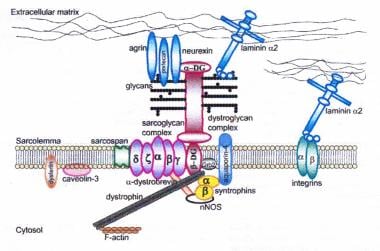
Dystrophin-glycoprotein complex bridges the inner cytoskeleton (F-actin) and the basal lamina. Mutations in all sarcoglycans, dysferlin, and caveolin-3, as well as mutations that cause abnormal glycosylation of alpha-dystroglycan can result in limb-girdle muscular dystrophy syndrome.
Click to see larger pictureClick to see detailView Full Size Image
Media type:  Image

Media file 2:  
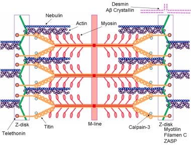
Schematic of the sarcomere with labeled molecular components that are known to cause limb-girdle muscular dystrophy or myofibrillar myopathy. Mutations in actin and nebulin cause the congenital myopathy nemaline rod myopathy, and the mutations in myosin cause familial hypertrophic cardiomyopathy.
Click to see larger pictureClick to see detailView Full Size Image
Media type:  Image

Media file 3:  Top, Photomicrograph shows normal alpha-sarcoglycan staining of a myopathic biopsy specimen. Note dark staining around the rims of the muscle fibers. Bottom, Alpha-sarcoglycan stain of a muscle biopsy specimen from a patient with alpha-sarcoglycan deficiency. Note the absence of staining at the rims of the muscle fibers. Patterns of staining similar to these are observed in all the sarcoglycanopathies, dysferlinopathy, calpainopathy and limb-girdle muscular dystrophy type 2I (LGMD2I, Fukutin-related proteinopathy). However, staining may be variably reduced or absent.
Click to see larger pictureClick to see detailView Full Size Image
Media type:  Photo

Media file 4:  Gomori trichrome–stained section in patient with myofibrillar myopathy. Note the abnormal accumulations of blue-red material in several muscle fibers.
Click to see larger pictureClick to see detailView Full Size Image
Media type:  Photo

Media file 5:  Immunohistochemical staining by using an anti-desmin antibody in a patient with a myofibrillar myopathy. Courtesy of Alan Pestronk.
Click to see larger pictureClick to see detailView Full Size Image
Media type:  Photo


  1. Walton JN, Nattrass FJ. On the classification, natural history and treatment of the myopathies. Brain. 1954;77(2):169-231. [Medline].
  2. Moore SA, Shilling CJ, Westra S. Limb-girdle muscular dystrophy in the United States. J Neuropath Exp Neurol. 2006;65:995-1003. [Medline].
  3. Sveen ML, Schwartz M, Vissing J. High prevalence and phenotype-genotype correlations of limb girdle muscular dystrophy type 2I in Denmark. Ann Neurol. 2006;59:808-15. [Medline].
  4. Godfrey C, Escolar D, Brockington M. Fukutin gene mutations in steroid-responsive limb girdle muscular dystrophy. Ann Neurol. 2006;60:603-10. [Medline].
  5. Fanin M, Nascimbeni AC, Angelini C. Screening of calpain-3 autolytic activity in LGMD muscle: a functional map of CAPN3 gene mutations. J Med Genet. 2007;44:38-43. [Medline].
  6. Angelini C, Fanin M, Freda MP, et al. The clinical spectrum of sarcoglycanopathies. Neurology. Jan 1 1999;52(1):176-9. [Medline].
  7. Balci B, Uyanik G, Dincer P, et al. An autosomal recessive limb girdle muscular dystrophy (LGMD2) with mild mental retardation is allelic to Walker-Warburg syndrome (WWS) caused by a mutation in the POMT1 gene. Neuromuscul Disord. Apr 2005;15(4):271-5. [Medline].
  8. Bar H. Mucke N. Ringler P. Muller SA. Kreplak L. Katus HA. Aebi U. Herrmann H. Impact of disease mutations on the desmin filament assembly process. J Molec Biol. Jul 2006;360:1031-42. [Medline].
  9. Bashir R, Britton S, Strachan T, et al. A gene related to Caenorhabditis elegans spermatogenesis factor fer-1 is mutated in limb-girdle muscular dystrophy type 2B. Nat Genet. Sep 1998;20(1):37-42. [Medline].
  10. Betz RC, Schoser BG, Kasper D, et al. Mutations in CAV3 cause mechanical hyperirritability of skeletal muscle in rippling muscle disease. Nat Genet. Jul 2001;28(3):218-9. [Medline].
  11. Boito CA, Melacini P, Vianello A, et al. Clinical and molecular characterization of patients with limb-girdle muscular dystrophy type 2I. Arch Neurol. 2005;62:1894-9. [Medline].
  12. Brockington M, Yuva Y, Prandini P, et al. Mutations in the fukutin-related protein gene (FKRP) identify limb girdle muscular dystrophy 2I as a milder allelic variant of congenital muscular dystrophy MDC1C. Hum Mol Genet. Dec 1 2001;10(25):2851-9. [Medline].
  13. Bönnemann CG, Bushby K. The limb-girdle muscular dystrophies. In: Engel AG, Franzini-Armstrong C. Myology. 3rd ed. New York, NY: McGraw Hill; 2004:1077-1121.
  14. Carbone I, Bruno C, Sotgia F, et al. Mutation in the CAV3 gene causes partial caveolin-3 deficiency and hyperCKemia. Neurology. Mar 28 2000;54(6):1373-6. [Medline].
  15. Carrie A, Piccolo F, Leturcq F, et al. Mutational diversity and hot spots in the alpha-sarcoglycan gene in autosomal recessive muscular dystrophy (LGMD2D). J Med Genet. Jun 1997;34(6):470-5. [Medline].
  16. D'Amico A, Tessa A, Bruno C. Expanding the clinical spectrum of POMT1 phenotype. Neurology. 2006;66:1564-7. [Medline].
  17. D'Amico A. Benedetti S. Petrini S. Sambuughin N. Boldrini R. Menditto I. Ferrari M. Verardo M. Goldfarb L. Bertini E. Major myofibrillar changes in early onset myopathy due to de novo heterozygous missense mutation in lamin A/C gene. Neuromuscular Disorders. Dec 2005;15:847-50. [Medline].
  18. den Dunnen JT. Leiden Muscular Dystrophy pages. Center for Human and Clinical Genetics. The Netherlands: Leiden University Medical Center; 2003. Available at http://www.dmd.nl. Accessed 2003.
  19. Ferreiro A. Ceuterick-de Groote C. Marks JJ. Goemans N. Schreiber G. Hanefeld F. Fardeau M. Martin JJ. Goebel HH. Richard P. Guicheney P. Bonnemann CG. Desmin-related myopathy with Mallory body-like inclusions is caused by mutations of the selenoprotein N gene. Ann Neurol. May 2004;55:676-86. [Medline].
  20. Fischer D, Walter MC, Kesper K. Diagnostic value of muscle MRI in differentiating LGMD2I from other LGMDs. J Neurol. 2005;252:538-47. [Medline].
  21. Foroud T, Pankratz N, Batchman AP, et al. A mutation in myotilin causes spheroid body myopathy. Neurology. Dec 27 2005;65(12):1936-40. [Medline].
  22. Frosk P, Weiler T, Nylen E, et al. Limb-girdle muscular dystrophy type 2H associated with mutation in TRIM32, a putative E3-ubiquitin-ligase gene. Am J Hum Genet. Mar 2002;70(3):663-72. [Medline].
  23. Fulizio L, Nascimbeni AC, Fanin M, et al. Molecular and muscle pathology in a series of caveolinopathy patients. Hum Mutat. Jan 2005;25(1):82-9. [Medline].
  24. Goudeau B. Rodrigues-Lima F. Fischer D. Casteras-Simon M. Sambuughin N. de Visser M. Laforet P. Ferrer X. Chapon F. Sjoberg G. Kostareva A. Sejersen T. Dalakas MC. Goldfarb LG. Vicart P. Variable pathogenic potentials of mutations located in the desmin alpha-helical domain. Human Mutation. Sep 2006;27:906-13. [Medline].
  25. Guglieri M, Magri F, Comi GP. Molecular etiopathogenesis of limb girdle muscular and congenital muscular dystrophies: boundaries and contiguities. Clinica Chimica Acta. 2005;361:54-79. [Medline].
  26. Hackman P, Vihola A, Haravuori H, et al. Tibial muscular dystrophy is a titinopathy caused by mutations in TTN, the gene encoding the giant skeletal-muscle protein titin. Am J Hum Genet. Sep 2002;71(3):492-500. [Medline].
  27. Hauser MA, Horrigan SK, Salmikangas P, et al. Myotilin is mutated in limb girdle muscular dystrophy 1A. Hum Mol Genet. Sep 1 2000;9(14):2141-7. [Medline].
  28. Holt KH, Campbell KP. Assembly of the sarcoglycan complex. Insights for muscular dystrophy. J Biol Chem. Dec 25 1998;273(52):34667-70. [Medline].
  29. Kramerova I, Beckmann JS, Spencer MJ. Molecular and cellular basis of calpainopathy (limb girdle muscular dystrophy type 2A). Biochim Biophys Acta. Feb 2007;1772(2):128-44. [Medline].
  30. Kramerova I, Kudryashova E, Tidball JG, Spencer MJ. Null mutation of calpain 3 (p94) in mice causes abnormal sarcomere formation in vivo and in vitro. Hum Mol Genet. Jul 1 2004;13(13):1373-88. [Medline].
  31. Laval SH, Bushby KM. Limb-girdle muscular dystrophies--from genetics to molecular pathology. Neuropathol Appl Neurobiol. Apr 2004;30(2):91-105. [Medline].
  32. Liu J, Aoki M, Illa I, et al. Dysferlin, a novel skeletal muscle gene, is mutated in Miyoshi myopathy and limb girdle muscular dystrophy. Nat Genet. Sep 1998;20(1):31-6. [Medline].
  33. Messina DN, Speer MC, Pericak-Vance MA, McNally EM. Linkage of familial dilated cardiomyopathy with conduction defect and muscular dystrophy to chromosome 6q23. Am J Hum Genet. Oct 1997;61(4):909-17. [Medline].
  34. Minetti C, Sotgia F, Bruno C, et al. Mutations in the caveolin-3 gene cause autosomal dominant limb-girdle muscular dystrophy. Nat Genet. Apr 1998;18(4):365-8. [Medline].
  35. Moreira ES, Wiltshire TJ, Faulkner G, et al. Limb-girdle muscular dystrophy type 2G is caused by mutations in the gene encoding the sarcomeric protein telethonin. Nat Genet. Feb 2000;24(2):163-6. [Medline].
  36. Muchir A, Bonne G, van der Kooi AJ, et al. Identification of mutations in the gene encoding lamins A/C in autosomal dominant limb girdle muscular dystrophy with atrioventricular conduction disturbances (LGMD1B). Hum Mol Genet. May 22 2000;9(9):1453-9. [Medline].
  37. Neuromuscular Disease Center. Dilated cardiomyopathy. St Louis, Mo: Washington University. Available at www.neuro.wustl.edu/neuromuscular/msys/cardiac2.htm#dilated. Accessed January 12, 2006.
  38. Neuromuscular Disease Center. Large or prominent muscles. Familial partial lipodystrophy (Kobberling-Dunnigan syndrome). St Louis, Mo: Washington University. Available at www.neuro.wustl.edu/neuromuscular/mother/mlarge.html#kds. Accessed September 19, 2005.
  39. Olive M, Goldfarb LG, Shatunov A, et al. Myotilinopathy: refining the clinical and myopathological phenotype. Brain. Oct 2005;128(Pt 10):2315-26. [Medline].
  40. Ozawa E, Mizuno Y, Hagiwara Y. Molecular and cell biology of the sarcoglycan complex. Muscle Nerve. 2005;32:563-76. [Medline].
  41. Palenzuela L, Andreu AL, Gamez J, et al. A novel autosomal dominant limb-girdle muscular dystrophy (LGMD 1F) maps to 7q32.1-32.2. Neurology. Aug 12 2003;61(3):404-6. [Medline].
  42. Passos-Bueno MR, Vainzof M, Moreira ES, Zatz M. Seven autosomal recessive limb-girdle muscular dystrophies in the Brazilian population: from LGMD2A to LGMD2G. Am J Med Genet. Feb 19 1999;82(5):392-8. [Medline].
  43. Penisson-Besnier I. Talvinen K. Dumez C. Vihola A. Dubas F. Fardeau M. Hackman