Practice Essentials
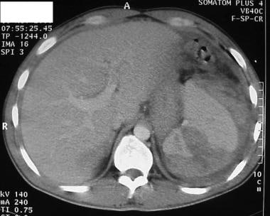
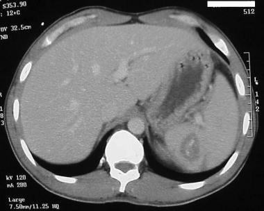
Although protected under the bony ribcage, the spleen remains the most commonly affected organ in blunt injury to the abdomen in all age groups. While some references occasionally document liver injuries as being more common, blunt injuries to the spleen are documented more frequently as the primary solid organ injury in the abdomen. These injuries are common in both rural and urban environments and result from motor vehicle crashes, domestic violence, sporting events, and accidents involving bicycle handlebars. See the images below.
A general surgeon in a community hospital is just as likely to observe and treat a splenic injury as the full-time trauma surgeon in an American College of Surgeons (ACS) –verified Level 1 or Level 2 trauma center. For this reason, all physicians involved in emergency care, especially surgeons, whether rural or urban, must keep up-to-date on issues regarding splenic injury diagnosis, splenic salvage techniques, indications for nonoperative treatment, and potential complications arising from both operative splenectomy and nonoperative management of this important organ.
History of the Procedure
In 1893, Reigner published the first documented successful splenectomy in the German literature. Operative mortality rates remained high until the 1950s, when new and rapid advancements in surgical and anesthesia sciences occurred. Nonoperative care during this period was predominantly fatal. Prior to the advent of CT scanning, physical examination and diagnostic procedures such as diagnostic peritoneal lavage (DPL) and radioisotope scans were the only diagnostic methods. Minor splenic injury was probably frequently missed, while major injury prompting laparotomy for hypotension or physical findings was the norm.
With the widespread availability of computed tomography surgeons began to focus on those needing surgery and those who could be observed safely. Starting with the pediatric population and expanding into the adult population, nonoperative observation became more prevalent for hemodynamically stable patients. Further improvements in CT sensitivity and specificity made vascular extravasation easier to diagnose, and interventional radiology became an integral part of the management of splenic injuries, in some institutions replacing emergency operation as the treatment of choice. Some authors have even noted that resident experience with splenectomy for trauma have been surpassed by medical indications for splenectomy and that emergent splenectomy may be an endangered species in training centers. [1]
Problem
The spleen, weighing 75-150 g, is a highly vascular organ that filters an estimated 10-15% of total blood volume every minute. The spleen may hold 40-50 mL of red cells in reserve on average; however, with changes in internal smooth muscle, it can pool significantly more blood. Historically, many early shock studies performed in canine models were invalidated when it was discovered that dogs could autotransfuse stored red cells from their spleen with smooth muscle contraction. Humans do not have this ability. As much as 25% of the circulating platelets are estimated to be held in reserve in the spleen. Although protected anatomically under the rib cage in the left upper quadrant of the abdomen, it is frequently injured by blunt external trauma. It can also be iatrogenically injured in emergency operations, especially when preexisting adhesions make mobilization of intra-abdominal structures difficult.
Because of the immunologic function of the spleen, interest over the last century has turned to salvage of the spleen rather than splenectomy. The advent of CT scanning has made conservative management more practical and safer for victims of splenic injury. CT scanning has facilitated safe, nonoperative management in young and old patients to an unprecedented degree, but deaths due to splenic rupture are still reported in hospital discharge statistics from both Level 1 trauma centers and community hospitals.
A thorough knowledge of splenic function, anatomy, and pathophysiology is necessary to continue the progress of the last decade and to decrease the mortality rate from this common injury in the United States and worldwide.
Epidemiology
Determining the actual frequency of splenic injuries with precision in the United States or worldwide is not possible. Hospital discharge data may not document the injury if there are numerous, more serious injuries or diseases. A general consensus of trauma admissions at Level 1 trauma centers across the country suggests splenic injury occurs in as many as 25% of the average 800-1200 admissions for blunt trauma per year. This is a select population of patients with multiple injuries and does not take into account isolated splenic injuries observed and treated at nontrauma centers.
Etiology
Splenic injury is most often observed in blunt trauma. While penetrating trauma (eg, gun shot wounds, knife wounds) may involve the spleen, the incidence of injury is well below that of the small and large intestine. A third mechanism that combines aspects of blunt and penetrating trauma occurs with explosive type injuries, as seen in warfare and civilian bombing.
Although the spleen is relatively protected under the ribcage, injury due to rapid deceleration, such as occurs in motor vehicle crashes, direct blows to the abdomen in domestic violence, or leisure and play activities such as bicycling, frequently result in a variety of splenic injuries.
Another cause of splenic injury has been gaining notice. There have been case reports of splenic injury following colonoscopy. [2] Ha and Minchin performed a literature search to identify the demographic profile, risk factors, clinical presentations, diagnosis and management of this rare complication. [3] The investigators found 66 patients (median age, 65 y) with a 4.5% mortality rate, the majority (n = 41, 62.1%) of which occurred in uneventful colonoscopies. Symptoms primarily (74%) appeared within 24 hours, and workup in the form of blood tests and CT scanning was performed in the majority (93.9%). [3]
In addition, over half of (56.1%) affected patients underwent laparotomy and splenectomy, with the most common findings of splenic hematoma (47%), laceration (47%), and rupture (33.3%). [3] Ha and Minchin concluded that recognition of postcolonoscopy splenic injury as an important complication will not only rise, but it will be necessary given the increasing numbers of colonoscopies being performed for colorectal diseases and the possibility of delayed diagnosis resulting in adverse outcomes.
Pathophysiology
Though normally protected by its anatomic position, preexisting illness or disease can markedly increase the risks and severity of splenic injury. Infectious mononucleosis, malaria, and hematologic abnormalities can lead to acute or chronic enlargement of the spleen. This is often accompanied by a thinning of the capsule, making the spleen more fragile as well as engendering a greater mass effect in decelerating trauma. Minor impact in patients with splenomegaly reportedly results in major injury and the need for splenectomy.
Severe acute respiratory syndrome coronavirus 2 can target the spleen, and cases of atraumatic splenic rupture have been reported in patients with coronavirus disease 2019 (COVID-19). [4, 5, 6] In the case of a 13-year-old boy who presented with left-sided abdominal pain, the splenic rupture was the sole manifestation of COVID-19. [4]
Presentation
The clinical presentation of splenic injury is highly variable. Most patients with minor focal injury to the spleen complain of left upper quadrant abdominal tenderness. Left shoulder tenderness may also be present as a result of subdiaphragmatic nerve root irritation with referred pain.
With free intraperitoneal blood, diffuse abdominal pain, peritoneal irritation, and rebound tenderness are more likely. If the intra-abdominal bleeding exceeds 5-10% of blood volume, clinical signs of early shock may manifest. Signs include tachycardia, tachypnea, restlessness, and anxiety. Patients may have a mild pallor noted only by friends and family. Clinical signs include decreased capillary refill and decreased pulse pressure. With increasing blood loss into the abdominal cavity, abdominal distension, peritoneal signs, and overt shock may be observed.
Hypotension in a patient with a suspected splenic injury, especially if young and previously healthy, is a grave sign and a surgical emergency. This should prompt immediate evaluation and intervention either in the OR or interventional radiology if a state of compensated shock can be maintained. Unstable patients have nearly exsanguinated in CT scanners while in the process of documenting splenic injury, when they would have been better served by exploration in the operating room or embolization in the IR suite.
Indications
In simple terms, unstable patients suspected of splenic injury and intra-abdominal hemorrhage should undergo exploratory laparotomy and splenic repair or removal. A blunt trauma patient with evidence of hemodynamic instability unresponsive to fluid challenge with no other signs of external hemorrhage should be considered to have a life-threatening solid organ (splenic) injury until proven otherwise. Transient responders, those patients who respond to an initial fluid bolus only to deteriorate again with a drop in blood pressure and increasing tachycardia, are also likely to have solid organ injury with ongoing hemorrhage. Patients with compensated shock may be managed by angioembolization but only if these services can be performed in a timely manner equivalent to that of operative intervention.
DPL may be a valuable adjunct if time permits and multiple other injuries are present. Focused abdominal sonographic technique (FAST) in experienced hands is helpful in documenting the presence or absence of blood in the peritoneal cavity, which highly suggests the possibility of splenic injury. However, bedside FAST in the resuscitation suite does not show actual splenic injury well enough to use as a diagnostic modality for solid organ injury imaging. Rozycki et al performed a pilot study using bedside organ assessment with sonography after trauma (BOAST) and documented its limitations in identifying solid organ injury, especially at lower grades of injury. FAST is excellent for documenting the presence or absence of intra-abdominal fluid but should not be viewed as an equivalent to CT scanning with regard to injury site determination. [7]
Sirlin et al showed that patterns of fluid accumulation on FAST may be used to improve identification of specific organ injuries, but this still does not approach the sensitivity and specificity of CT. [8]
In the stable trauma patient, commonly defined as a patient with systolic blood pressure greater than 90 mm Hg with a heart rate less than 120 beats per minute (bpm), CT scanning provides the most ideal noninvasive means for evaluating the spleen. Helical or spiral scanners may provide even more information and may clarify the degree of injury. In the cases of CT scan–documented splenic injury, the decision for operative intervention is determined by the grade of the injury, the patient's current and preexisting medical conditions, and the facilities available at the hospital, including the intensive care unit and the availability of operating and anesthesia services.
The availability of interventional angiographic services also impacts a surgeon's decision for or against operative intervention. The use of MRI has also been reported in the literature as an option in the patient with an elevated creatinine level. [9]
The major determining factors in operative intervention in the stable patient with a splenic injury include grade of injury (American Association for the Surgery of Trauma [AAST] scale), presence of intraperitoneal blood, presence of a blush on CT scan, calculated risk of rebleeding, presence and severity of concomitant injuries, and options regarding blood transfusion.
Signs of persistent bleeding and hemodynamic instability unresponsive to fluid and blood administration are clear indications for surgery. The decision for operative intervention in other cases requires the thoughtful consideration of the surgeon. Angioembolization, once contraindicated in compensated shock, has now been reported as a safe method of splenic salvage when immediately available in the treating facility. [10] A healthy 25-year-old patient who has a CT scan grade 4 laceration with stable vital signs and minimal fluid requirements may be safe to observe under controlled conditions, while a 55-year-old patient who is a Jehovah's Witness and who has a CT scan grade 2 oozing splenic injury and pelvic fracture would probably benefit more from early surgical intervention.
Relevant Anatomy
The spleen sits in the left upper quadrant of the abdomen under the diaphragm and lateral to the stomach. Left shoulder pain, also known as the Kehr sign, results when blood from an injured spleen irritates the diaphragm and creates referred pain. The spleen is completely encircled and covered with peritoneum except for the insertion of the splenic artery and vein. This capsule around the spleen, especially the thicker layer in young patients, provides added protection against blunt injury. The spleen is primarily fixated to the posterior aspect of the left upper quadrant by gastrosplenic and splenorenal ligaments. The size and thickness of these ligaments vary greatly, with some spleens appearing to be very mobile, while others appear fixed in the left upper quadrant.
The major arterial supply to the spleen is through the splenic artery, which branches off the celiac artery and runs superior and posterior to the pancreas. The artery commonly bifurcates externally to the spleen, supplying upper and lower poles separately, a finding that may make splenorrhaphy much easier for the operating surgeon. The splenic vein courses with the artery but empties into the superior mesenteric vein and then into the portal vein. The arterial supply and venous drainage of the spleen is augmented by the short gastric vessels that branch from the left gastroepiploic artery. These vessels may be as short as 1 mm, thus creating a challenge during emergency operative intervention. Notably, the splenic artery and vein may have small branches feeding the body and tail of the pancreas, so care should be taken in dissecting these vessels away from the splenic hilum.
The tail of the pancreas is often intimately positioned near the splenic hilum and can be easily damaged during splenectomy if adequate care is not taken to identify and protect the organ.
Contraindications
No contraindications to operative intervention exist in a hemodynamically unstable patient with a splenic injury. However, hypotension or unstable vital signs are a contraindication to CT scanning, and deaths due to splenic rupture and ongoing bleeding have occurred in the radiology suite while trying to document a splenic injury. Unstable patients can be assessed by FAST or DPL in addition to clinical examination but should not undergo CT scanning of the abdomen for diagnosis.
-
Hemisplenectomy (splenorrhaphy) with preservation of greater than 50% of splenic parenchyma.
-
Intra-parenchymal blush observed on helical CT scan.
-
Physical findings in postsplenectomy sepsis with peripheral thrombosis and disseminated intravascular coagulation (DIC).
-
Grade 4-5 splenic laceration on helical CT scan.