Practice Essentials
Pyelolithotomy is a surgical procedure used in cases involving a stone in the renal pelvis. Indications for pyelolithotomy include minimally branched staghorn stones in the renal pelvis of complex collecting systems and excessive morbid obesity. Pyelolithotomy is also appropriate in patients who are undergoing major open abdominal or retroperitoneal surgical procedures for other indications; the most common concomitant procedure is open pyeloplasty for ureteropelvic junction (UPJ) obstruction. See the image below.
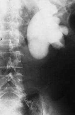 Intravenous pyelogram demonstrating ureteropelvic junction obstruction with dilatation of the collecting system and lack of excretion of contrast.
Signs and symptoms
Patients may be asymptomatic or may present with following symptoms:
-
Renal colic
-
Flank pain
-
Sepsis
-
Hematuria
Diagnosis
Laboratory studies
The usual preoperative laboratory studies include the following:
-
Complete blood cell count
-
Blood type
-
Activated partial thromboplastin time and prothrombin time
-
Electrolytes
-
Blood urea nitrogen
-
Creatinine
In addition, a urinalysis (with culture and sensitivity) is performed.
Imaging studies
The following imaging studies are usually performed to confirm the diagnosis:
-
Radiograph of the kidneys, ureters, and bladder (KUB)
-
CT scan
-
Intravenous pyelogram (IVP)
See Workup for more detail.
Management
A pyelolithotomy can be performed as an open, laparoscopic, or robotic procedure. The approach can be transperitoneal or retroperitoneal.
See Treatment for more detail.
Background
The term pyelo means renal pelvis, and the term lithotomy means removal of stone. Since the advent of extracorporeal shockwave lithotripsy (ESWL) and percutaneous nephropyelolithotomy (PCN), pyelolithotomy has become an uncommon surgery in most developing countries. However, before these newer technologies, pyelolithotomy was the procedure of choice for stones within the renal pelvis, including stones that demonstrated minimal invasion into calyces and infundibulum. Pyelolithotomy differs from an anatrophic nephrolithotomy, as the anatrophic nephrolithotomy allows for greater access to calyces and allows for repair of infundibulum and calyces. Anatrophic nephrolithotomy is indicated for large multiple-branched staghorn calculi with infundibular stenosis.
ESWL is clearly noninvasive, but it may necessitate (1) a cystoscopy and the insertion of a stent to drain the kidney or (2) a nephrostomy in some cases involving infection. ESWL is associated with less morbidity than pyelolithotomy, but the overall failure rate and the amount of residual stone fragments are higher. Lower pole stones fragments do not flush out of the renal unit as readily as midpole and upper pole fragments.
PCN is a highly technical procedure and requires some experience for optimal results. At some facilities, these procedures require the teamwork of a radiologist and a urologist. Morbidity is higher than with ESWL, but residual stone fragments are less common. The stone-free rate associated with percutaneous nephrolithotomy (PNL) is 90%; ESWL, 54%.
The 2004 American Urological Association (AUA) guidelines recommend that staghorn smaller than 2500 mm2 with normal renal anatomy should be treated with PNL as first-line treatment and with ESWL as a follow-up procedure.
Pyelolithotomy continues to have a role in the management of renal pelvic stones in areas where ESWL and PNL are not feasible because of the lack of equipment or expertise.
Indications for pyelolithotomy include minimally branched staghorn stones in the renal pelvis of complex collecting systems and excessive morbid obesity. Pyelolithotomy is also appropriate in patients who are undergoing major open abdominal or retroperitoneal surgical procedures for other indications; the most common concomitant procedure is open pyeloplasty for ureteropelvic junction (UPJ) obstruction.
History of the Procedure
On October 8, 1872, Ingalls performed a nephrotomy at Boston City Hospital. In 1880, Henry Morris, an English surgeon, performed the first pyelolithotomy on a 31-year-old woman. Vincenz Czerny also performed a pyelolithotomy in 1880. These initial operations were performed without regard for renal vasculature, anatomy, or functionality. The technique was refined after Gil-Vernet better described renal vascularity and function of the collecting system musculature. [1] The incision of the renal pelvis was initially taken vertically but, after Gil-Vernet's description, became a transverse incision, therefore preserving anatomic musculature and blood supply.
Presentation
Patients may be asymptomatic or may present with symptoms that include renal colic, flank pain, sepsis, and/or hematuria. In addition, incidental findings of stones on a CT scan or during laboratory workup studies that demonstrated an elevated creatinine level have followed with a finding of partially obstructive stones within the renal pelvis.
Indications
Pyelolithotomy is a surgical procedure in cases involving a stone in the renal pelvis. This was a common procedure until the development of extracorporeal shockwave treatment, PNL, and ureteroscopic laser lithotripsy. However, pyelolithotomy continues to be performed when other modalities fail or when proper facilities are unavailable.
Although it is now considered overly invasive for routine use, pyelolithotomy continues to have a role in select cases. Criteria include the size of the stone, [2] the need for concomitant open surgery, and an inaccessibility to ESWL or PCN. Current guidelines advocate pyelolithotomy or anatrophic nephrolithotomy when the stone burden is greater than 2500 mm2, in cases of extreme morbid obesity, or when the patient presents with a complex collecting system.
Other indications are relative and include failure of stone clearance via PCN, ureteroscopy, or ESWL owing to difficult extraction, stone composition (ie, cystine), or anatomy (ie, ectopic, pelvic, or horseshoe kidney). Pyelolithotomy is also indicated in combination with pyeloplasty, without increasing morbidity or decreasing the success rate. [3]
Indications for stone removal (possible pyelolithotomy) include sepsis, severe flank pain, obstruction with impending parenchymal renal loss, and hematuria. Patients who present for pyelolithotomy also meet the criteria as outlined above.
Relevant Anatomy
The renal pelvis is posterior to the hilum of the kidney. From anterior to posterior, the relationship of the structures is renal vein, renal artery, and pelvis.
The pelvis can be extrarenal or intrarenal. In an intrarenal pelvis, the pelvis is embedded in the parenchyma of the kidney. An extrarenal pelvis is exposed outside of the parenchyma and is easily reachable. The renal pelvis joins the ureter at the UPJ. Normal pelvis volume is 3-5 mL.
On the left side, the ovarian vein or testicular vein is adjacent to the ureter and pelvis. Recognize and identify these veins during surgery to avoid injury and bleeding.
Contraindications
Pyelolithotomy is absolutely contraindicated in patients in a poor general medical condition. Only consider this surgery when all other options fail.
Relative contraindications include branched staghorn calculi with infundibular stenosis and stones in the calices. These conditions may be approached using the Boyce anatrophic nephrolithotomy or calycelectomy.
-
Intravenous pyelogram demonstrating ureteropelvic junction obstruction with dilatation of the collecting system and lack of excretion of contrast.
-
Retrograde pyelogram demonstrating ureteropelvic junction obstruction secondary to annular stricture.


