Continually Updated Clinical Reference
 
 
  All Sources     eMedicine     Medscape     Drug Reference     MEDLINE
 
eMedicine - Papillary Necrosis : Article by

Quick Find
Authors & Editors
Introduction
Indications
Relevant Anatomy
Contraindications
Workup
Treatment
Complications
Outcome and Prognosis
Future and Controversies
Multimedia
References


Patient Education
Kidneys and Urinary System Center

Blood in Urine Overview

Causes of Blood in Urine

Blood in Urine Symptoms

Blood in Urine Treatment


Author: Jeffrey M Donohoe, MD, Assistant Professor of Pediatric Urology, Department of Surgery, Division of Urology, Medical College of Georgia

Jeffrey M Donohoe is a member of the following medical societies: American Academy of Pediatrics and American Urological Association

Coauthor(s): Jack H Mydlo, MD, Chief, Department of Urology, Woodhull Hospital; Chair and Professor, Department of Urology, Temple University School of Medicine

Editors: Gamal Mostafa Ghoniem, MD, FACS, Fellowship Program Director, Clinical Professor of Surgery, Head, Section of Voiding Dysfunction, Female Urology and Reconstruction, Cleveland Clinic Florida; Francisco Talavera, PharmD, PhD, Senior Pharmacy Editor, eMedicine; J Stuart Wolf, Jr, MD, FACS, David A Bloom Professor of Urology, Director, Division of Minimally Invasive Urology, Department of Urology, University of Michigan Medical Center; Bradley Fields Schwartz, DO, FACS, Associate Professor of Urology, Director, Center for Laparoscopy and Endourology, Department of Surgery, Southern Illinois University School of Medicine

Author and Editor Disclosure

Synonyms and related keywords: papillary necrosis, renal papillary necrosis, RPN, renal medullary necrosis, medullary necrosis, interstitial nephritis, sloughed papilla, urinary obstruction, ureteropelvic junction obstruction, UPJ obstruction, ureteral obstruction, ureterovesical junction obstruction, analgesic nephropathy, analgesic-induced nephropathy, analgesic abuse, pyelonephritis, tubulointerstitial nephritis, urinary tract infection, UTI, sickle cell hemoglobinopathy, sickle cell trait, sickle cell disease, tuberculosis, cirrhosis of the liver, cirrhosis, chronic alcoholism, renal transplant rejection, diabetes mellitus, diabetes complications, systemic vasculitis, phenacetin

Renal papillary necrosis (RPN) is characterized by coagulative necrosis of the renal medullary pyramids and papillae brought on by several associated conditions and toxins that exhibit synergism toward the development of ischemia. The clinical course of renal papillary necrosis varies depending on the degree of vascular impairment, the presence of associated causal factors, the overall health of the patient, the presence of bilateral involvement, and, specifically, the number of affected papillae.

Renal papillary necrosis can lead to secondary infection of desquamated necrotic foci, deposition of calculi, and/or separation and eventual sloughing of papillae, with impending acute urinary tract obstruction. Multiple sloughed papillae can obstruct their respective calyces or can congregate and embolize to more distal sites (eg, ureteropelvic junction, ureter, ureterovesical junction). Previously undiagnosed congenital anomalies (eg, partial ureteropelvic junction obstruction) can provide a narrowed area where the sloughed papilla can nest and obstruct.

Renal papillary necrosis is potentially disastrous and, in the presence of bilateral involvement or an obstructed solitary kidney, may lead to renal failure. The infectious sequelae of renal papillary necrosis are more serious if the patient has multiple medical problems, particularly diabetes mellitus.

History of the Procedure

In 1877, Friedrich first described renal papillary necrosis in a patient with urinary obstruction resulting from hypertrophy of the prostate.1 Later, Gunther2 and Edmondson et al3 described renal papillary necrosis as a lesion associated with diabetes. Since then, researchers have reported that 17-90% of all patients with renal papillary necrosis have diabetes and that 25-73% of patients have severe urinary tract obstructions. In 1945, Spuhler and Zollinger documented the first description of analgesic nephropathy.4 Since then, analgesic abuse has been increasingly significant in the development of papillary necrosis, particularly in Australia, England, and Scandinavia.

Ludwig von Beethoven's 1827 autopsy report predates all of these descriptions. The original Latin autopsy protocol by Johann Wagner was translated into German by von Seyfried in 1832 and has only recently been rediscovered.

Reports indicate that Beethoven, the highly regarded musical genius and arguably the best composer of all time, was a long-term abuser of alcohol and analgesics. Beethoven was prone to headaches, back pain, and attacks of rheumatism or gout. His physician often prescribed salicin, a commonly used analgesic substance at that time, which was made from dried and powdered willow bark. His alcohol abuse was compounded by a viral hepatitis infection that led to liver cirrhosis and chronic pancreatitis. Davies postulated that Beethoven developed diabetes mellitus secondary to chronic pancreatitis. Beethoven had no reported history of urinary obstruction, nor was it mentioned in his autopsy report.

Wagner's autopsy, as translated by von Seyfried into German, reported "calcareous concretions" filling every calyx of both kidneys. He described the concretions as "symmetrical and soft like a pea cut across the middle." According to Davies and Schwarz, this finding is so typical of renal papillary necrosis that the diagnosis is as near to certain as possible without a histological examination. Researchers postulate that Beethoven's renal papillary necrosis was most likely a consequence of analgesic abuse and decompensated liver cirrhosis, which ultimately caused his death. Davies contests that Beethoven also had diabetes and that this illness was the primary risk factor for him developing renal papillary necrosis. Indeed, all 3 conditions may have been synergistic factors. In any case, Wagner's protocol from his autopsy of Beethoven represents the first description of papillary necrosis recorded in the literature.

Problem

Renal papillary necrosis is sometimes classified as one end of a spectrum of changes associated with pyelonephritis and tubulointerstitial nephritis. Renal papillary necrosis is often considered a complication or extension of severe pyelonephritis that is more devastating than usual because of associated disease states, particularly diabetes and urinary tract obstructions. Other sources believe this consideration is inaccurate and archaic because renal papillary necrosis does not usually occur with florid pyelonephritis.

Indeed, pyelonephritis and an ascending urinary tract infection are conditions that are commonly associated with renal papillary necrosis. Infection is a frequent and important finding in most cases, contributing significantly to the clinical presentation of renal papillary necrosis (ie, fever and chills in approximately two thirds of patients, positive urine culture results in 70%). However, renal papillary necrosis can occur in the absence of infection, indicating that infection may not be the primary process in the pathogenesis. Conversely, infection is likely a complication of renal papillary necrosis; the necrotic papillae act as a nidus for infection and lithogenesis. Infection within necrotic material and calculi is often difficult to definitively treat with antibiotics alone, and infection often recurs as renal papillary necrosis progresses to chronic pyelonephritis.

Renal papillary necrosis is considered a sequela of ischemia occurring in the renal papillae and the medulla. Various insults generate this ischemia, one of which may be infection. The boggy inflammatory interstitium of the pyelonephritic kidney compresses the medullary vasculature and, thus, predisposes the patient to ischemia and renal papillary necrosis. This vasculature can become compressed, attenuated, or impaired from several other associated diseases, most notably diabetes mellitus, urinary tract obstruction, and analgesic nephropathy. Therefore, renal papillary necrosis is a distinct clinical and pathophysiological entity primarily caused by ischemia that can develop without pyelonephritis or urinary tract infection and is likely a focus for infectious complications. Renal papillary necrosis has a well-documented association with several diseases that predispose a patient to ischemia.

Renal papillary necrosis is primarily a bilateral process, as is expected considering the systemic nature of the associated diseases and the ischemic pathophysiologic mechanism of renal papillary necrosis. Reports indicate that, among patients in whom a single kidney is involved at initial presentation, the contralateral kidney will develop renal papillary necrosis within 4 years. Physicians diagnose true unilateral renal papillary necrosis when the patient's predisposing factor is infection or obstruction that is limited to one kidney, as is the scenario in a patient with congenital ureteropelvic junction obstruction.

Frequency

Renal papillary necrosis generally affects individuals who are in the middle decades of life or older. The typical patient is aged 53 years, with nearly half of cases occurring in individuals older than 60 years and more than 90% of cases occurring in individuals older than 40 years. Renal papillary necrosis is uncommon in individuals younger than 40 years and in the pediatric population, except in patients with sickle cell hemoglobinopathies, hypoxia, dehydration, and septicemia.

In general, renal papillary necrosis is more common in women than in men. Mandel organized the first comprehensive review to focus attention on renal papillary necrosis.5 In his series, which examined 160 cases of renal papillary necrosis from the world literature, 96 patients (60%) had diabetes mellitus, 48 patients (30%) had urinary tract obstruction, and 15 patients (9.4%) had both. In the group with diabetes, women outnumbered men, and in the group of patients without diabetes, men outnumbered women, further reflecting the frequency and significance of urinary tract obstructions in elderly men with renal papillary necrosis. An autopsy report from 1957 by Simon and associates documented the presence of acute or chronic pyelonephritis in 95%.6

Analgesic nephropathy, another associated condition and causal factor of renal papillary necrosis, is also more common in women than in men. Analgesic nephropathy is particularly prevalent in individuals with recurrent headaches or chronic, unremitting muscle and joint pain and in patients with psychoneuroses.

Occupational risks are not associated with developing renal papillary necrosis.

Etiology

Generally, any condition associated with ischemia predisposes an individual to papillary necrosis. Important general considerations include shock, massive fluid sequestration (eg, as in pancreatitis), dehydration, hypovolemia, and hypoxia. Certain conditions have a known association with renal papillary necrosis, and the underlying mechanism of these conditions is ischemia, which ultimately leads to renal papillary necrosis.

A useful mnemonic device for the conditions associated with renal papillary necrosis is POSTCARDS, which stands for the following:

  • Pyelonephritis
  • Obstruction of the urinary tract
  • Sickle cell hemoglobinopathies, including sickle cell trait
  • Tuberculosis
  • Cirrhosis of the liver, chronic alcoholism
  • Analgesic abuse
  • Renal transplant rejection, radiation
  • Diabetes mellitus
  • Systemic vasculitis

More than half the patients with renal papillary necrosis have 2 or more of these causative factors. Thus, renal papillary necrosis in most patients is multifactorial in origin, and physicians must consider the pathogenesis of renal papillary necrosis a combination of detrimental factors that overlap and operate in concert to cause renal papillary necrosis (see Image 1).

One of the most common and most preventable etiologic factors is the use of analgesics. A classic factor is phenacetin, with its highly toxic metabolite, p-phenetidin. Recently, however, the rising popularity of nonsteroidal anti-inflammatory drugs (NSAIDs), particularly those that inhibit cyclooxygenases (ie, COX-1, COX-2) has led to a relatively high frequency of adverse events in patients at risk for renal papillary necrosis.

In healthy individuals in whom renal arterial blood flow is not compromised, NSAIDs have little effect unless they are used in excess. This is mostly true because the kidney is not relying on the vasodilatory effects of prostaglandin to supply adequate perfusion. However, in patients who are predisposed to renal hypoperfusion, local prostaglandin synthesis protects the glomeruli and tubules from ischemia. The inhibition of prostaglandin synthesis by NSAIDs that inhibit COX-1 and, as recently reported, COX-2, removes this protective mechanism and predisposes the kidney to further renal hypoperfusion and, ultimately, ischemia. An extremely important precaution is to strictly monitor patients with prior renal disease or any of the above-mentioned etiologic conditions when prescribing NSAIDs.

Additionally, note that a short course of NSAIDs has caused papillary necrosis and nonoliguric renal failure in otherwise healthy individuals as young as age 17 years. A case such as this may be an anomaly, but caution is warranted when prescribing NSAIDs, and adequate hydration is recommended.

These precautions should also be extended to patients receiving specific COX-2 inhibitors. Touted as being safer than COX-1 inhibitors because they spare the gastrointestinal tract, COX-2 inhibitors have been shown to significantly decrease renal medullary prostaglandin levels. Therefore, care should be exercised when administering even the COX-2 inhibitors to patients with a predisposition to renal disease or renal papillary necrosis, and otherwise healthy patients should maintain adequate hydration and avoid physiologic stress while on these medications.

Pathophysiology

Renal papillary necrosis is classified as focal (ie, involving only the tip of the papilla) or diffuse (ie, involving the whole papilla and areas of the medulla), depending primarily on the patient's degree of impaired vasculature. Renal papillary necrosis may simply affect a single papilla, or the entire kidney may be grossly involved. Once again, renal papillary necrosis is more often a bilateral process; many of the predisposing factors are systemic. Renal papillary necrosis never involves the entire medulla; the disease is always strictly limited to the inner, more distal zone of the medulla and the papilla.

Researchers recognize 2 pathologic forms of renal papillary necrosis—the medullary form and the papillary form. The pathogenic form is dictated by the degree of vascular impairment. The medullary form is characterized by intact fornices, discrete grain-sized necrotic areas, and later defects in the papillae. Clinicians often observe sinus tracts extruding from irregular medullary cavities. In the papillary form, the calyceal fornices and the entire papillary surface are destroyed, demarcated, and sequestered. If these fornices and papillary surfaces are not sloughed, they reepithelialize and acquire a smoother appearance.

Patients with medullary ischemia develop decreased glomerular filtration rates, salt wasting, an impaired ability to concentrate, and polyuria because the vasa rectae supply the medulla and serve the countercurrent exchange mechanism.

The pathologic findings on a cut section include gray-white to yellow necrosis that resembles infection on the tips or distal two thirds of the pyramids. Microscopically, the tissue shows characteristic coagulative infarct necrosis, with preserved tubule outlines. The leukocytic response is limited to the junctions between preserved and destroyed tissue. After the acute phase, scars that can be observed on the cortical surface as fibrous depressions replace the inflammatory foci. This pyelonephritic scar is usually associated with inflammation, fibrosis, and a deformation of the underlying calyces and pelvis.

Papillary necrosis is easily induced experimentally by administering a combination of aspirin and phenacetin, usually to subjects in a water-depleted state. The phenacetin metabolite p-phenetidin exhibits high renal toxicity; it injures cells by covalent binding and oxidative damage. These papillotoxins target interstitial cells via hydroperoxidase-mediated activation. Aspirin induces its potentiating effect by inhibiting the vasodilatory effects of prostaglandin, thus predisposing the papilla to ischemia. Therefore, papillary damage may be the result of a combination of the direct toxic effects of phenacetin metabolites and ischemic injury to both tubular cells and vessels. Grossly, the cortex exhibits depressed areas that represent cortical atrophy overlying the raised necrotic papillae. The papillae within the kidney show various stages of necrosis with calcification, fragmentation, desquamation, and sloughing.

This development contrasts with the papillary necrosis observed in patients with diabetes, whose papillae are generally at the same stage of acute necrosis at any given time. Microscopically, the individual papillary changes range from a patchy appearance to the advanced form, wherein the entire papilla is necrotic. When the papillae remain attached, they are structureless masses with ghosts of tubules and foci of dystrophic calcification. These tubules and calcifications subsequently fragment and slough, becoming potential obstructive entities. The cortical columns of Bertin that contain the glomeruli characteristically remain uninvolved. Certain papillotoxins target interstitial cells, resulting in hydroperoxidase-mediated activation with subsequent inflammatory, degradative, and necrotic cell changes.

Clinical

Renal papillary necrosis has a variable clinical course that ranges from a chronic, protracted, and relapsing form to an acute, rapidly progressive form. The acute progressive form is particularly rare, but the effects are devastating, resulting in death from septicemia and renal failure. Patients with the more common chronic form may remain asymptomatic until diagnosed incidentally through the appearance of a ring shadow on a radiographic image, by the passage of sloughed papillae in the urine, or during autopsy. The symptomatic form manifests as episodes of pyelonephritis and hydronephrosis, and it mimics nephrolithiasis.

The most common presenting symptoms in symptomatic patients include fever and chills, flank and/or abdominal pain, and hematuria. Acute renal failure with oliguria or anuria is rare; when these symptoms develop, the disease may be fulminant, requiring dialysis and potentially resulting in death.

If renal function deteriorates suddenly in a patient with confirmed diabetes or in a patient with a known history of chronic obstruction and/or pyelonephritis, consider the diagnosis of papillary necrosis, even if the patient is asymptomatic.

Acute ureteral obstruction from sloughed papillae manifests as flank pain and colic due to hydronephrosis or pyonephrosis; hematuria is invariably present. Pyonephrosis or secondary acute or relapsing pyelonephritis compounds the presentation with fever, chills, prostration, and sepsis.


The differential diagnoses of flank pain or hematuria includes a plethora of conditions with infectious, neoplastic, congenital, metabolic, and hematogenous sources. Intraluminal and extraluminal lesions may result in ureteral obstruction.

If acute ureteral obstruction manifests as hematuria, colic, and flank pain, consider that nephrogenic calculi may have embolized to the ureter. Approximately 15% of all calculi, particularly uric acid stones, are not radiopaque (ie, visible on radiographs); therefore, the examining clinician needs to perform further imaging studies for an accurate identification.

CT scanning is an excellent imaging modality for identifying all calculi (including uric acid stones), except for the rare indinavir (Crixivan) crystal concretions that form in the ureters of patients with HIV infection who are taking this protease inhibitor. If calculi are not seen on a CT scan, consider the possibility of an obstructing urothelial tumor; cytology and endoscopic procedures help to determine this diagnosis.

Blood coagulum downstream from a bleeding source in the kidney (eg, renal cell carcinoma, angioma, angiomyolipoma, arteriovenous malformation, trauma to a pyelonephritic or multicystic kidney) can also manifest with a similar clinical picture. If a patient has acute ureteral obstruction with fever and leukocytosis, the preferred treatment is drainage of the system.

Pyonephrosis may or may not be present; therefore, the initial short-term management of an obstructed system should be conservative, with resuscitation, drainage, and intravenous antibiotics. Further diagnostic studies can be performed later, once the physician is certain that the system is sterile. This strategy prevents further clinical deterioration.

Extraureteric causes of ureteral obstruction include retroperitoneal lymphadenopathy from metastatic tumors or lymphoma, retroperitoneal fibrosis, uterine myoma, bladder masses, and high-stage pelvic tumors (eg, cervical cancer).

Anything that causes severe bladder outlet obstruction leads to bilateral hydronephrosis or hydronephrosis of a solitary kidney. Congenital anomalies can also directly obstruct or provide stasis that may lead to pyelonephritis and papillary necrosis. Duplicate collecting systems, vesicoureteral reflux, and ureteroceles are important offenders to consider.

In the clinical scenario of fever, chills, prostration, and sepsis without hydronephrosis, the differential diagnoses include pyelonephritis, perinephric abscess, renal hemorrhage, tubulointerstitial nephritis, and glomerulonephritis.

In asymptomatic patients who present with acute renal failure characterized by azotemia, the physician must consider bilateral obstruction or the presence of a solitary obstructed kidney. Renal ultrasonography is a wise first test in this scenario, along with tests for urine and plasma urea, creatinine, sodium, and osmolality. After excluding postrenal causes, consider prerenal and renal sources.


Briefly reviewing basic renal anatomy and histology allows a clear understanding of the pathophysiology of the underlying ischemia of renal papillary necrosis and how this ischemia is distributed.

The renal papilla is the rounded apex of each medullary pyramid and represents the confluence of the collecting ducts from each nephron within that pyramid. An individual minor calyx cups each papilla; these calyces represent the most proximal aspect of the renal collecting system and are lined with transitional cells.

The renal papillary blood supply is derived from 2 sources: the vasa rectae, which disperse blood from the efferent arteriole, and the interlobar branches of the renal artery, which run within the adventitia of the minor calyces. The vasa rectae arise from the efferent arteriole within the renal cortex and form wide and plentiful vascular bundles at the base of the medullary pyramid. The bundles taper as they continue distally toward the apex and papilla. This process results in the papillary tip receiving only a marginal supply of blood, which appears to be a predisposing factor for the central role of ischemia in the development of renal papillary necrosis.

The already tenuous vasculature is further impaired by several pathophysiologic states, including the above-described boggy inflammatory interstitium of pyelonephritis, the microangiopathy of diabetes mellitus, the increased pyelovenous pressure of urinary obstruction, the chronic hyperbilirubinemia of liver cirrhosis, the oxidative damage of analgesic nephropathy, and the intraluminal stasis of sickle cell disease.


Patients with known or suspected renal papillary necrosis should limit or completely avoid the use of analgesics. Other nephrotoxic medications should also be avoided. The routine use of indwelling catheters should be discouraged except when clinically indicated. Clean intermittent catheterization, although more time-consuming, is equally effective for the purpose of monitoring output is less likely to cause nosocomial urinary tract infection. 

In patients with obstructed collecting systems who are hemodynamically unstable, obtunded, and floridly septic, avoiding any retrograde instrumentation of the ureter, such as stent placement, should be seriously considered. Unnecessary retrograde instrumentation is discouraged because of the risk of irrigating purulent or contaminated urinary inoculum from the lower urinary tract into the renal pelvis and pyelovenous system, exacerbating urosepsis.

In general, patients who are more ill should preferably be treated with antegrade percutaneous nephrostomy placement. However, an uncorrectable concomitant coagulopathy may preclude any percutaneous procedures, considering the potential morbidity of renal hemorrhage. Attempt correction of any coexisting coagulopathy prior to percutaneous nephrostomy. In patients with hemodynamic instability who have a grossly infected and obstructed collecting system with an uncorrectable coagulopathy, the problem is more dire and a truly effective treatment remains elusive. These patients are probably best treated with retrograde stent placement to avoid a potentially lethal renal hemorrhage. In the severely ill patient, perform the quickest and safest procedure to establish drainage of the hydronephrotic section. This means that carefully considered retrograde procedures are sometimes the preferred modalities.

Broad-spectrum intravenous antibiotics administered immediately prior to any retrograde study may have a protective effect.


Lab Studies

  • The general diagnostic studies include a urinalysis (ie, routine, microscopic), a complete blood cell count, a complete metabolic panel, and prothrombin time and activated partial thromboplastin time determinations. If patients have concomitant fever, obtain urine and blood cultures under sterile conditions. If patients are prostrate and obtunded, measure arterial blood gases and perform standard electrocardiography and chest radiography. If acute obstruction is suspected, perform renal ultrasonography or another radiographic evaluation and request an immediate consultation with a urologist.
  • Patients who present with hematuria, even if diagnostic interventions indicate papillary necrosis, require a full urologic workup for their hematuria because they may have a concomitant bladder tumor or similar lesion.
    • Perform a routine and microscopic urinalysis from properly collected specimens (ie, sterile catheterization, clean-catch midstream).
    • Perform a urine culture obtained via sterile catheterization or clean-catch midstream.
    • Obtain a urine cytology study on a voided specimen.
    • Perform an imaging study, preferably with intravenous contrast, to evaluate the upper urinary tract. Use CT scanning or intravenous urography (IVU), depending on preference. Perform the imaging study prior to cystoscopy because if the study is limited or incomplete, a urologist may need to perform bilateral retrograde pyelography (RPG) in addition to routine cystoscopy. The test of choice to evaluate the upper tracts of patients with contraindications to intravenous contrast is a bilateral RPG.
    • If necessary, perform a cystoscopy (ie, flexible or rigid) with bilateral RPGs.
    • Ureteroscopy may be indicated if the RPG reveals a filling defect in either collecting system. Do not dismiss any persistent collecting system filling defect as a sloughed papilla or blood clot until certain it is not a urothelial papillary tumor or radiolucent stone.
  • The most common urinalysis findings include proteinuria, pyuria, bacteriuria, and low urine-specific gravity. More than 50% of patients develop leukocytosis and azotemia.
  • An acutely elevated serum creatinine level indicates that the offending process is either bilateral or unilateral. This process can be obstructive or may be the manifestation of some toxic, metabolic, or inflammatory insult.
  • Patients with known or possible obstruction require an urgent consultation with a urologist.
  • If the clinical picture is suggestive, investigate for any of the conditions associated with renal papillary necrosis, including pyelonephritis, obstructed urinary tract, hemoglobinopathies, tuberculosis, liver cirrhosis, analgesic abuse, renal transplant rejection, and diabetes mellitus.
  • Clinical findings may also prompt performing hemoglobin electrophoresis, a subdermal tuberculin test, liver function tests, serum ammonium measurements, serum and urine salicylate and acetaminophen levels, a hemoglobin A1c measurement, and cyclosporin or tacrolimus levels.

Imaging Studies

  • For representative images, refer to Papillary Necrosis in the Radiology section of the eMedicine journal.
  • Radiography
    • Standard radiography of the abdomen that visualizes the span of the kidneys, ureters, and bladder is very good for visualizing radiopaque calculi and may offer hints as to whether the patient has 2 kidneys. However, this imaging modality neither yields information on the integrity of the urinary tract nor helps to diagnose hydronephrosis or to elucidate kidney function.
    • Thus, plain radiography is not paramount because it is generally not diagnostic for renal papillary necrosis. Much better radiographic tools are available for this purpose.
  • CT scanning
    • If the clinical scenario suggests acute obstruction, CT scanning is the imaging modality of choice, mostly because it is extremely accurate for diagnosing calculi—one of the prime differential diagnoses of a sloughed papilla.
    • A CT scan also shows the entire anatomy of the collecting system and easily reveals hydronephrosis, inflammatory changes, and purulent collections, all without the administration of intravenous contrast. With the administration of contrast and delayed films, if necessary, clinicians can easily visualize filling defects. Contrast images also provide a good, albeit unquantified, estimate of cortical function.
    • CT scans can also be used to accurately diagnose renal papillary necrosis. Historically, subsequent verification via IVU was required. However, Lang et al have shown that they can identify papillary and medullary necrosis at an early and reversible stage using multiphasic helical CT scanning.7, 8 When adequately treated with antibiotics, reperfusion improved in approximately 60% of patients within 3 months.
    • When intravenous contrast is contraindicated, CT scanning without contrast may be ideal for diagnosing acute obstruction, estimating renal function, and, most importantly, excluding nephrolithiasis or ureterolithiasis. Ultrasonography has similar capabilities but, without high-grade obstruction, is not as sensitive for diagnosing calculi. Although less expensive and less invasive (ie, no radiation exposure), ultrasonography is operator-dependent and less sensitive for diagnosing calculi. A bilateral RPG is preferred in patients with contraindications to intravenous contrast and in those in whom the urinary tract must indispensably be opacified.
    • CT findings include (1) small kidneys, (2) ring shadows in the medullae, (3) contrast-filled clefts in the renal parenchyma, and (4) renal pelvic filling defects.
    • Lang et al describe the ischemic changes of early medullary and papillary necrosis as "a circumscribed, yet often poorly marginated area of diminished enhancement in the tip of the medullary period." They claim these changes can be seen on scans taken in the early corticomedullary phase but are best seen on scans taken in the nephrographic phase.7, 8
  • Intravenous urography with nephrotomography
    • This modality provides an excellent display of the anatomy; even very minor morphological changes in the urinary tract are precisely documented.
    • IVU is typically the imaging method of choice for diagnosing renal papillary necrosis, although it has its limitations. Clear IVU imaging largely depends on a paucity of stool or bowel gas, which is usually not the case, meaning that images can be obscured. Additionally, approximately 15% of calculi are not radiopaque; thus, IVU is not the best initial test in patients who present with colic, in whom stones are more common and who require a different workup and treatment plan. In addition, in severe cases, renal function may be so poor that diagnostic changes cannot be demonstrated. Lastly, IVU is contraindicated in patients with azotemia and in patients with coexisting diseases, particularly allergy, asthma, dehydration, diabetes mellitus, thyrotoxicosis, and plasmocytoma. With the advances in CT imaging and the limitations of IVU, many clinicians and radiologists consider CT scan the imaging modality of choice for renal papillary necrosis.
    • If, for any reason, IVU is not the best choice, contrast-enhanced CT scanning, with its far superior contrast resolution, may demonstrate necrotic detached papillae within medullary cavities, thus establishing the diagnosis.
    • IVU findings include (1) shrinkage and irregularity of papillae, with consequent widening of calyceal fornices, creating what are described as hooks and spurs; (2) desquamated papilla in situ, demarcated by contrast material as a ring shadow, often in a triangular shape (commonly referred to as the ring sign); (3) a calix without a papilla; (4) a partially calcified filling defect in the renal pelvis (ie, sequestered papilla); and (5) contrast-containing rice-grain–sized cavities in the papilla, which are pathognomonic for the medullary form of renal papillary necrosis.
  • Renal ultrasonography
    • This imaging modality is safe, quick, inexpensive, noninvasive, and diagnostic for hydronephrosis, certain anomalies, and stones large enough to provide a shadow. It is also operator-dependent, which should be taken into consideration.
    • Ulreich could not duplicate his IVU-confirmed diagnosis of renal papillary necrosis when reviewing the sonograms of the same patients.9
    • Vijayaraghavan et al describe sonographic features of necrotic sloughed papillae representing filling defects in the ureter.10 In one third of their patients, necrosed papillae were visualized in cavities in the medullary region communicating with the calyces.
    • Ultrasonography findings may suggest the diagnosis late in the course of the disease but is not sensitive enough to be confirmatory in the earlier, more reversible phases of renal papillary necrosis.
  • Retrograde pyelography
    • Images may reveal a clubbed calyx or a filling defect in the ureter.
    • This test is more invasive because it requires endoscopic access.
    • Precautions such as intravenous antibiotic prophylaxis must be taken because this procedure involves retrograde introduction of contrast, which can increase intrapelvic pressure and may lead to pyelovenous backflow of infectious material, thus predisposing the patient to sepsis. Gentle slow introduction of contrast decreases the likelihood of this complication, but intravenous antibiotics are warranted nonetheless.

Diagnostic Procedures

  • Although CT scanning, IVU, and ultrasonography findings can suggest the diagnosis of renal papillary necrosis, urologic intervention confirms the diagnosis and excludes other obstructing agents (ie, tumors, stones, blood clots). See Surgical therapy.

Histologic Findings

The characteristic pathologic finding is coagulative infarct necrosis. See Pathophysiology.


Medical therapy

Because ischemia is such a prominent underlying factor in the development of renal papillary necrosis, promptly resuscitate patients and treat their hypoxia, if present. In addition, patients with acute disease may require broad-spectrum intravenous antibiotics, hydration, glycemic control, and urinary alkalinization. Cessation of analgesic abuse stabilizes and may improve renal function.

In patients without acute ureteral obstruction, treat the infectious and metabolic complications of renal papillary necrosis by replacing insensible losses, maintaining hydration, alkalinizing the urine, and administering antibiotics directed toward the pathogen (as revealed by culture or Gram stain and by observing for the development of obstruction or sepsis). Patients with hematuria significant enough to cause an acute drop in their hematocrit level may require blood transfusions. Patients with sickle cell disease may require exchange transfusions, and patients with diabetes who have acute infectious complications and refractory hyperglycemia may require insulin therapy. Basically, ameliorate the ischemia with hydration and alkalinization, treat the underlying cause of the renal papillary necrosis (eg, maintain normal glycemic state), and institute targeted antibiotic therapy.

Surgical therapy

Patients with renal papillary necrosis may require diagnostic and therapeutic urologic intervention. The urologist is responsible for evaluating any obstruction, hematuria, overwhelming infection, and associated malignancies and for preventing recurrences of these sequelae.

Acute obstruction with concomitant urinary tract infection is a urologic emergency that requires immediate percutaneous nephrostomy to relieve the obstruction, ureteral stent placement, or endoscopic retrieval of the obstructing sloughed papillae. Endoscopic retrieval is not recommended unless the offending papillae are crowning or extruding from the ureteral orifice; even then, the procedure is challenging. Retrograde pyelography and ureteroscopy are useful diagnostic tools, but consider these only when the patient is afebrile and after intravenous administration of antibiotics. Otherwise, a ureteral stent would suffice, delaying retrograde instrumentation until the patient is afebrile.

The recommended treatment is to drain the dilated collecting system either endoscopically or percutaneously. In patients with severe disease who are febrile and have smoldering sepsis, percutaneous nephrostomy is preferred because it does not require general anesthesia and carries a smaller risk of pyelovenous reflux and worsening sepsis. Cystoscopy and ureteral stent placement allow cystoscopic surveillance of the bladder, which is necessary if hematuria is the presenting symptom. However, in a patient with hydronephrosis, high fever, leukocytosis, and overt sepsis, the preferred treatment is to percutaneously drain the kidney. Perform diagnostic cystoscopy and RPGs (if necessary) later, when the patient's situation is not so dire.

Nephrectomy may be life-saving in patients with overwhelming infection (ie, emphysematous pyelonephritis). Consider that papillary necrosis is primarily a bilateral disease, and these patients must be informed that this may result in progressive renal failure and possible dialysis dependency in the future.

In selected patients, ureteroscopic investigation of a ureteral filling defect may be warranted. A basket catheter can be introduced through the ureteroscope to extract the offending sloughed papilla. This is performed only in afebrile patients, after broad-spectrum intravenous antibiotics have been administered.

Patients who present with hematuria, even if all the diagnostic interventions indicate papillary necrosis, require a full urologic workup for their hematuria. A thorough evaluation of the urinary tract, as outlined in Lab Studies, limits the differential diagnoses of hematuria, excluding other possible causes. Attribute the hematuria to papillary necrosis only after performing the studies listed in Lab Studies and deeming the results negative.

Keep in mind that, if the patient's system is acutely obstructed with possible pyonephrosis, retrograde studies such as RPG and ureteroscopy are contraindicated because they are likely to cause or exacerbate sepsis from pyelovenous reflux of purulent material from the lower urinary tract. If this clinical scenario occurs, decompress the system with either a double-J ureteral stent or, preferably, a nephrostomy tube. Send any urine or pus obtained from these procedures for microscopic analysis, Gram stain, and culture. After proper decompression, administer systemic antibiotics with empiric coverage until the Gram stain and culture results are received. Once the patient responds systemically, with stable hemodynamics, no fever, no acidosis, and no leukocytosis, the urologist can proceed with the diagnostic workup.

If the infection rages and the patient does not improve despite supportive measures and proper antibiotic coverage, a nephrectomy may be life-saving. However, remember that the disease is usually bilateral.

Surgery may be indicated for associated anatomic anomalies that predispose patients to urinary stasis and recurrent urinary tract infections. Treatable conditions include calculi, ureteropelvic junction obstruction, vesicoureteral reflux, ureteral strictures, and ureteroceles.

If transitional cell carcinoma of the collecting system is identified, thoroughly evaluate the patient for metastatic disease. If metastases are not found, the proper treatment for presumed invasive transitional cell carcinoma of the upper urinary tract is radical nephroureterectomy, removing the entire transmural ureter and a cuff of bladder mucosa. Recently, some physicians are resecting and staging tumors endoscopically and are treating selected patients more conservatively (ie, surveillance, if the tumor does not invade the muscle layer). Nevertheless, nephroureterectomy remains the criterion standard.

Preoperative details

Give the patient intravenous hydration and withhold food for 8 hours. Obtain informed consent; the patient must be aware that ureteroscopy and ureteral stent placement are possibilities.

Ensure that the patient has medical clearance to undergo a procedure that requires general anesthesia.

Postoperative details

Common complications after any instrumentation of the ureter include infection, extravasation and urinoma formation, bleeding, ureteral stricture, and urosepsis due to pyelovenous backflow. Persistent postoperative fever or failure to thrive may be harbingers of the complications listed above.

Ensure that patients clearly understand that, if they require an indwelling ureteral stent, these devices are associated with a host of unique complications.

Follow-up

Proper follow-up includes a visit with a general practitioner to prevent further exacerbations and to manage any associated conditions. Follow-up may include a referral to specialists, as deemed necessary by the primary care doctor.

Stopping analgesic intake and controlling blood pressure help to preserve renal function, and preventing symptomatic urinary infections with long-term, low-dose medical therapy reduces the morbidity associated with renal papillary necrosis. If analgesic use is indispensable to certain patients, instruct them to hydrate accordingly. Reports indicate that adequate hydration may help prevent lesions in persons who must take analgesics long-term.

Physicians may find prophylactic antibiotics useful for treating patients with obstructed urinary tracts who are not surgical candidates. Patients who receive urinary tract intervention require follow-up evaluations with a urologist, particularly if they require further treatment. In any case, hematuria in these patients requires a complete evaluation by the urologist.

For excellent patient education resources, visit eMedicine's Kidneys and Urinary System Center. Also, see eMedicine's patient education article Blood in the Urine.


Necrotic papillae represent a fertile environment for the deposition of both infectious organisms and lithogenic sediment. This necrotic deposition can lead to the development of florid pyelonephritis, perirenal abscesses, and sepsis. Calculous formation compounds the necrosis because certain bacteria thrive within the calculi. Calculi can also propagate, which may lead to further obstruction, increased pyelovenous pressure, and worsened ischemia.

Always consider sloughed papillae as a cause of ureteral obstruction in the differential diagnoses of flank pain, colic, and hematuria, especially when no calculi are visible and particularly in patients with diabetes.

The development of transitional cell carcinoma of the renal pelvis or calyces is a serious complication, particularly in patients with papillary necrosis associated with analgesic abuse.


The prognosis of renal papillary necrosis depends on the etiology of the ischemic insult, the number of associated pathologic factors, the dispersal of the necrosis, the involvement of one or both kidneys, and the overall health of the patient. Elderly debilitated patients with multiple medical problems have a poor prognosis, as do patients with overwhelming sepsis and multiple comorbidities. The prognosis is generally worse in patients with diabetes, specifically those who are not compliant and who are prone to severe episodes of hyperglycemia because of the systemic nature of their disease. 

Considering the synergistic nature of its predisposing factors, papillary necrosis may be avoided by controlling chronic diseases such as sickle cell disease, diabetes, and cirrhosis. Patients with such conditions should be careful to avoid excessive use of analgesics that are known to be associated with papillary necrosis. Patients who use such analgesics should be screened for signs and symptoms of urinary tract infections and/or urinary obstruction and treated accordingly. When papillary necrosis arises unexpectedly (ie, in a patient with sepsis), the treatment focus should be to prevent urinary tract infections (eg, by avoiding unnecessary use of indwelling catheters), to maintain adequate hydration and homeostasis, to avoid analgesics and other nephrotoxic medications, and to maintain tight glycemic control in patients with diabetes.


Significant contributions aimed at improving the prevention, diagnosis, and treatment of renal papillary necrosis include preliminary studies by Falkenberg et al, who are investigating monoclonal antibodies that may provide direct diagnostic access to the renal papilla and may allow for early detection of papillary damage.11 Monoclonal antibodies specific for papillary antigens have been used to detect these antigens in urine following toxic insults to the kidney.

Studies by Garber et al have revealed that the angiotensin-converting enzyme (ACE) inhibitor enalapril has a protective and therapeutic effect in rats with bromoethylamine-induced renal papillary necrosis, which is characterized by marked interstitial fibrosis, impressive decreases in the glomerular filtration rate, and albuminuria.12 Histologic examination of rats treated with enalapril reveals a 67-88% decrease in renal papillary necrosis. These studies also demonstrate renoprotective effects of enalapril, including a significant improvement in the glomerular filtration rate and elimination of albuminuria.

A report from Abe et al described the treatment of renal papillary necrosis in a patient with diabetes.13 Prostaglandin E1 was infused intravenously at a dose of 40 mg/d for 14 days. This attempt at improving renal circulation increased both creatinine clearance and renal plasma flow, with a concomitant decrease in proteinuria. Vasodilatory agents such as prostaglandin E1 may improve renal circulation and hemodynamics and should be considered as possible therapy for renal papillary necrosis, particularly in patients with diabetes.

Finally, although they are a reasonable course of preventive treatment, prophylactic antibiotics are by no means standard treatment in patients with renal papillary necrosis. The utility of antibiotics requires further study. More importantly, prevention of nosocomial urinary tract infection should take precedence. If indwelling catheters are necessary or if the patients has risk factors for urinary stasis or frank obstruction, prophylactic antibiotics may prove useful.


Media file 1:  
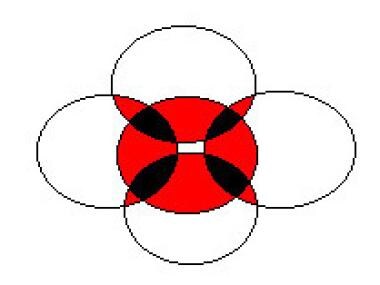
In this figure, the multifactorial nature of renal papillary necrosis is represented by 5 of the disease's most frequently associated conditions: infection, obstruction, diabetes mellitus, analgesic abuse, and sickle cell disease. Each circle represents a condition. Note how the conditions overlap; the red areas show the coexistence of 2 conditions, and the black areas represent 3 coexistent conditions. Multiple conditions exhibit synergism and, therefore, worsen both the severity of the disease and the prognosis.
Click to see larger pictureClick to see detailView Full Size Image
Media type:  Graph

Media file 2:  Cystoscopic photograph of sloughed papilla extruding from the ureteral orifice. The patient was a 51-year-old man with poorly controlled diabetes and a history of microhematuria and an acute onset of severe left flank pain. Findings of upper tract imaging with a renal ultrasonography and intravenous pyelography were remarkable only for mixed heterogeneity consistent with medical renal disease. Urine cytology results were negative, and culture showed no growth. In-office flexible cystoscopy revealed the mass extruding from the left ureteral orifice, which required sedation and rigid cystoscopy to extract. Gross examination yielded a tan, friable, irregular, wedge-shaped soft tissue mass 1.7 cm X 1.6 cm X 1.5 cm. Bilateral retrograde pyelography revealed a clubbed left upper pole calyx and no other filling defects. The pathology was necrotic epithelial tissue.
Click to see larger pictureClick to see detailView Full Size Image
Media type:  Photo


  1. Friedrich N. Ueber necrose der nierenpapillen bei hydronephrose. Virchows Arch A Path Anat. 1877;69:308-12.
  2. Gunther GW. Die papillennekrosen der niere bei diabetes. Munchen Med Wochenschr. 1937;84:1695-9.
  3. Edmondson HA, Martin HE, Evans N. Necrosis of renal papillae and acute pyelonephritis in diabetes mellitus. Arch Intern Med. 1947;79:148.
  4. Spuhler O, Zollinger HU. [Chronic interstitial nephritis.]. Z Klin Med. 1953;151(1):1-50. [Medline].
  5. Mandel EE. Renal medullary necrosis. Am J Med. Sep 1952;13(3):322-7. [Medline].
  6. Simon HB, Bennett WA, Emmett JL. Renal papillary necrosis: a clinicopathologic study of 42 cases. J Urol. Apr 1957;77(4):557-67. [Medline].
  7. Lang EK, Macchia RJ, Thomas R, Davis R, Ruiz-Deya G, Watson RA. Detection of medullary and papillary necrosis at an early stage by multiphasic helical computerized tomography. J Urol. Jul 2003;170(1):94-8. [Medline].
  8. Lang EK, Macchia RJ, Thomas R, Davis R, Ruiz-Deya G, Watson RA, et al. Multiphasic helical CT diagnosis of early medullary and papillary necrosis. J Endourol. Feb 2004;18(1):49-56. [Medline].
  9. Ulreich S. Ultrasound in the evaluation of renal papillary necrosis [letter]. Radiology. Sep 1983;148(3):864. [Medline].
  10. Vijayaraghavan SB, Kandasamy SV, Mylsamy A, Prabhakar M. Sonographic features of necrosed renal papillae causing hydronephrosis. J Ultrasound Med. Sep 2003;22(9):951-6; quiz 957-8. [Medline].
  11. Falkenberg FW, Hildebrand H, Lutte L, Schwengberg S, Henke B, Greshake D, et al. Urinary antigens as markers of papillary toxicity. I. Identification and characterization of rat kidney papillary antigens with monoclonal antibodies. Arch Toxicol. 1996;71(1-2):80-92. [Medline].
  12. Garber SL, Mirochnik Y, Desai SS, Arruda JA, Dunea G. Angiotensin-converting enzyme inhibition reduces the effect of bromoethylamine-induced papillary necrosis and renal fibrosis. J Am Soc Nephrol. Jun 1998;9(6):1052-9. [Medline].
  13. Abe K, Ozono Y, Miyazaki M, Furusu A, Shioshita K, Sasaki O, et al. Prostaglandin E1 for renal papillary necrosis in a patient with diabetes mellitus. J Int Med Res. Mar-Apr 1999;27(2):90-5. [Medline].
  14. Akhund L, Quinet RJ, Ishaq S. Celecoxib-related renal papillary necrosis. Arch Intern Med. Jan 13 2003;163(1):114-5. [Medline].
  15. Barnes DJ. Beethoven's final illness. Lancet. Mar 16 1996;347(9003):766. [Medline].
  16. Bing RJ. Cyclooxygenase-2 inhibitors: is there an association with coronary or renal events?. Curr Atheroscler Rep. Mar 2003;5(2):114-7. [Medline].
  17. Breyer MD, Hao C, Qi Z. Cyclooxygenase-2 selective inhibitors and the kidney. Curr Opin Crit Care. Dec 2001;7(6):393-400. [Medline].
  18. Cotran R, Kumar V, Robbins SL. The kidney. In: Cotran R, Kumar V, Robbins SL, eds. Pathologic Basis of Disease. 5th ed. Philadelphia, Pa: WB Saunders; 1994:964-72.
  19. Davies PJ. Beethoven's nephropathy and death: discussion paper. J R Soc Med. Mar 1993;86(3):159-61. [Medline].
  20. Eknoyan G. Chronic glomerulonephritis and chronic interstitial nephritis. In: Schrier RW, Gottschalk CW. Diseases of the Kidney. Vol. 2. 5th ed. Little Brown and Co: Boston, Mass; 1993:1976-9.
  21. Eknoyan G, Qunibi WY, Grissom RT, Tuma SN, Ayus JC. Renal papillary necrosis: an update. Medicine (Baltimore). Mar 1982;61(2):55-73. [Medline].
  22. Gower PE. A prospective study of patients with radiological pyelonephritis, papillary necrosis and obstructive atrophy. Q J Med. Apr 1976;45(178):315-49. [Medline].
  23. Hagiwara N, Fujihiro S, Deguchi T. [Renal papillary necrosis managed by transurethral procedures: a case report]. Hinyokika Kiyo. Jun 2003;49(6):329-31. [Medline].
  24. Kamath S, Moody MP, Hammonds JC, Wells IP. Papillary necrosis causing hydronephrosis in renal allograft treated by percutaneous retrieval of sloughed papilla. Br J Radiol. Apr 2005;78(928):346-8. [Medline].
  25. Kankuri E, Solatunturi E, Vapaatalo H. Effects of phenacetin and its metabolite p-phenetidine on COX-1 and COX-2 activities and expression in vitro. Thromb Res. Jun 15 2003;110(5-6):299-303. [Medline].
  26. Kovacevic L, Bernstein J, Valentini RP, Imam A, Gupta N, Mattoo TK. Renal papillary necrosis induced by naproxen. Pediatr Nephrol. Aug 2003;18(8):826-9. [Medline].
  27. Lange S. Diseases. In: Lange S, ed. Teaching Atlas of Urologic Radiology. First ed. New York, NY: Thieme Medical; 1995:88-9.
  28. Lange S. Differential Diagnosis: Urographic findings. In: Lange S, ed. Teaching Atlas of Urologic Radiology. First ed. New York, NY: Thieme Medical; 1995:198-203.
  29. Lauler DP, Schreiner GE, David A. Renal medullary necrosis. Am J Med. Jul 1960;29:132-56. [Medline].
  30. Lindvall N. Radiological changes of renal papillary necrosis. Kidney Int. Jan 1978;13(1):93-106. [Medline].
  31. Saifuddin A, Bark M. Case report: computed tomography demonstration of renal papillary necrosis. Clin Radiol. Oct 1991;44(4):275-6. [Medline].
  32. Salo JO, Talja M, Lehtonen T. Ureteroscopy in the treatment of ureteral obstruction caused by papillary necrosis. Eur Urol. 1987;13(1-2):140-1. [Medline].
  33. Sargent JC, Sargent JW. Unilateral renal papillary necrosis. J Urol. May 1955;73(5):757-64. [Medline].
  34. Schaeffer AJ. Infections of the urinary tract. In: Walsh PC, Retik AB, Vaughan ED Jr, eds. Campbell's Urology. Vol. 1. 7th ed. Philadelphia, Pa: WB Saunders; 1998:536-7, 568.
  35. Schwarz A. Beethoven's renal disease based on his autopsy: a case of papillary necrosis. Am J Kidney Dis. Jun 1993;21(6):643-52. [Medline].
  36. Stamm WE. Urinary tract infections and pyelonephritis. In: Isselbacher KJ, Braunwald E, Wilson JD, Martin JB, Fauci AS, Kasper DL, eds. Harrison's Principles of Internal Medicine. Vol. 1. 13th ed. New York, NY: McGraw-Hill; 1994:553.
  37. von Seyfried I. Autopsy protocol of Ludwig van Beethoven, translated from Latin. In: Ludwig van Beethovens Studien im Generalba. 2nd ed. Contrapunkt und in der Compositions: 1852-1853.
  38. Voulgarelis M, Ziakas PD. Images in clinical medicine. Renal papillary necrosis unmasking sickle cell disease. N Engl J Med. Mar 24 2005;352(12):1237. [Medline].
  39. Wen SF. Nephrotoxicities of nonsteroidal anti-inflammatory drugs. J Formos Med Assoc. Mar 1997;96(3):157-71. [Medline].
  40. Zadeii G, Lohr JW. Renal papillary necrosis in a patient with sickle cell trait. J Am Soc Nephrol. Jun 1997;8(6):1034-9. [Medline].

Papillary Necrosis excerpt

Article Last Updated: Jan 9, 2008