| Patient Education |
|
Click here for patient education.
|
|
You are in: eMedicine Specialties >
Cardiology > Pericardial Disease
Cardiac Tamponade
Article Last Updated: May 24, 2006
AUTHOR AND EDITOR INFORMATION
Section 1 of 11
Author: Chakri Yarlagadda, MD, FACC, Consulting Staff, Section of Cardiology, St Elizabeth Hospital
Chakri Yarlagadda is a member of the following medical societies: American College of Cardiology, American College of Physicians, American Society of Echocardiography, American Society of Nuclear Cardiology, and Heart Rhythm Society
Coauthor(s):
Wahoub M Hout, MD, FACC, Cath Lab Co-Director, Clinical Assistant Professor of NEOUCOM, Department of Medicine, St Elizabeth Hospital
Editors: Russell F Kelly, MD, Program Director, Assistant Professor, Department of Internal Medicine, Division of Cardiology, Cook County Hospital, Rush Medical College; Francisco Talavera, PharmD, PhD, Senior Pharmacy Editor, eMedicine; Ronald J Oudiz, MD, Director of Pulmonary Hypertension, Associate Professor, Department of Medicine, Division of Cardiology, Harbor-UCLA Medical Center, David Geffen School of Medicine at UCLA; Amer Suleman, MD, Consultant in Electrophysiology and Cardiovascular Medicine, Department of Internal Medicine, Division of Cardiology, Medical City Dallas Hospital; Michael E Zevitz, MD, Assistant Professor of Medicine, Finch University of the Health Sciences, The Chicago Medical School; Consulting Staff, Private Practice
Author and Editor Disclosure
Synonyms and related keywords:
cardiac tamponade, pericardial tamponade, reduced ventricular filling, hemodynamic compromise, malignant disease, pericarditis, pericardial effusion, accumulation of fluid in the pericardial space, postmyocardial infarction, free wall ventricular rupture, Dressler syndrome, systemic lupus erythematosus, rheumatoid arthritis, dermatomyositis, radiation therapy, antireflux surgery, pneumopericardium
Background
Cardiac tamponade is a clinical syndrome caused by the accumulation of fluid in the pericardial space, resulting in reduced ventricular filling and subsequent hemodynamic compromise. Cardiac tamponade is a medical emergency. The overall risk of death depends on the speed of diagnosis, the treatment provided, and the underlying cause of the tamponade.
Pathophysiology
The pericardium, which is the membrane surrounding the heart, is composed of 2 layers. The parietal pericardium is the outer fibrous layer; the visceral pericardium is the inner serous layer. The pericardial space normally contains 20-50 mL of fluid. Pericardial effusions can be serous, serosanguineous, hemorrhagic, or chylous.
Reddy et al describe 3 phases of hemodynamic changes in tamponade.
- Phase I: The accumulation of pericardial fluid causes increased stiffness of the ventricle, requiring a higher filling pressure. During this phase, the left and right ventricular filling pressures are higher than the intrapericardial pressure.
- Phase II: With further fluid accumulation, the pericardial pressure increases above the ventricular filling pressure, resulting in reduced cardiac output.
- Phase III: A further decrease in cardiac output occurs, which is due to equilibration of pericardial and left ventricular (LV) filling pressures.
The underlying pathophysiologic process for the development of tamponade is markedly diminished diastolic filling because transmural distending pressures are insufficient to overcome the increased intrapericardial pressures.
Systemic venous return is also altered during tamponade. Because the heart is compressed throughout the cardiac cycle due to the increased intrapericardial pressure, systemic venous return is impaired and right atrial collapse occurs. During inspiration, intrapericardial and right atrial pressures decrease because of negative intrathoracic pressure. This results in augmented systemic venous return to right-sided chambers and a marked increase in the right ventricular volume. Because the pulmonary vascular bed is a vast and compliant circuit, blood preferentially accumulates in the venous circulation, at the expense of LV filling. This results in a reduced cardiac output.
The amount of pericardial fluid needed to impair the diastolic filling of the heart depends on the rate of fluid accumulation and the compliance of the pericardium. Rapid accumulation of as little as 150 mL of fluid can result in a marked increase in pericardial pressure and can severely impede cardiac output, whereas 1000 mL of fluid may accumulate over a longer period without any significant effect on diastolic filling of the heart. This is due to adaptive stretching of the pericardium over time. A more compliant pericardium can allow considerable fluid accumulation over a longer period without hemodynamic insult.
Frequency
United States
The incidence of cardiac tamponade is 2 cases per 10,000 population in the United States. Approximately 2% of penetrating injuries are reported to result in cardiac tamponade.
Mortality/Morbidity
Cardiac tamponade is a medical emergency. Early diagnosis and treatment are crucial to reduce morbidity and mortality. Untreated, it is rapidly and universally fatal.
Sex
In children, cardiac tamponade is more common in boys than in girls, with a male-to-female ratio of 7:3. In adults, cardiac tamponade appears to be slightly more common in men than in women. The male-to-female ratio of 1.25:1 noted at the authors� referral center is based on the International Classification of Diseases (ICD) code 423.9. However, a ratio of 1.7:1 is observed at another level 1 trauma center.
Age
Cardiac tamponade related to trauma or HIV is more common in young adults, whereas tamponade due to malignancy and/or renal failure occurs more frequently in elderly individuals.
History
Symptoms vary with the underlying cause and the acuteness of the tamponade. Patients with acute tamponade may present with dyspnea, tachycardia, and tachypnea. Cold and clammy extremities from hypoperfusion are also observed in some patients.
- A comprehensive review of the patient's history usually helps identify the probable etiology of a pericardial effusion, which may result in cardiac tamponade.
- Patients with systemic or malignant disease present with weight loss, fatigue, or anorexia.
- Chest pain may be the presenting symptom in patients with pericarditis or myocardial infarction.
- Musculoskeletal pain or fever may be present in patients with an underlying connective tissue disorder.
- A history of renal failure can lead to a consideration of uremia as a cause of pericardial effusion.
- Careful review of a patient's medications may indicate drug-related lupus leading to a pericardial effusion.
- Recent cardiovascular surgery, coronary intervention, or trauma can lead to the rapid accumulation of pericardial fluid and tamponade.
- Recent pacemaker lead implantation or central venous catheter insertion can lead to the rapid accumulation of pericardial fluid and tamponade.
- Consider HIV-related pericardial effusion and tamponade if the patient has a history of intravenous drug abuse or opportunistic infections.
- Inquire about chest wall radiation (ie, for lung, mediastinal, or esophageal cancer).
- Inquire about symptoms of night sweats, fever, and weight loss, which may be indicative of tuberculosis.
Physical
Distended neck veins are a common feature in patients with tamponade. Evidence of chest wall injury may be present in trauma patients. Tachycardia, tachypnea, and hepatomegaly are observed in more than 50% of patients with cardiac tamponade, and diminished heart sounds and a pericardial friction rub are present in approximately one third of patients.
- The Beck triad or acute compression triad
- Described in 1935, this complex of physical findings refers to increased jugular venous pressure, hypotension, and diminished heart sounds.
- These findings result from a rapid accumulation of pericardial fluid. However, this classic triad is usually observed in patients with acute cardiac tamponade.
- Pulsus paradoxus or paradoxical pulse:
- This is an exaggeration (>12 mm Hg or 9%) of the normal inspiratory decrease in systemic blood pressure.
- To measure the pulsus paradoxus, patients are often placed in a semirecumbent position; respirations should be normal. The blood pressure cuff is inflated to at least 20 mm Hg above the systolic pressure and slowly deflated until the first Korotkoff sounds are heard only during expiration. At this pressure reading, if the cuff is not further deflated and a pulsus paradoxus is present, the first Korotkoff sound is not audible during inspiration. As the cuff is further deflated, the point at which the first Korotkoff sound is audible during both inspiration and expiration is recorded. If the difference between the first and second measurement is greater than 12 mm Hg, an abnormal pulsus paradoxus is present.
- The paradox is that while listening to the heart sounds during inspiration, the pulse weakens or may not be palpated with certain heartbeats, while S1 is heard with all heartbeats.
- A pulsus paradoxus can be observed in patients with other conditions, such as constrictive pericarditis, severe obstructive pulmonary disease, restrictive cardiomyopathy, pulmonary embolism, rapid and labored breathing, and right ventricular infarction with shock.
- A pulsus paradoxus may be absent in patients with markedly elevated LV diastolic pressures, atrial septal defect, pulmonary hypertension, and aortic regurgitation.
- Kussmaul sign
- This was described by Adolph Kussmaul as a paradoxical increase in venous distention and pressure during inspiration.
- This sign is usually observed in patients with constrictive pericarditis but occasionally is observed in patients with effusive-constrictive pericarditis and cardiac tamponade.
- Ewart sign
- Also known as the Pins sign, this is observed in patients with large pericardial effusions.
- It is described as an area of dullness, with bronchial breath sounds and bronchophony below the angle of the left scapula.
- The y descent
- The y descent is abolished in the jugular venous or right atrial waveform.
- This is due to an increase in intrapericardial pressure, preventing diastolic filling of the ventricles.
Causes
For all patients, malignant diseases are the most common cause of pericardial tamponade. Tamponade can occur as a result of any type of pericarditis.
- HIV infection
- Infection - Viral, bacterial (tuberculosis), fungal
- Drugs - Hydralazine, procainamide, isoniazid, minoxidil
- Postcoronary intervention (ie, coronary dissection and perforation)
- Trauma
- Cardiovascular surgery (postoperative pericarditis)
- Postmyocardial infarction (free wall ventricular rupture, Dressler syndrome)
- Connective tissue diseases - Systemic lupus erythematosus, rheumatoid arthritis, dermatomyositis
- Radiation therapy
- Iatrogenic - After sternal biopsy, transvenous pacemaker lead implantation, pericardiocentesis, or central line insertion
- Uremia
- Idiopathic pericarditis
- Complication of surgery at the esophagogastric junction such as antireflux surgery
- Pneumopericardium (due to mechanical ventilation or gastropericardial fistula)
Cardiogenic Shock
Pericarditis, Constrictive
Pericarditis, Constrictive-Effusive
Pulmonary Embolism
Tension Pneumothorax
Other Problems to be Considered
Large pleural effusion: Cases of cardiac tamponade have been reported with large pleural effusions. The increased intrapleural pressure resulting from large pleural effusions can be transmitted to the pericardial space and impair ventricular filling, thus simulating the hemodynamic equivalent of cardiac tamponade.
Tension pneumopericardium: The hemodynamic changes simulate acute cardiac tamponade. Clinically, distant heart sounds, bradycardia, and shifting tympany occur over the precordium and a characteristic murmur is heard, termed bruit de la roue de moulin. This is usually observed in infants with mechanical ventilation but is also observed after sternal bone marrow aspiration, penetrating chest wall injury, esophageal rupture, and bronchopericardial fistula.
Rapid and labored breathing: Large decreases in intrathoracic pressure with deep inspirations, often observed during respiratory failure, can accentuate the pulsus paradoxus, simulating pericardial tamponade.
Lab Studies
- Creatine kinase and isoenzymes: Levels are elevated in patients with myocardial infarction and cardiac trauma.
- Renal profile and CBC count with differential: These tests are useful in the diagnosis of uremia and certain infectious diseases associated with pericarditis.
- Coagulation panel: The prothrombin time and activated partial thromboplastin time are useful for determining bleeding risk during interventions, such as pericardial drainage, the placement of pericardial windows, or both.
- Antinuclear antibody assay, erythrocyte sedimentation rate, and rheumatoid factor: Although nonspecific, results from these tests may give clues to a connective tissue disease predisposing to the development of pericardial effusion.
- HIV testing: Approximately 24% of all pericardial effusions are reported to be associated with HIV infection.
- Purified protein derivative testing: This is used to diagnose tuberculosis, which is an important and not uncommon cause of pericardial effusion and tamponade.
Imaging Studies
- Chest radiography findings may show cardiomegaly, water bottle–shaped heart, pericardial calcifications, or evidence of chest wall trauma (see Image 1).
- Although echocardiography provides useful information, cardiac tamponade is a clinical diagnosis (see Clinical and Image 3). The following may be observed with 2-dimensional echocardiography:
- An echo-free space posterior and anterior to the left ventricle and behind the left atrium: After cardiac surgery, a localized posterior fluid collection without significant anterior effusion may occur and may readily compromise cardiac output.
- Early diastolic collapse of the right ventricular free wall
- Late diastolic compression/collapse of the right atrium
- Swinging of the heart in its sac
- LV pseudohypertrophy
- A greater than 40% relative inspiratory augmentation of right-side flow
- A greater than 25% relative decrease in inspiratory flow across the mitral valve
- Conditions that may simulate pericardial effusion on 2-dimensional echocardiography findings include the following:
- A large left pleural effusion
- Any tumor surrounding the heart
- Mitral annular calcification
- A descending thoracic aorta
- A catheter in the right ventricle
- An enlarged left atrium
- An annular subvalvular LV aneurysm
- A bronchogenic cyst
Other Tests
- With a 12-lead electrocardiogram (see Image 2), the following findings are suggestive but not diagnostic of pericardial tamponade.
- Sinus tachycardia
- Low-voltage QRS complexes
- Electrical alternans (also observed during supraventricular and ventricular tachycardia): Alternation of QRS complexes, usually in a 2:1 ratio, on electrocardiogram findings is called electrical alternans. This is due to movement of the heart in the pericardial space. Electrical alternans is also observed in patients with myocardial ischemia, acute pulmonary embolism, and tachyarrhythmias.
- PR segment depression
Procedures
- Swan-Ganz catheterization
- Before or after insertion of the Swan-Ganz catheter, the system must be zeroed after positioning the transducer at the midpoint of the left atrium. Then calibrate the monitoring system. Prior to insertion, test the balloon and flush all the ports. Then insert the catheter into one of the major veins. At a depth of 20 cm, inflate the balloon and slowly advance the catheter, while continuously monitoring the pressure from the distal lumen. Always deflate the balloon before withdrawing the Swan-Ganz catheter. The waveforms help indicate the position of the catheter tip if fluoroscopy is not readily accessible. At approximately the 40- to 50-cm mark, the wedge pressure is usually recorded. Secure the catheter position, and obtain a chest x-ray film to confirm the position.
- In tamponade, near equalization (within 5 mm Hg) of the right atrial, right ventricular diastolic, pulmonary arterial diastolic, and pulmonary capillary wedge pressure (reflecting left atrial pressure) occurs. The right atrial pressure tracings display a prominent systolic x descent and abolished systolic y descent.
- Boltwood et al described the diastolic equalization of pulmonary capillary and right atrial pressures as predominantly inspiratory, known as the inspiratory traction sign. This is due to inspiratory traction of the taut pericardium by the diaphragm.
Histologic Findings
Occasionally, a pericardial biopsy is performed when the etiology of the pericardial effusion that caused the tamponade is unclear. This is especially useful in cases of tuberculous pericardial effusions because cultures of the pericardial fluid in these cases rarely yield a positive result for mycobacteria. However, granulomas seen on pericardial biopsy specimens are often seen in patients with tuberculous pericarditis. In general, cytopathologic findings from pericardial fluid and histologic findings from pericardial biopsy specimens depend on the underlying pathology.
Medical Care
Cardiac tamponade is a medical emergency. Preferably, patients should be monitored in an intensive care unit.
- All patients should receive the following:
- Oxygen
- Volume expansion with blood, plasma, dextran, or isotonic sodium chloride solution, as necessary to maintain adequate intravascular volume
- Bed rest with leg elevation: This may help increase venous return.
- Inotropic drugs (eg, dobutamine): These can be useful because they do not increase systemic vascular resistance while increasing cardiac output.
- Positive-pressure mechanical ventilation: This should be avoided because it may decrease venous return.
- Further medical care includes the following:
- Pericardiocentesis: Removal of pericardial fluid is the definitive therapy for tamponade.
- Emergency subxiphoid percutaneous drainage: This is a life-saving bedside procedure. The subxiphoid approach is extrapleural; hence, it is the safest for blind pericardiocentesis. A 16- or 18-gauge needle is inserted at an angle of 30-45° to the skin, near the left xiphocostal angle, aiming towards the left shoulder. When performed emergently, this procedure is associated with a reported mortality rate of approximately 4% and a complication rate of 17%.
- Echocardiographically guided pericardiocentesis (often performed in the cardiac catheterization laboratory): This is usually performed from the left intercostal space. First, mark the site of entry based on the area of maximal fluid accumulation closest to the transducer. Then, measure the distance from the skin to the pericardial space. The angle of the transducer should be the trajectory of the needle during the procedure. Avoid the inferior rib margin while advancing the needle to prevent neurovascular injury. Leave a 16-gauge catheter in place for continuous drainage.
- Percutaneous balloon pericardiotomy: This can be performed using an approach similar to that for echo-guided pericardiocentesis, in which the balloon is used to create a pericardial window.
- Treatment of the underlying cause to prevent recurrence
Surgical Care
- For a hemodynamically unstable patient or one with recurrent tamponade, provide the following care:
- Surgical creation of a pericardial window: This involves the surgical opening of a communication between the pericardial space and the intrapleural space. Open thoracotomy and/or pericardiotomy may be required in some cases, and these should be performed by an experienced surgeon.
- Pericardiodesis or sclerosing the pericardium: This is a therapeutic option for patients with recurrent pericardial effusion or tamponade. Through the intrapericardial catheter, corticosteroids, tetracycline, or antineoplastic drugs (eg, anthracyclines, bleomycin) can be instilled into the pericardial space.
- Pericardio-peritoneal shunt: In some patients with malignant pericardial effusions, creation of a pericardio-peritoneal shunt helps prevent recurrent tamponade.
- Pericardiectomy: Resection of the pericardium (pericardiectomy) through a median sternotomy or left thoracotomy is rarely required to prevent recurrent pericardial effusion and tamponade.
Consultations
- Hemodynamically stable patients - Cardiologist
- Hemodynamically unstable patient - Cardiologist, cardiothoracic surgeon
The role of medication therapy in cardiac tamponade is limited. Occasionally, inotropic agents that do not increase the peripheral vascular resistance, such as dobutamine, may be used.
Drug Category: Adrenergic agonist agents
By stimulating beta-1 receptors in the heart, stroke volume and cardiac output are increased.
| Drug Name | Dobutamine (Dobutrex) |
| Description | Synthetic catecholamine and a direct inotropic agent that stimulates cardiac beta-receptors with minimal increase in systemic vascular resistance. |
| Adult Dose | 0.5-1 mcg/kg/min IV initially; titrate until desired therapeutic effect attained |
| Pediatric Dose | Administer as in adults |
| Contraindications | Documented hypersensitivity; idiopathic hypertrophic subaortic stenosis and atrial fibrillation or flutter |
| Interactions | Beta-adrenergic blockers antagonize effects; general anesthetics may increase toxicity |
| Pregnancy | B - Usually safe but benefits must outweigh the risks.
|
| Precautions | Following myocardial infarction, use with extreme caution; hypovolemic state should be corrected before using this drug; ECG and blood pressure should be monitored continuously; pulmonary wedge pressure and cardiac output should be monitored, if possible; marked mechanical obstruction (severe valvular aortic stenosis) may prevent observation of improvement |
Further Inpatient Care
- After pericardiocentesis, leave the intrapericardial catheter in place after securing it to the skin using sterile procedure and attaching it to a closed drainage system via a 3-way stopcock. Periodically check for reaccumulation of fluid, and drain as needed. The catheter can be left in place for 1-2 days and can be used for pericardiodesis. Serial fluid cell counts can be useful for helping discover an impending bacterial catheter infection, which could be catastrophic. If the WBC count rises significantly, the pericardial catheter must be removed immediately.
- A Swan-Ganz catheter can be left in place for continuous monitoring of hemodynamics and to assess the effect of reaccumulation of pericardial fluid.
- A repeat echocardiogram should be performed within 24 hours.
- A repeat chest radiograph should be performed within 24 hours.
Further Outpatient Care
- A follow-up echocardiogram and chest radiograph should be performed at a monthly follow-up examination to check for recurrent fluid accumulation.
Complications
- Pulmonary edema
- Shock
- Death
Prognosis
- Prognosis depends on prompt recognition and management of the condition and the underlying cause of the tamponade.
Medical/Legal Pitfalls
- Be aware of clinical conditions that may simulate tamponade or pericardial effusion on 2-dimensional echocardiography findings.
- Pulsus paradoxus may be absent in patients with low-pressure tamponade, right heart tamponade, or atrial septal defects.
- Pulsus paradoxus can be observed in patients with nontamponade conditions involving labored breathing (eg, asthma, severe chronic obstructive pulmonary disease) and in those with right ventricular infarction.
- Tension pneumothorax may clinically simulate cardiac tamponade.
- Early diagnosis with a high index of suspicion is necessary to minimize the morbidity and mortality from tamponade.
| Media file 1:
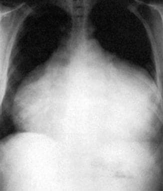
This anteroposterior-view chest radiograph shows a massive bottle-shaped heart and conspicuous absence of pulmonary vascular congestion. Reproduced with permission from Chest, 1996: 109:825. |
 | View Full Size Image | |
Media type: X-RAY
|
| Media file 2:
A 12-lead ECG showing sinus tachycardia with electrical alternans. Reproduced with permission from Chest, 1996; 109:825. |
 | View Full Size Image | |
Media type: ECG
|
| Media file 3:
Two-dimensional echocardiogram (diastolic still frame) in parasternal long-axis view showing pericardial effusion and early diastolic collapse of the right ventricular free wall. Reproduced with permission from Chest, 1996; 109:825 |
 | View Full Size Image | |
Media type: Image
|
- Aikat S, Ghaffari S. A review of pericardial diseases: clinical, ECG and hemodynamic features and management. Cleve Clin J Med. Dec 2000;67(12):903-14. [Medline].
- Alam HB, Levitt A, Molyneaux R, et al. Can pleural effusions cause cardiac tamponade?. Chest. Dec 1999;116(6):1820-2. [Medline].
- Allen KB, Faber LP, Warren WH, Shaar CJ. Pericardial effusion: subxiphoid pericardiostomy versus percutaneous catheter drainage. Ann Thorac Surg. Feb 1999;67(2):437-40. [Medline].
- Beauchamp KA. Pericardial tamponade: an oncologic emergency. Clin J Oncol Nurs. Jul 1998;2(3):85-95. [Medline].
- Boltwood C, Rieders D, Gregory KW. Inspiratory tracking sign in pericardial disease. Circulation. 1984;(suppl II) 70:103.
- Chen Y, Brennessel D, Walters J, et al. Human immunodeficiency virus-associated pericardial effusion: report of 40 cases and review of the literature. Am Heart J. Mar 1999;137(3):516-21. [Medline].
- Eisenberg MJ, de Romeral LM, Heidenreich PA, et al. The diagnosis of pericardial effusion and cardiac tamponade by 12-lead ECG. A technology assessment. Chest. Aug 1996;110(2):318-24. [Medline].
- Goldstein JA. Cardiac tamponade, constrictive pericarditis, and restrictive cardiomyopathy. Curr Probl Cardiol. Sep 2004;29(9):503-67. [Medline].
- Griffin S, Fountain W. Pericardio-peritoneal shunt for malignant pericardial effusion. J Thorac Cardiovasc Surg. Dec 1989;98(6):1153-4. [Medline].
- Guberman BA, Fowler NO, Engel PJ, et al. Cardiac tamponade in medical patients. Circulation. Sep 1981;64(3):633-40. [Medline].
- Kaplan LM, Epstein SK, Schwartz SL, et al. Clinical, echocardiographic, and hemodynamic evidence of cardiac tamponade caused by large pleural effusions. Am J Respir Crit Care Med. Mar 1995;151(3 Pt 1):904-8. [Medline].
- Medary I, Steinherz LJ, Aronson DC, La Quaglia MP. Cardiac tamponade in the pediatric oncology population: treatment by percutaneous catheter drainage. J Pediatr Surg. Jan 1996;31(1):197-9; discussion 199-200. [Medline].
- Muller-Stich BP, Linke G, Leemann B, et al. Cardiac tamponade as a life-threatening complication in antireflux surgery. Am J Surg. Jan 2006;191(1):139-41.
- Patel MR, Meine TJ, Lindblad L, et al. Cardiac tamponade in the fibrinolytic era: analysis of >100,000 patients with ST-segment elevation myocardial infarction. Am Heart J. Feb 2006;151(2):316-22.
- Paunovic B, Sharma S, Miller A. Swan-Ganz Catheterization. eMedicine Journal [serial online]. 2005. [Full Text].
- Reddy PS, Curtiss EI. Cardiac tamponade. Cardiol Clin. Nov 1990;8(4):627-37. [Medline].
- Reddy PS, Curtiss EI, Uretsky BF. Spectrum of hemodynamic changes in cardiac tamponade. Am J Cardiol. Dec 15 1990;66(20):1487-91. [Medline].
- Spodick DH. The normal and diseased pericardium: current concepts of pericardial physiology, diagnosis and treatment. J Am Coll Cardiol. Jan 1983;1(1):240-51. [Medline].
- Spodick DH. Pericarditis, pericardial effusion, cardiac tamponade, and constriction. Crit Care Clin. Jul 1989;5(3):455-76. [Medline].
- Spodick DH. Diseases of the pericardium. In: Chatterjee K, ed. Cardiology: An Illustrated Text/Reference. Vol 2. New York, NY: Gower Medical; 1991:. 10.38-10.64.
- Tsang TS, Oh JK, Seward JB. Diagnosis and management of cardiac tamponade in the era of echocardiography. Clin Cardiol. Jul 1999;22(7):446-52. [Medline].
- Tsang TS, Freeman WK, Sinak LJ, Seward JB. Echocardiographically guided pericardiocentesis: evolution and state-of-the-art technique. Mayo Clin Proc. Jul 1998;73(7):647-52. [Medline].
- Walensky P. Pericardial diseases. In: Linhart JW, Joyner CR, eds. Diagnostic Echocardiography. St. Louis, Mo: Mosby-Year Book; 1982:. 109.
Cardiac Tamponade excerpt Article Last Updated: May 24, 2006
|