Practice Essentials
Urinary tract obstruction is a common problem encountered by urologists, primary care physicians, and emergency medicine physicians. Obstruction can develop secondary to calculi, tumors, strictures, anatomical abnormalities, or functional abnormalities.
Obstruction to urinary flow can occur at any point in the urinary tract, from the kidneys to the urethral meatus, but certain sites are more susceptible to obstruction. The 3 points of narrowing along the ureter include the ureteropelvic junction (UPJ), the crossing of the ureter over the area of the pelvic brim at the level of the iliac vessels, and the ureterovesical junction (UVJ).
When urinary tract obstruction develops, subsequent accumulation of urine distends the urinary tract proximal to the point of obstruction and can result in pain, which may be the first symptom of obstruction. Distortion of the urinary tract and acute kidney injury can develop; the severity depends on the degree and duration of obstruction. In addition, urinary stasis in an obstructed urinary tract may predispose to urinary tract infection.
A patient with any of the following needs immediate attention by a urologist:
-
Complete urinary tract obstruction
-
Any type of obstruction in a solitary kidney
-
Obstruction with fever, infection, or both
-
Kidney failure
Patients with pain that is uncontrolled by oral medications or with persistent nausea and vomiting that causes dehydration also need immediate attention, as well as hospital admission.
Note the computed tomography (CT) image below.
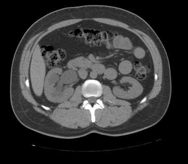 A noncontrast, axial CT image showing right-sided hydronephrosis. In this particular case, the patient had a distal ureteral stricture secondary to prior ureterolithiasis.
As time goes on, new procedures emerge and old procedures are modified to relieve urinary tract obstruction. In addition, with newer cameras and equipment and the use of laparoscopy, surgical intervention is becoming more advanced.
Background
Use of the urethral catheter to relieve urinary tract obstruction dates back to the time of Hippocrates. The first catheters were made of metal; by the Middle Ages, more flexible catheters were developed. Rubber catheters were developed in the 19th century. Currently, various sizes, compositions (eg, latex, silicone), and tips (coude, straight, council tip) of catheters are available.
Suprapubic access to the bladder can be traced back to the 16th century. It was initially considered a procedure of last resort but was refined in the 20th century. At present, it is a fairly common mode for relief of lower urinary tract obstruction.
Pathophysiology
Chronic urinary tract obstruction can lead to permanent damage to the urinary tract. Infravesical obstruction can lead to changes in the bladder, such as the following:
-
Trabeculation
-
Cellule formation
-
Diverticula
-
Bladder wall thickening
-
Ultimately, detrusor muscle decompensation
Progressive back pressure on the ureters and kidneys can occur and can cause hydroureter and hydronephrosis. The ureter can then become dilated and tortuous, with the inability to adequately propel urine forward. Hydronephrosis can cause permanent nephron damage and renal failure. Urinary stasis along any portion of the urinary tract increases the risk of stone formation and infection, and, ultimately, upper urinary tract injury. Urinary tract obstruction can have long-lasting effects on the physiology of the kidney, including its ability to concentrate urine. [1]
In the setting of an acute urinary tract obstruction, an increase in intraluminal pressure causes smooth muscle cells to increase contractions and ureteral wall pressure. As the duration of the obstruction lengthens, smooth muscle cells contract with less force and ureteral wall dilation increases. With a superimposed urinary infection, as often occurs in chronic obstruction, the loss of muscle tone is even more dramatic and progressive dilation occurs with no further increase or decrease in wall tension. [2]
Etiology
Obstruction of urinary flow can occur anywhere from the kidneys to the urethral meatus. Dividing the urinary tract into the upper urinary tract, defined as the kidney and ureter to the hiatus with the bladder, and the lower urinary tract, defined as the bladder and urethra to the urethral meatus, allows for further delineation of the cause of obstruction.
Certain points along the upper urinary tract are more susceptible to obstruction. The 3 points of narrowing along the ureter include the ureteropelvic junction (UPJ), the crossing of the ureter over the area of the pelvic brim (the iliac vessels), and the ureterovesical junction (UVJ).
Obstruction can be extrinsic, from compressive or restrictive force, or intrinsic, from a multitude of factors. The most common causes of intraluminal obstruction are calculi, blood clots, tumors, or sloughed papilla. These obstructions present acutely, leading to severe renal colic with flank pain, hematuria, nausea, vomiting, and fever. Ureteral strictures, which are caused by stone disease, cancer, maldevelopment, or iatrogenic causes such as ureteroscopy, tend to develop over time, causing chronic obstruction and renal atrophy.
Women may have an additional area of ureteral narrowing as the distal ureter crosses posterior to the pelvic blood vessels and the broad ligament in the posterior pelvis. Women can also experience urinary tract obstruction when the ureters become externally compressed by pelvic tumors or by advanced cervical or gynecologic malignancies.
In older women, prolapse of pelvic structures, such as the uterus and bladder, can lead to urinary tract obstruction. In younger women, pregnancy can result in ureteral obstruction from the gravid uterus. [3] Gynecologic malignancies should always be considered when upper tract obstruction is present.
In men, an enlarged prostate (benign prostatic hyperplasia [BPH]) can result in bladder outlet obstruction. In addition, urinary stasis from obstruction due to BPH may promote the formation of bladder calculi, which themselves may produce obstruction; such bladder calculi are more likely to form in men with a history of renal stone disease or gout. [4] Calculi may also form secondary to radical prostatectomy, due to migration of clips into the bladder or bladder neck stenosis from hypertrophic scars. [5] Rare cases of bladder outlet obstruction caused by giant prostatic calculi have been reported. [6]
Anterior urethral stricture, which may be secondary to genitourinary tract infection or trauma, including iatrogenic trauma, can also lead to urinary tract obstruction in males. [7, 8]
Other extrinsic causes of ureteral obstruction can occur. Although less common, these can still cause significant obstruction by inhibiting ureteral peristalsis or applying external pressure to the ureter. Vascular causes such aberrant lower pole renal arteries oriented anterior to the ureter, known as crossing vessels, can apply pressure at the level of the UPJ or proximal ureter and cause obstruction. Abdominal aortic aneurysms and common iliac artery aneurysms can externally compress the ureter along its natural path. Vascular graft placement has been shown to cause hydronephrosis in up to 10-20% of patients from a mechanical obstruction of the ureter; these occasionally resolve spontaneously. [1]
Retroperitoneal fibrosis can trap the ureters in fibrotic tissue, inhibiting peristalsis. This can occur in a unilateral or bilateral fashion and can be caused by a coexistent malignancy in 8-10% of cases.
Persistence of the posterior subcardinal vein in utero may cause obstruction by coursing the ureter behind the inferior vena cava. This is known as a retrocaval ureter and occurs on the right side, with a male predominance. Obstruction of the ureter typically becomes symptomatic in the third or fourth decade of life. [1]
Fungal (eg, Candida) bezoars can cause urinary tract obstruction, mainly in immunocompromised patients. Other risk factors for the development of fungal bezoars in the bladder include the following [9] :
-
Anatomical abnormalities of the urinary tract
-
Diabetes mellitus
-
Indwelling urinary catheters
-
Increased use of broad-spectrum antibiotics
-
Corticosteroid therapy
In children, more common causes of obstruction include the following:
-
UPJ or UVJ obstruction
-
Ectopic ureter
Prenatal screening with ultrasonography is important in early identification of obstruction. In addition, children with incontinence or urinary tract infection need a workup because they may also have some type of urinary tract obstruction.
Epidemiology
Obstructive uropathy has an annual incidence of 1.7 per 1000 population. It causes approximately 10% of all cases of acute kidney injury and chronic kidney disease. [10]
In an autopsy series of 59,064 patients aged 0-80 years, the frequency of hydronephrosis was 3.1%. In women with uterine prolapse, hydronephrosis occurs in approximately 5% with first-degree prolapse and in 40% with third-degree prolapse.
Hydronephrosis is found in 2-2.5% of children. Prenatal hydronephrosis is the most common urinary tract anomaly, occurring in 1-5% of pregnancies. Approximately 25% of these cases are caused by urinary obstruction. [11]
In women, hydronephrosis is more likely develop during the third to seventh decade of life, secondary to pregnancy and gynecologic malignancies. In men, hydronephrosis occurs most often after age 60 years, secondary to prostatic obstruction.
Prognosis
The prognosis of urinary tract obstruction depends on the cause, location, degree, and duration of obstruction, as well as the presence of a urinary tract infection. The longer the duration of obstruction, the greater the severity of obstruction, and the presence of a concomitant infection can lead to a worse prognosis. The prognosis is favorable if the patient's kidney function is normal, the infection is cleared, and the obstruction is relieved in a timely manner.
Hydronephrosis from obstructive uropathy can lead to the destruction of kidney function, and unless decompression is performed, the mortality rate is 40%. In contrast, with successful decompression the mortality rate is less than 5%. [10]
Complications of obstructive uropathy include:
-
Infection, including cystitis (bladder infection), pyelonephritis (kidney infection), abscess formation, and urosepsis
-
Urinary extravasation with urinoma formation
-
Urinary fistula formation
-
Kidney insufficiency or failure
-
Bladder dysfunction secondary to a defunctionalized bladder
-
Pain
-
Longitudinal image of right kidney displaying moderate hydronephrosis.
-
A noncontrast, axial CT image showing right-sided hydronephrosis. In this particular case, the patient had a distal ureteral stricture secondary to prior ureterolithiasis.
-
Flexible cystoscope; Gyrus ACMI ICN-2.
-
Axial CT images with intravenous contrast, revealing right-sided hydronephrosis (left image) and an obstructing right ureteropelvic junction stone (right image).
-
Coronal CT image with intravenous contrast, displaying (left) delayed contrast excretion and bilateral hydronephrosis secondary to (right) bladder outlet obstruction from benign prostatic hyperplasia, and an extremely distended bladder.
-
Intravenous pyelogram, 1-hour delayed image showing left-sided hydroureteronephrosis secondary to distal ureteral obstruction.
-
Intravenous pyelogram displaying right-sided ureteropelvic junction obstruction and normal excretory image of the left collecting system.
-
T2-weighted MRI, coronal image, displaying a right-sided duplicated system with obstruction of the lower pole moiety.
-
T2-weighted MRI, coronal image, displaying left-sided ureteropelvic junction obstruction.
-
Mercaptoacetyltriglycine (MAG3) renal scan with furosemide (Lasix); delayed emptying of left-sided collecting system consistent with obstructive hydronephrosis.
-
Mercaptoacetyltriglycine (MAG3) renal scan with furosemide (Lasix); , delayed emptying of left-sided collecting system consistent with obstructive hydronephrosis.
-
Retrograde urethrogram displaying complete obstruction of prostatic urethra.
-
Cystoscopic image of vapor-resection of an obstructing prostate. Obstructing lateral lobes can be seen proximal to the verumontanum.
-
Kidney-ureter-bladder (KUB) image displaying a large right-sided renal stone and an indwelling ureteral stent.


