You are in: eMedicine Specialties > Medicine, Ob/Gyn, Psychiatry, and Surgery > Oncology
|
Pancreatic Cancer Last Updated: December 15, 2005 |
|
| Synonyms and related keywords: pancreatic cancer, pancreas cancer, pancreatic carcinoma, pancreas carcinoma, gastrointestinal cancer, GI cancer, gastrointestinal carcinoma, GI carcinoma, pancreas tumor, pancreatic tumor, malignancy, exocrine, endocrine, pancreatic adenocarcinoma, chronic pancreatitis from alcohol
|
|   |
AUTHOR INFORMATION
| Section 1 of 11  |
|
| Author: Richard A Erickson, MD, FACP, FACG, Professor of Medicine, Texas A&M Health Science Center; Director, Division of Gastroenterology, Department of Internal Medicine, Scott and White Clinic and Hospital |
| Richard A Erickson, MD, FACP, FACG, is a member of the following medical societies:
American College of Gastroenterology,
American College of Physicians,
American Gastroenterological Association, and
American Society for Gastrointestinal Endoscopy |
| Editor(s): Lodovico Balducci, MD, Professor of Oncology and Medicine, University of South Florida College of Medicine; Division Chief, Senior Adult Oncology Program, H Lee Moffitt Cancer Center and Research Institute; Francisco Talavera, PharmD, PhD, Senior Pharmacy Editor, eMedicine;
Benjamin Movsas, MD, Vice-Chairman, Department of Radiation Oncology, Fox Chase Cancer Center;
Rajalaxmi McKenna, MD, FACP, Consulting Staff, Department of Medicine, Southwest Medical Consultants, SC, Good Samaritan Hospital, Advocate Health Systems;
and John S Macdonald, MD, Professor of Medicine, New York Medical College; Chief, Division of Medical Oncology, St Vincent's Hospital and Medical Center; Medical Director, Saint Vincent's Comprehensive Cancer Center |
Disclosure
|   |
INTRODUCTION
| Section 2 of 11  |
|
Background: In the United States, approximately 30,000 people die of pancreatic cancer each year. Among cancers of the gastrointestinal tract, it is the third most common malignancy and the fifth leading cause of cancer-related mortality. The disease is difficult to diagnose in its early stages, and most patients have incurable disease by the time they present with symptoms. The overall 5-year survival rate for this disease is less than 5%. Pathophysiology: Pancreatic cancers can arise from both the exocrine and endocrine portions of the pancreas. Of pancreatic tumors, 95% develop from the exocrine portion of the pancreas, including the ductal epithelium, acinar cells, connective tissue, and lymphatic tissue. Approximately 75% of all pancreatic carcinomas occur within the head or neck of the pancreas, 15-20% occur in the body of the pancreas, and 5-10% occur in the tail. Typically, pancreatic cancer first metastasizes to regional lymph nodes, then to the liver, and less commonly, to the lungs. It can also directly invade surrounding visceral organs such as the duodenum, stomach, and colon.
The molecular genetics of pancreatic adenocarcinoma have been well studied. Of these tumors, 80-95% have mutations in the KRAS2 gene, and 85-98% have mutations, deletions, or hypermethylation in the CDKN2 gene. Of these cancers, 50% have mutations in TP53 and about 55% have homozygous deletions or mutations of Smad4. Some of these mutations can also be found in high-risk precursors of pancreatic cancer. For example, in chronic pancreatitis, 30% of patients have detectable mutations in TP16 and 10% have K-ras mutations. Although studies are underway, the genetic mutations associated with pancreatic adenocarcinoma are not yet clinically useful in screening for or diagnosing the disease.
As in other organs, chronic inflammation is a predisposing factor in the development of pancreatic cancer. Patients with chronic pancreatitis from alcohol, especially those with familial forms, have much higher incidence and an earlier age of onset of pancreatic carcinoma. Frequency:
- In the US: Approximately 30,000 new cases of pancreatic cancer are diagnosed each year in the United States. Because of the lethality of the disease, the number of deaths per year from pancreatic cancer is almost identical. The overall incidence of pancreatic cancer is approximately 8-10 cases per 100,000 persons per year. The incidence of pancreatic cancer in males has been slowly dropping over the past 2 decades, while the incidence in females has increased slightly. These trends probably represent the effect of changing smoking rates for men and women.
- Internationally: Worldwide, pancreatic cancer ranks thirteenth in incidence but eighth as a cause of cancer death. The highest incidence rate is approximately 13 cases per 100,000 persons per year in black males in the United States. Native Hawaiian males and men of Korean, Czech, Latvian, and New Zealand Maori ancestry also have high incidence rates, ie, 11 cases per 100,000 persons per year. Most other countries have incidence rates of 8-12 cases per 100,000 persons per year. In some areas of the world, pancreatic cancer is quite infrequent; for example, the incidence in India is less than 2 cases per 100,000 persons per year.
Mortality/Morbidity:
- Pancreatic carcinoma is unfortunately usually a fatal disease. Most patients eventually succumb to the consequences of local invasion and metastatic cancer, and true long-term cures are rare. Endocrine and cystic neoplasms of the pancreas have much better survival rates than pancreatic adenocarcinoma. These tumors are discussed in separate articles.
- Overall, fewer than 5% of all patients are still alive 5 years after initial diagnosis. The collective median survival time of all patients is 4-6 months.
- In patients able to undergo a successful curative resection (only 20% of patients), median survival time ranges from 12-19 months, and the 5-year survival rate is 15-20%. The best predictors of long-term survival after surgery are a tumor diameter of less than 3 cm, no nodal involvement, negative resection margins, and diploid tumor DNA content.
Race:
- The incidence of pancreatic cancer for black males in the United States is 11.9-13.7 cases per 100,000 persons per year, and the incidence for black females is 10.5-11.9 cases per 100,000 persons per year.
- For white males in the United States, incidence is 8.2 cases per 100,000 persons per year, and for females the incidence is 6 cases per 100,000 persons per year.
- The reasons for the significant racial differences observed in the frequency of pancreatic cancer are unclear. Certainly, differences in dietary habits and the frequency of cigarette smoking are recognized among different groups. Obesity has been identified as a possible risk factor for pancreatic cancer and may contribute to the higher incidence of this disease among blacks. However, these variations may also be related to the underlying frequency of predisposing genetic mutations for pancreatic cancer or to other unknown factors.
Sex: The male-to-female ratio for pancreatic cancer is 1.2-1.5:1.
Age:
- Age is the most significant risk factor for pancreatic cancer.
- In the absence of predisposing conditions, such as familial pancreatic cancer and chronic pancreatitis, pancreatic cancer is unusual in persons younger than 45 years. After age 50 years, the frequency of pancreatic cancer increases linearly. At age 70, mortality incidence due to pancreatic cancer is approximately 60 deaths per 100,000 persons per year.
- The median age at diagnosis is 69 years in whites and 65 years in blacks.
|   |
CLINICAL
| Section 3 of 11  |
|
History: The early clinical diagnosis of pancreatic cancer is fraught with difficulty. Unfortunately, the initial symptoms are often quite nonspecific and subtle in onset. - Patients typically report the gradual onset of nonspecific symptoms such as anorexia, malaise, nausea, fatigue, and midepigastric or back pain.
- Significant weight loss is a characteristic feature of pancreatic cancer.
- These initial symptoms can be easily attributed to other processes unless a physician has a high index of suspicion for the possibility of underlying pancreatic carcinoma. Delayed diagnosis is a common problem in patients with pancreatic cancer, with fewer than a third of patients being diagnosed within 2 months of the onset of their symptoms.
- Pain is the most common presenting symptom in patients with pancreatic cancer. Typically, it is midepigastric in location, with radiation of the pain sometimes occurring to the mid- or lower-back region. Back radiation of the pain is a worrisome sign indicating retroperitoneal invasion of the splanchnic nerve plexus by the tumor.
- Weight loss may be related to anorexia and/or subclinical malabsorption from pancreatic exocrine insufficiency caused by pancreatic duct obstruction by the cancer.
- The onset of diabetes mellitus within the previous year is sometimes associated with pancreatic carcinoma. However, only about 1% of cases of new-onset diabetes mellitus in adults are actually related to occult pancreatic cancer.
- The most characteristic sign of pancreatic carcinoma of the head of the pancreas is painless obstructive jaundice.
- Patients with this sign may come to medical attention before their tumor grows large enough to cause abdominal pain.
- These patients usually notice a darkening of their urine and lightening of their stools before they or their families notice the change in skin pigmentation. Physicians can usually recognize clinical jaundice when the total bilirubin reaches 2.5-3 mg%. Patients and their families do not usually notice clinical jaundice until the total bilirubin reaches 6-8 mg%. Urine darkening, stool changes, and pruritus are often noticed by patients before clinical jaundice.
- Pruritus may accompany obstructive jaundice.
- Depression is reported to be more common in patients with pancreatic cancer than in patients with other abdominal tumors. In some patients, depression may be the most prominent presenting symptom. This may be, in part, secondary to the high frequency of delayed diagnosis with this disease.
- Migratory thrombophlebitis (ie, Trousseau sign) and venous thrombosis also occur with higher frequency in patients with pancreatic cancer.
Physical: The physical examination findings in a patient with pancreatic cancer are usually limited to evidence of significant weight loss and some mild-to-moderate midepigastric tenderness. - Patients with jaundice may have a palpable gallbladder (ie, Courvoisier sign) and may have evidence of skin excoriations from pruritus.
- Patients presenting with end-stage disease may have ascites, a palpable abdominal mass, hepatomegaly from liver metastases, or splenomegaly from portal vein obstruction.
Causes: Overall, estimates indicate that 40% of pancreatic cancer cases are sporadic in nature. Another 30% are related to smoking, and 20% are associated with dietary factors. Only 5-10% are hereditary in nature. Fewer than 5% of all pancreatic cancers are related to underlying chronic pancreatitis. - Smoking is the most common environmental risk factor for pancreatic carcinoma.
- People who smoke have at least a 2-fold increased risk for pancreatic cancer.
- Alcohol consumption does not appear to be an independent risk factor for pancreatic cancer unless it is associated with chronic pancreatitis.
- The incidence of pancreatic cancer appears to be higher in people with increased energy consumption and lower in those with a diet rich in fresh fruits and vegetables.
- Despite early reports to the contrary, coffee consumption does not appear to be an independent risk factor for pancreatic carcinoma.
- Numerous studies have examined the relative risk of pancreatic cancer in persons with diabetes mellitus.
- Meta-analysis of 30 studies concluded that patients with diabetes mellitus of at least 5-years' duration have a 2-fold increased risk of developing pancreatic carcinoma.
- Long-standing chronic pancreatitis is a substantial risk factor for the development of pancreatic cancer. A multicenter study of more than 2000 patients with chronic pancreatitis showed a 26-fold increase in the risk of developing pancreatic cancer. This risk increased linearly with time, with 4% of patients who had chronic pancreatitis for 20 years' duration developing pancreatic cancer.
- The risk of pancreatic cancer is even higher in patients with hereditary pancreatitis. The mean age of development of pancreatic cancer in these patients is approximately 57 years. The relative risk of pancreatic cancer in hereditary pancreatitis is increased more than 50-fold, and the cumulative risk rate of pancreatic cancer by age 70 years is 40%. This cumulative risk increases to 75% in those families with a paternal inheritance pattern.
- Approximately 5-10% of patients with pancreatic carcinoma have some genetic predisposition to developing the disease.
- The inherited disorders that increase the risk of pancreatic cancer include hereditary pancreatitis, multiple endocrine neoplasia, hereditary nonpolyposis colorectal cancer, familial adenomatous polyposis and Gardner syndrome, familial atypical multiple mole melanoma syndrome, von Hippel-Lindau syndrome, and germline mutations in the BRCA2 gene.
|   |
DIFFERENTIALS
| Section 4 of 11  |
|
Abdominal Aortic Aneurysm Ampullary Carcinoma Bile Duct Strictures Bile Duct Tumors Cholangitis Cholecystitis Choledochal Cysts Choledocholithiasis Cholelithiasis
Duodenal Ulcers Gastric Cancer Gastric Ulcers Neoplasms of the Endocrine Pancreas Pancreatitis, Acute Pancreatitis, Chronic
Other Problems to be Considered:
Intestinal ischemia
Gastric lymphoma
Pancreatic lymphoma
Hepatocellular carcinoma (hepatoma) |
|
|   |
WORKUP
| Section 5 of 11  |
|
Lab Studies:
- General laboratory studies
- The laboratory findings in patients with pancreatic cancer are usually nonspecific. As with many chronic diseases, a mild normochromic anemia may be present.
- Thrombocytosis is also sometimes observed in patients with cancer.
- Patients presenting with obstructive jaundice show significant elevations in bilirubin (conjugated and total), alkaline phosphatase, gamma-glutamyl transpeptidase, and to a lesser extent, aspartate aminotransferase and alanine aminotransferase.
- Interestingly, amylase and lipase are infrequently elevated in pancreatic carcinoma unless the patient is presenting with acute pancreatitis secondary to the pancreatic cancer. Only 5% of patients with pancreatic cancer initially present with an episode of pancreatitis.
- Liver metastases alone do not usually cause jaundice and usually result in relatively low-grade elevations of alkaline phosphatase and transaminase levels.
- Patients may also have laboratory evidence of malnutrition (eg, low serum albumin or cholesterol level).
- The major useful tumor marker for pancreatic carcinoma is carbohydrate antigen 19-9 (CA 19-9).
- CA 19-9 is a murine monoclonal antibody originally made against colorectal cancer cells. The CA 19-9 antigen is a sialylated oligosaccharide that is most commonly found on circulating mucins in cancer patients. It is also normally present within the cells of the biliary tract and can be elevated in acute or chronic biliary disease. In healthy patients, 5-10% lack the enzyme to produce CA 19-9.
- The reference range of CA 19-9 is less than 33-37 U/mL. Of patients with pancreatic carcinoma, 75-85% have elevated CA 19-9 levels. In the absence of biliary obstruction or benign pancreatic disease, a CA 19-9 value greater than 100 U/mL is highly specific for malignancy, usually pancreatic.
- Evaluation of CA 19-9 levels has been used as an adjunct to imaging studies for helping determine the resectability potential of pancreatic carcinoma. Fewer than 4% of patients with a CA 19-9 level of more than 300 U/mL have been found to have resectable tumors.
- Unfortunately, CA 19-9 is least sensitive for small early-stage pancreatic carcinomas and thus has not proven to be effective for the early detection of pancreatic cancer or as a screening tool.
- An elevated CA 19-9 level is found in 0.2% of an asymptomatic population older than 40 years. Of these elevations, 80% are false-positive results. If only symptomatic patients are studied, 4.3% have elevated CA 19-9 levels. Two thirds of these results are false positive. To date, no standardized role has been found for CA 19-9 measurements in pancreatic carcinoma, and the usefulness of this practice must still be classified as only a supplement to other diagnostic modalities.
- Carcinoembryonic antigen (CEA) is a high molecular weight glycoprotein found normally in fetal tissues. It has commonly been used as a tumor marker in other gastrointestinal malignancies. However, it has minimal utility in pancreatic carcinoma.
- The reference range is less than or equal to 2.5 mg/mL.
- Only 40-45% of patients with pancreatic carcinoma have elevations in CEA levels.
- Multiple other benign and malignant conditions can lead to elevated CEA levels; thus, CEA is not a sensitive or specific marker for pancreatic cancer.
Imaging Studies:
- A number of continually evolving imaging modalities are available to help diagnose pancreatic carcinoma in patients in whom the disease is suggested clinically. These include computed tomography (CT) scanning, transcutaneous ultrasonography (TUS), endoscopic ultrasonography (EUS), magnetic resonance imaging (MRI), endoscopic retrograde cholangiopancreatography (ERCP), and positron emission tomography (PET) scanning. Which of these modalities is used in any particular institution depends largely on local availability and expertise with the procedure.
- Additional considerations in the choice of diagnostic modality include the accuracy of the imaging procedure for providing staging information, its ability to simultaneously obtain tissue for a biopsy, and its capacity to facilitate therapeutic procedures such as biliary stent placement or celiac neurolysis.
- Computed tomography scanning
- Because of its ubiquitous availability and ability to image the whole abdomen and pelvis, abdominal CT scanning is usually the mainstay of initial diagnostic modalities used for assessing patients suspected to have pancreatic carcinoma.
- The quality of CT scanners has been rapidly evolving. Older generation scanners can detect 70-80% of pancreatic carcinomas. Unfortunately, 40-50% of tumors smaller than 3 cm are missed, and these are the tumors most likely to be resectable.
- Newer models using spiral (ie, helical) CT scanning with multiple detectors and dual-phase contrast enhancement have significantly improved the sensitivity and specificity of abdominal CT findings in patients with pancreatic carcinoma. Dual-phase spiral CT findings are approximately 80% accurate for helping determine the resectability potential of pancreatic carcinoma. However, small tumors can still be missed even with the most advanced CT scanning currently available.
- CT scanning can be used to direct fine-needle aspiration of pancreatic masses.
- Transcutaneous ultrasonography
- Even though it is less expensive and generally more readily available than CT scanning, TUS has less utility in pancreatic carcinoma than CT scanning because the pancreas is often obscured by overlying gas.
- Additionally, the depth of the pancreas from the abdominal wall limits transcutaneous ultrasonic imaging to lower frequency (2-5 MHz), and thus, a lower-resolution ultrasonographic image is obtained. Therefore, TUS can help detect only 60-70% of pancreatic carcinomas, and similar to CT scanning, more than 40% of the lesions smaller than 3 cm are missed.
- TUS is very useful as an initial screening test in evaluating patients who present with possible obstructive jaundice. By helping to detect intrahepatic or extrahepatic bile duct dilation, abdominal ultrasonography can help rapidly and accurately assess whether or not a patient has a biliary obstruction. However, other studies, such as abdominal CT scanning, EUS, ERCP, or magnetic resonance cholangiopancreatography (MRCP), usually should then be performed to definitively diagnose the source of biliary obstruction.
- Endoscopic ultrasonography
- EUS obviates the physical limitations of TUS by placing a high-frequency ultrasonographic transducer on an endoscope, which is then positioned in the stomach or duodenum endoscopically to help visualize the head, body, and tail of the pancreas.
- Additionally, because of the proximity of the pancreas to the EUS transducer, high-frequency ultrasonography (7.5-12 MHz) can be used to produce very high-resolution (submillimeter) images. Where expert EUS is available, it has proven to be the most sensitive and specific diagnostic test for pancreatic cancer.
- In numerous series, EUS has detection rates of 99-100% for all pancreatic carcinomas, including those smaller than 3 cm. EUS is as accurate as ERCP or MRCP for assessing the etiology of obstructive jaundice.
- An additional significant diagnostic advantage is EUS-guided fine-needle aspiration, which allows for the simultaneous cytologic confirmation of pancreatic carcinoma at the time of EUS diagnosis.
- EUS appears to be equivalent to dual-phase spiral CT scanning for assessing tumor resectability potential.
- Endoscopic retrograde cholangiopancreatography
- ERCP is highly sensitive for helping detect pancreatic carcinoma. Of patients with pancreatic adenocarcinoma, 90-95% have abnormalities on ERCP findings. However, the changes observed on ERCP are not always highly specific for pancreatic carcinoma and can be difficult to differentiate from changes observed in patients with chronic pancreatitis.
- ERCP is more invasive than the other diagnostic imaging modalities available for pancreatic carcinoma. ERCP also carries a 5-10% risk of significant complications with the procedure.
- Brush cytology and forceps biopsy at the time of ERCP have been used to diagnose pancreatic carcinoma histologically; however, in most series, the yield of a cytologic diagnosis with these procedures has been less than 50%.
- ERCP findings provide only limited staging information, but ERCP does have the advantage of allowing for therapeutic palliation of obstructive jaundice with either a plastic or metal biliary stent.
- Magnetic resonance imaging
- The role of MRI in pancreatic cancer has been less well studied than the role of CT scanning. It does not appear to be superior to spiral CT scanning. However, in patients with jaundice, MRCP can be used as a noninvasive method for imaging the biliary tree and pancreatic duct.
- Whether MRCP is as sensitive and specific for pancreaticobiliary pathology as other procedures is still being investigated. Because of the difficulty of working within intense magnetic fields, MRI is limited by the inability to perform MRI-directed needle aspirations; however, this technology is undergoing rapid change.
- Positron emission tomography scanning
- PET scanning uses 18F-fluorodeoxyglucose (FDG) to image both the primary tumor and metastatic disease.
- PET scanning can be especially useful in looking for occult metastatic disease. Its role in pancreatic cancer evaluation management is still under investigation. False-positive PET scans have been reported in pancreatitis.
- Most patients suspected of having pancreatic carcinoma are initially studied with transcutaneous abdominal ultrasonography and/or spiral CT scanning (usually not with dual-phase contrast thin-cut pancreatic protocols). Patient management thereafter can vary from institution to institution depending on local expertise and interest.
- If patients have obvious hepatic metastatic disease based on initial TUS or CT findings, they undergo a CT- or TUS-guided biopsy of one of the liver metastases and then proceed to palliative therapy.
- Patients with a suggested or definite pancreatic mass observed on abdominal CT scanning or TUS or those who are still considered to have pancreatic cancer but do not have an obvious pancreatic mass need to have more definitive imaging studies. This can be done using high-quality thin-cut CT scanning with dual phase contrast and/or by other procedures such as endoscopic ultrasonography.
- In the author's institution, where high-quality endoscopic ultrasonography and EUS-guided fine-needle aspiration is readily available, EUS plays a central role in the definitive diagnosis and staging of patients with pancreatic carcinoma (see Staging).
- If a pancreatic mass is observed on EUS images, EUS-guided fine-needle aspiration is performed to confirm the disease cytologically. At the same time, the patient is staged using EUS to determine resectability potential. Patients thought to have resectable tumors based on EUS findings proceed directly to operative intervention. If tumors are deemed unresectable based on EUS findings and patients have obstructive jaundice, they proceed directly to therapeutic stent placement with ERCP while under the same endoscopic sedation. Most patients undergo dedicated pancreas protocol CT scanning to complete preoperative staging if the initial CT scan was not of adequate quality.
- Patients with unresectable disease are offered chemotherapy for their disease. In institutions without EUS and EUS-guided fine-needle aspiration capabilities, spiral CT scanning with CT-guided pancreatic fine-needle aspiration or biopsy plays the central role in evaluation.
- Abdominal TUS can also be used as an initial diagnostic study. However, this approach rarely obviates eventually performing abdominal CT scanning or EUS in patients in whom disease is a strong possibility.
- ERCP is also used frequently for evaluating patients with jaundice or patients with possible pancreatic masses based on findings from imaging modalities if EUS is not available.
- The most difficult clinical situation in which to diagnose pancreatic carcinoma is in the patient with underlying chronic pancreatitis. In this situation, all of the above imaging studies may show abnormalities that may not help differentiate between pancreatic carcinoma and chronic pancreatitis. Even tumor markers can be elevated in patients with chronic pancreatitis. In these patients, one must often combine multiple imaging modalities, close clinical follow-up, serial imaging studies, and occasionally empiric resection to diagnose an underlying pancreatic carcinoma.
Other Tests:
- The necessity of obtaining a cytologic or tissue diagnosis of pancreatic cancer prior to operation remains controversial.
- Some centers advocate the practice of aggressively operating on all patients thought to have pancreatic cancer and argue against a preoperative tissue diagnosis.
- The contention in these centers is that negative findings after preoperative fine-needle aspiration may just be sampling error and, thus, should not stop a pancreaticoduodenectomy if a potentially resectable pancreatic neoplasm is strongly suggested based on preoperative testing.
- Additionally, preoperative attempts at fine-needle aspiration or biopsy of the pancreas might contaminate the peritoneum with tumor cells.
- Other surgeons are hesitant to perform an operation with as much potential for morbidity as a pancreaticoduodenectomy on patients without a positive tissue or cytologic diagnosis of cancer. Additionally, tissue diagnosis is almost always required prior to initiation of chemotherapy, radiation therapy (whether palliative or neoadjuvant), or nonoperative palliation of obstructive jaundice using permanent metallic stents.
- Studies of the risk of peritoneal contamination with CT-guided biopsy have suggested that this risk is actually very low. EUS-guided fine-needle aspiration provides the additional advantage of aspiration through tissue that would ultimately be included in the operative field should the patient undergo resection.
- Additionally, the histology of a pancreatic tumor can change the surgical approach to the tumor. For example, a pancreatic lymphoma should be treated medically rather than with operative resection.
- Finally, patients and their families usually want a definitive diagnosis prior to making major therapeutic decisions.
- EUS-guided fine-needle aspiration has proven to be the most effective means for making a definitive cytologic diagnosis of pancreatic carcinoma. Using EUS-guided fine-needle aspirations, a cytologic diagnosis can be made in 85-95% of patients. A recent study has also suggested that transcutaneous aspiration may be associated with a higher risk of peritoneal tumor spread than aspiration with EUS. Thus, for potentially resectable tumors, EUS-guided fine-needle aspiration is the preferred biopsy technique, if it is available and if a biopsy needs to be obtained.
- The yield of CT-guided fine-needle aspiration or biopsy findings is approximately 50-85% in the lesions that are visible on CT scanning.
- Staging laparoscopy or laparotomy
- Some centers advocate performing a staging laparoscopy or laparotomy before proceeding to attempted resection. A few centers also advocate performing intraoperative laparoscopic ultrasonography to help further assess the tumor stage and to look for occult metastases.
- Using this approach, a significant number of patients are found to have previously unsuspected peritoneal or liver metastases. The operations avoided by staging laparoscopy largely depend on how aggressively and accurately the patient was staged preoperatively.
Histologic Findings: Of all pancreatic cancers, 80% are adenocarcinomas of the ductal epithelium. Only 2% of tumors of the exocrine pancreas are benign. Less common histologic appearances of exocrine pancreatic cancers include giant cell carcinoma, adenosquamous carcinoma, microglandular adenocarcinoma, mucinous carcinoma, cystadenocarcinoma, papillary cystic carcinoma, acinar cystadenocarcinoma, and acinar cell cystadenocarcinoma. Very rarely, primary connective tissue cancers of the pancreas can occur. The most common of these is primary pancreatic lymphoma.
Cystic neoplasms of the pancreas account for fewer than 5% of all pancreatic tumors. These consist of benign serous cystadenomas, premalignant mucinous cystadenomas, and cystadenocarcinomas.
Patients can also develop tumors of the islet cells of the pancreas. These can be functionally inactive islet cell carcinomas or benign or malignant functioning tumors such as insulinomas, glucagonomas, and gastrinomas. Staging: Once an imaging modality has helped establish a probable diagnosis of pancreatic cancer, the next issue is whether the lesion is amenable to surgical resection. Only 20% of all patients presenting with pancreatic cancer are ultimately found to have easily resectable tumors with no evidence of local advancement. No survival benefit is achieved for patients undergoing noncurative resections for pancreatic carcinoma. Thus, to avoid operating on patients who cannot benefit from the operation, accurate preoperative staging is very important.
Cancer of the exocrine pancreas is classified by the tumor, nodal, metastases (TNM) staging system. The staging for pancreatic cancer has recently been modified by the American Joint Committee on Cancer (AJCC). - Staging of pancreatic tumors is as follows (AJCC, 2002):
- Tumor (T)
- TX - Primary tumor cannot be assessed
- T0 - No evidence of primary tumor
- Tis - Carcinoma in situ
- T1 - Tumor limited to the pancreas, 2 cm or smaller in greatest dimension
- T2 - Tumor limited to the pancreas, larger than 2 cm in greatest dimension
- T3 - Tumor extension beyond the pancreas (eg, duodenum, bile duct, portal or superior mesenteric vein) but not involving the celiac axis or superior mesenteric artery
- T4 - Tumor involves the celiac axis or superior mesenteric arteries
- Regional lymph nodes (N)
- NX - Regional lymph nodes cannot be assessed
- N0 - No regional lymph node metastasis
- N1 - Regional lymph node metastasis
- Distant metastasis (M)
- MX - Distant metastasis cannot be assessed
- M0 - No distant metastasis
- M1 - Distant metastasis
- Stage grouping for pancreatic cancer is as follows:
- Stage 0 - Tis, N0, M0
- Stage IA - T1, N0, M0
- Stage IB - T2, N0, M0
- Stage IIA - T3, N0, M0
- Stage IIB - T1-3, N1, M0
- Stage III - T4, Any N, M0
- Stage IV - Any T, Any N, M1
- At initial presentation, only 20% of patients present with stage I disease, 40% present with locally advanced disease, and 40% present with disease metastatic to nodes or distant sites.
- To date, studies show that EUS is approximately 70-80% accurate for correctly staging pancreatic carcinoma. EUS appears to better assess involvement of the portal vein/superior mesenteric vein. Spiral CT scanning with dual-phase contrast probably has similar or better overall accuracy and is especially good at assessing major arterial involvement. EUS is better than CT scanning to help detect abnormal lymph nodes around the pancreas and celiac axis. Furthermore, with the addition of EUS-guided fine-needle aspiration, EUS can help cytologically document metastatic disease in suggestive lymph nodes.
|   |
TREATMENT
| Section 6 of 11  |
|
Medical Care: The only therapy that has definitively been shown to increase the survival of patients with pancreatic cancer is surgical resection. For patients with disease not amenable to curative resection, little has been shown to significantly impact survival. The mean survival for patients with unresectable disease remains 4-6 months. Other therapies for pancreatic cancer should include palliation of the major symptoms of disease.
Chemotherapy
- Pancreatic carcinoma is inherently resistant to chemotherapeutic regimens, either alone or in combinations. The most active agents have been 5-fluorouracil (5-FU) and the more recently approved gemcitabine.
- Current studies focus on biologic agents, such as antagonists to the epidermal growth factor receptor, farnesyl transferase inhibitors, and antiangiogenic agents, and on newer chemotherapeutics such as Taxotere.
- Radiation therapy alone has little impact on pancreatic carcinoma. Some centers advocate combining radiation therapy with the radiosensitizing agents 5-FU or gemcitabine.
- A number of major oncologic centers recommend using neoadjuvant chemoradiation therapy for either all patients with potentially operable disease or for patients with locally advanced disease. After finishing this therapy, the patient is restaged, and if the disease still appears resectable, pancreatic resection is undertaken.
Palliative therapy - Patients not undergoing resection for pancreatic cancer should have therapy focused on palliating their major symptoms. Pain relief is crucial in these patients. Narcotic analgesics should be used early and in adequate dosages. Combining narcotic analgesics with tricyclic antidepressants or antiemetics can sometimes potentiate their analgesic effects. In some patients, narcotics are insufficient and other approaches must be considered.
- Neurolysis of the celiac ganglia may provide significant long-term pain relief in patients with refractory abdominal pain. This can be performed either transthoracically or transabdominally by invasive radiology or anesthesiology, transgastrically using EUS-guided fine-needle injection, or intraoperatively when assessing the patient's potential for resection.
- Radiation therapy for pancreatic cancer can palliate pain but does not affect the patient's survival.
- Some patients may be experiencing pain from the obstruction of the pancreatic or biliary ducts, especially if the pain significantly worsens after eating. These patients may benefit from endoscopic decompression with stents.
- Obstructive jaundice warrants palliation if the patient has pruritus or right upper quadrant pain or has developed cholangitis. Some patient's anorexia also seems to improve after relief of biliary obstruction. Biliary obstruction from pancreatic cancer is usually best palliated by the endoscopic placement of plastic or metal stents. The more expensive and permanent metallic stents appear to have a longer period of patency and are preferable in patients with an estimated lifespan of more than 3 months. Plastic stents usually need to be replaced every 3-4 months.
- Patients can also undergo operative biliary decompression, either by choledochojejunostomy or cholecystojejunostomy, at the time of an operation for resectability assessment.
- Duodenal obstruction: Approximately 5% of patients develop duodenal obstruction secondary to pancreatic carcinoma. These patients can be palliated operatively with a gastrojejunostomy or an endoscopic procedure. Endoscopic stenting of duodenal obstruction is usually reserved for patients who are poor operative candidates. Some surgeons empirically palliate patients with a gastrojejunostomy at the time of an unsuccessful attempt at pancreatic resection in an effort to prevent the later need for this operation.
Surgical Care: Only 15-20% of patients with pancreatic carcinoma present with easily resectable disease with no evidence of local advancement (no nodal invasion or invasion into major venous structures). No additional survival benefit is achieved if the patient undergoes incomplete resection because of tumor extension into unresectable structures. Thus, the major goal of accurate preoperative staging for pancreatic cancer is to avoid performing a nonbeneficial abdominal operation that entails a 6- to 8-week recovery period on a patient with a median survival time of only 4-6 months.
The vast majority of patients ultimately found to have resectable disease have tumors of the head of the pancreas. Unfortunately, patients presenting with cancer of the body or tail of the pancreas have almost uniformly unresectable disease at the time of presentation and rarely survive over the long term, even if they undergo resection. Pancreatic body and tail lesions often manifest later in the disease, with larger lesions that have invaded deeply into the retroperitoneum or spread distantly into the peritoneum or liver.
The major determinant of resectability is the local invasion of the tumor into the vascular structures surrounding the pancreatic head. In cancers of the pancreatic head, the relationships among the cancer and the portal vein, superior mesenteric vein, and the celiac or superior mesenteric artery are critical. If the cancer has invaded into the portal or superior mesenteric vein, then a potentially curative resection may require either excision of part of the vessels or venous grafting. In expert hands, this can be accomplished with minimal additional morbidity; however, it may require the assistance of a vascular surgeon. Not all pancreatic surgeons advocate attempts at venous resection. In high-volume centers, curative resections with or without venous resection carry similar prognoses. Invasion or encasement of the superior mesenteric or celiac arteries makes an attempt at curative resection impossible.
In most series, documented metastases to peripancreatic lymph nodes are considered a poor prognostic finding and may be a relative contraindication to attempting a curative resection. As discussed in Imaging Studies, dual-phase spiral CT scanning and EUS appear to be the most accurate imaging tools for determining the potential resectability of pancreatic carcinomas. Preresection laparoscopy or exploratory laparotomy and intraoperative ultrasonography have also been advocated to determine which patients will fare best with attempts at curative resection. - Pancreaticoduodenectomy (Whipple operation)
- The standard operation for carcinoma of the head of the pancreas is a pancreaticoduodenectomy (Whipple procedure). This operation involves en bloc resection of the pancreatic head; the first, second, and third portions of the duodenum; the distal antrum; and the distal common bile duct.
- The patient's gastrointestinal tract is reconstructed with a gastrojejunostomy.
- The common bile duct and residual pancreas are anastomosed into a segment of small bowel.
- A more recent variation of the operation spares the pylorus, allowing for a more natural physiologic emptying of the stomach. Some surgeons prefer total pancreatectomy to avoid the risks of anastomotic leaks and pancreatic fistulas. This has the disadvantage of leaving the patient with brittle diabetes mellitus postoperatively.
- Historically, pancreaticoduodenectomy has been associated with significant mortality and morbidity. However, in high-volume referral centers, the mortality rate for this operation has consistently been less than 5%, with a morbidity rate of approximately 30%. Unfortunately, even in patients in whom all of the visible tumor has been successfully resected, the 5-year survival rate averages only 15-20%, although some recent series report survival rates up to 36%. This is still markedly better than the less than 3% 5-year survival rate with unresectable pancreatic cancer.
- Median survival time is approximately 12-19 months in patients who undergo successful resection versus 4-6 months of survival for unresectable pancreatic carcinoma.
- Neoadjuvant chemoradiation
- Some institutions use neoadjuvant chemotherapy and radiation therapy to try to improve the resectability potential of locally advanced cancers.
- Early results are promising; however, no prospective, randomized, controlled trials have been conducted to offer support for this approach.
Consultations: The management of pancreatic carcinoma is a multidisciplinary process. Most patients initially present to their primary care practitioner with general symptoms such as abdominal pain, weight loss, or fatigue. Patients may also be seen initially by a gastroenterologist if they present with obstructive jaundice. Typically, the management of pancreatic cancer would entail consultations with a gastroenterologist, medical oncologist, general surgeon or surgical oncologist, and possibly a radiation oncologist. - A gastroenterologist would usually be involved either for evaluation of the cause of the patient's presenting symptoms (eg, abdominal pain, nausea, weight loss, diarrhea) or for definitive diagnosis of the cause of jaundice by EUS and/or ERCP. Consultation with a gastroenterologist is needed if an endoscopically placed stent is needed for palliation of obstructive jaundice. If a gastroenterologist is able to provide EUS-guided fine-needle aspiration, then this is the preferred biopsy technique for pancreatic neoplasms, especially if resection is considered an option. Consultation with a gastroenterologist may also be required to place an enteral stent for palliation of duodenal obstruction by tumor.
- Consultation with a medical oncologist is often needed to select and administer neoadjuvant, adjuvant, or primary chemotherapy for the disease. Consultation with a medical oncologist is also useful for management of other common cancer symptoms such as pain and nausea.
- Consultation with a surgeon is needed when the patient's imaging studies suggest that operative resection may be feasible. The surgeon may perform diagnostic laparoscopy or even laparoscopic ultrasonography prior to an attempt at definitive resection. If curative resection is not possible, consultation with a surgeon may still be useful to consider operative palliation of biliary and/or duodenal obstruction. Consult with a surgeon or surgical oncologist who is very experienced in performing pancreaticoduodenectomies.
- Consultation with a radiologist may be needed for special issues such as obstructive jaundice that is difficult to manage where percutaneous transhepatic cholangiography may be needed.
- Consultation with a radiation oncologist is usually considered at the discretion of a medical oncologist when combined chemoradiation may be beneficial.
Diet: - As with most patients with advanced cancer, patients with pancreatic carcinoma are often anorexic. Usually, pharmacologic stimulation of appetite is not successful, but it may be tried.
- Patients may have some degree of malabsorption secondary to exocrine pancreatic insufficiency caused by the cancer obstructing the pancreatic duct. Patients with malabsorption diarrhea and weight loss may benefit from pancreatic enzyme supplementation. Their diarrhea may also be improved with avoidance of high-fat or high-protein diets.
|   |
MEDICATION
| Section 7 of 11  |
|
The most active single agents for pancreatic cancer have been 5-fluorouracil (5-FU) and gemcitabine. Gemcitabine appears to be slightly more active than 5-FU. Objective responses, meaning actual regression of tumor, have been 20% or less.
Drug Category: Antineoplastic agents -- These agents are used for chemotherapy. Drug Name
| Gemcitabine (Gemzar) -- A frequently quoted trial showed a small but statistically significant improvement in overall survival with gemcitabine versus 5-FU (5.7 vs 4.4 mo). Additionally, gemcitabine improved the quality of life in approximately 25% of patients. |
|---|
| Adult Dose | Multiple regimens are used, most common is 1000 mg/m2 once weekly for up to 7 wk or until toxic effects not tolerated; follow with 1 wk rest with subsequent cycles of once weekly infusion for 3 consecutive wk out of every 4 wk; the dose is dramatically reduced if combined with radiation therapy |
|---|
| Pediatric Dose | Not established |
|---|
| Contraindications | Documented hypersensitivity |
|---|
| Interactions | None reported |
|---|
| Pregnancy |
D - Unsafe in pregnancy
|
|---|
| Precautions | May cause myelosuppression (particularly thrombocytopenia); toxicities include flulike syndrome, LFT abnormality, maculopapular rash, pruritus, nausea, vomiting, dyspnea, hematuria, proteinuria, and hemolytic uremic syndrome |
|---|
Drug Name
| Fluorouracil (Adrucil, Efudex, Fluoroplex) -- Fluorinated pyrimidine antimetabolite that inhibits thymidylate synthase (TS) and also interferes with RNA synthesis and function. Has some effect on DNA. Useful in symptom palliation for patients with progressive disease. Commonly used in patients with gastrointestinal malignancies. Response rates are typically less than 20% in pancreatic cancer. |
|---|
| Adult Dose | 15 mg/kg/d IV continuous infusion (24 h) for 5 consecutive d |
|---|
| Pediatric Dose | Not established |
|---|
| Contraindications | Documented hypersensitivity; bone marrow suppression; serious infection; topical administration in pregnancy |
|---|
| Interactions | Increased risk of bleeding with anticoagulants, NSAIDs, platelet inhibitors, and thrombolytic agents; enhanced bone marrow toxicity with other immunosuppressive agents |
|---|
| Pregnancy |
D - Unsafe in pregnancy
|
|---|
| Precautions | Nausea, oral and GI ulcers, depression of immune system, and hemopoiesis failure (bone marrow suppression) may occur; adjust dosage in renal impairment |
|---|
Drug Name
| Erlotinib (Tarceva) -- Pharmacologically classified as a Human Epidermal Growth Factor Receptor Type 1/Epidermal Growth Factor Receptor (HER1/EGFR) tyrosine kinase inhibitor. EGFR is expressed on the cell surface of normal cells and cancer cells. FDA approved in combination with gemcitabine for first-line treatment of locally advanced, unresectable, or metastatic pancreatic cancer. |
|---|
| Adult Dose | 100 mg PO qd 1 h ac or 2 h pc |
|---|
| Pediatric Dose | Not established |
|---|
| Contraindications | None known |
|---|
| Interactions | Predominantly metabolized by CYP3A4; potent CYP3A4 inhibitors may decrease clearance (eg, ketoconazole increased AUC by two-thirds), caution with other strong CYP3A4 inhibitors (eg, atazanavir, clarithromycin, indinavir, itraconazole, nefazodone, nelfinavir, ritonavir, saquinavir, telithromycin, troleandomycin [TAO], voriconazole); CYP3A4 inducers may decrease AUC (ie, rifampin decreased AUC by two-thirds) |
|---|
| Pregnancy |
D - Unsafe in pregnancy
|
|---|
| Precautions | Caution with hepatic impairment; may cause interstitial lung disease (including fatalities), elevated INR and bleeding; instruct patient to immediately seek medical attention for severe or persistent diarrhea, nausea, anorexia, vomiting, onset or worsening of unexplained shortness of breath or cough, or eye irritation; commonly causes rash and diarrhea (diarrhea unresponsive to loperamide may require dose reduction or temporary therapy interruption) |
|---|
|   |
FOLLOW-UP
| Section 8 of 11  |
|
Deterrence/Prevention:
- Smoking is the most significant reversible risk factor for pancreatic cancer. Estimates indicate that smoking accounts for up to 30% of cases of pancreatic cancer.
- A diet high in energy intake and low in fresh fruits and vegetables increases the risk of pancreatic cancer.
- Alcohol consumption does not increase the risk of pancreatic cancer unless it leads to chronic pancreatitis. A multicenter study of more than 2000 patients with chronic pancreatitis showed a 26-fold increase in the risk of developing pancreatic cancer.
Prognosis:
- The mean survival for patients with unresectable disease remains 4-6 months, with a 5-year survival rate of less than 3%.
- The median survival for patients who undergo successful resection (only 20% of patients) is approximately 12-19 months, with a 5-year survival rate of 15-20%. Although discouraging, these results are still markedly better than those for patients with unresectable pancreatic carcinoma.
Patient Education:
|   |
MISCELLANEOUS
| Section 9 of 11  |
|
Medical/Legal Pitfalls:
- Pancreatic cancer in its early stages is a protean disease that can be difficult to distinguish from other much more common disorders. Early diagnosis is therefore often a problem. Many patients have sought care for symptoms for weeks to months before a definitive diagnosis of pancreatic cancer is made.
- Failure to accurately stage patients preoperatively can also be a problem. Patients with advanced disease should avoid fruitless attempts at resection. However, patients with potentially resectable disease should be offered this option because complete resection is the only therapy for pancreatic carcinoma associated with a significant improvement in survival.
- Failure to provide adequate pain control for patients with unresectable disease.
|   |
PICTURES
| Section 10 of 11  |
|
| Caption: Picture 1. 
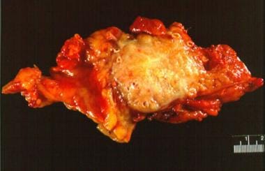
Pancreatic cancer. Gross section of an adenocarcinoma of the pancreas measuring 5 X 6 cm resected from the pancreatic body and tail. Although the tumor was considered to have been fully resected and had not spread to any nodes, the patient died of recurrent cancer within 1 year. |  | View Full Size Image |
|
Picture Type: Photo |
| Caption: Picture 2. Pancreatic cancer. Hematoxylin and eosin stain of a pancreatic carcinoma. Note the intense desmoplastic response around the neoplastic cells. The large amount of fibrotic reaction in these tumors can make obtaining adequate tissue by fine-needle aspiration difficult. |  | View Full Size Image |
|
Picture Type: Photo |
| Caption: Picture 3. Pancreatic cancer. T staging for pancreatic carcinoma. T1 and T2 stages are confined to the pancreatic parenchyma. T3 lesions invade local structures such as the duodenum, bile duct, and/or major peripancreatic veins, and T4 lesions invade surrounding organs (eg, stomach, colon, liver) or invade major arteries such as the superior mesenteric or celiac arteries.
|  | View Full Size Image |
|
Picture Type: Image |
| Caption: Picture 4. Pancreatic cancer. Computerized tomographic scan showing a pancreatic adenocarcinoma of the pancreatic head. The gallbladder (gb) is distended because of biliary obstruction. The superior mesenteric artery (sma) is surrounded by tumor, making this an unresectable T4 lesion. |  | View Full Size Image |
|
Picture Type: CT |
| Caption: Picture 5. Pancreatic cancer. Abdominal CT scan of a small, vaguely seen, 2-cm pancreatic adenocarcinoma (mass) causing obstruction of both the common bile duct (cbd) and pancreatic duct (pd).
|  | View Full Size Image |
|
Picture Type: CT |
| Caption: Picture 6. Pancreatic cancer. Endoscopic ultrasound of a 2.2-cm pancreatic adenocarcinoma of the head of the pancreas obstructing the common bile duct (CBD) but not invading the portal vein (PV) or superior mesenteric vein (SMV). Findings from endoscopic ultrasound–guided fine-needle aspiration revealed a moderately to poorly differentiated adenocarcinoma. Abdominal CT findings did not show this mass, and an attempt at endoscopic retrograde cholangiopancreatography at another institution was unsuccessful. |  | View Full Size Image |
|
Picture Type: Image |
| Caption: Picture 7. Algorithm for evaluation of a patient with suspected pancreatic cancer. CT scanning for definitive diagnosis and staging must be with thin-cut, multidetector, spiral CT scanning using dual-phase contrast imaging to allow for maximal information. This schema varies among institutions depending on local expertise, research interest, and therapeutic protocols for pancreatic carcinoma.
|  | View Full Size Image |
|
Picture Type: Image |
| Caption: Picture 8. Pancreatic cancer. Tip of linear array echoendoscope (Pentax FG 36UX) with 22-gauge aspiration needle exiting from biopsy channel. Insert shows magnification of aspiration needle tip. Note that the needle exits from the biopsy channel such that it appears continuously in the view of the ultrasonic transducer on the tip of the echoendoscope.
|  | View Full Size Image |
|
Picture Type: Photo |
| Caption: Picture 9. Pancreatic cancer. Cytologic samples from fine-needle aspirations (rapid Papanicolaou stain) of pancreatic adenocarcinomas. (A) Well differentiated, (B) moderately differentiated, (C) moderate to poorly differentiated, (D) poorly differentiated tumor.
|  | View Full Size Image |
|
Picture Type: Photo |
|   |
BIBLIOGRAPHY
| Section 11 of 11 |
|
-
Barkin JS, Goldstein JA: Diagnostic approach to pancreatic cancer. Gastroenterol Clin North Am 1999 Sep; 28(3): 709-22, xi[Medline].
-
Bueno de Mesquita HB, Maisonneuve P, Runia S, Moerman CJ: Intake of foods and nutrients and cancer of the exocrine pancreas: a population-based case-control study in The Netherlands. Int J Cancer 1991 Jun 19; 48(4): 540-9[Medline].
-
Burris HA, Moore MJ, Andersen J, et al: Improvements in survival and clinical benefit with gemcitabine as first-line therapy for patients with advanced pancreas cancer: a randomized trial. J Clin Oncol 1997 Jun; 15(6): 2403-13[Medline].
-
Chang KJ, Nguyen P, Erickson RA, et al: The clinical utility of endoscopic ultrasound-guided fine-needle aspiration in the diagnosis and staging of pancreatic carcinoma. Gastrointest Endosc 1997 May; 45(5): 387-93[Medline].
-
Cleary SP, Gryfe R, Guindi M, et al: Prognostic factors in resected pancreatic adenocarcinoma: analysis of actual 5-year survivors. J Am Coll Surg 2004 May; 198(5): 722-31[Medline].
-
De Bellis M, Sherman S, Fogel EL, et al: Tissue sampling at ERCP in suspected malignant biliary strictures (Part 1). Gastrointest Endosc 2002 Oct; 56(4): 552-61[Medline].
-
de Bellis M, Sherman S, Fogel EL, et al: Tissue sampling at ERCP in suspected malignant biliary strictures (Part 2). Gastrointest Endosc 2002 Nov; 56(5): 720-30[Medline].
-
Delcore R, Rodriguez FJ, Forster J, et al: Significance of lymph node metastases in patients with pancreatic cancer undergoing curative resection. Am J Surg 1996 Nov; 172(5): 463-8; discussion 468-9[Medline].
-
Erickson RA: Endoscopic ultrasonography: a new diagnostic imaging modality. Am Fam Physician 1997 May 1; 55(6): 2219-28[Medline].
-
Erickson RA, Garza AA: EUS with EUS-guided fine-needle aspiration as the first endoscopic test for the evaluation of obstructive jaundice. Gastrointest Endosc 2001 Apr; 53(4): 475-84[Medline].
-
Evans DB, Staley CA, Lee JE: Adenocarcinoma of the pancreas: recent controversies, current management, and future therapies. Gastrointest Cancer 1996; 1: 149-61.
-
Everhart J, Wright D: Diabetes mellitus as a risk factor for pancreatic cancer. A meta-analysis. JAMA 1995 May 24-31; 273(20): 1605-9[Medline].
-
Flanders TY, Foulkes WD: Pancreatic adenocarcinoma: epidemiology and genetics. J Med Genet 1996 Nov; 33(11): 889-98[Medline].
-
Goggins M, Schutte M, Lu J, et al: Germline BRCA2 gene mutations in patients with apparently sporadic pancreatic carcinomas. Cancer Res 1996 Dec 1; 56(23): 5360-4[Medline].
-
Gress F, Gottlieb K, Sherman S, Lehman G: Endoscopic ultrasonography-guided fine-needle aspiration biopsy of suspected pancreatic cancer. Ann Intern Med 2001 Mar 20; 134(6): 459-64[Medline].
-
Hahn SA, Kern SE: Molecular genetics of exocrine pancreatic neoplasms. Surg Clin North Am 1995 Oct; 75(5): 857-69[Medline].
-
Hayakawa T, Naruse S, Kitagawa M, et al: A prospective multicenter trial evaluating diagnostic validity of multivariate analysis and individual serum marker in differential diagnosis of pancreatic cancer from benign pancreatic diseases. Int J Pancreatol 1999 Feb; 25(1): 23-9[Medline].
-
Horton KM, Fishman EK: Adenocarcinoma of the pancreas: CT imaging. Radiol Clin North Am 2002 Dec; 40(6): 1263-72[Medline].
-
Hunt GC, Faigel DO: Assessment of EUS for diagnosing, staging, and determining resectability of pancreatic cancer: a review. Gastrointest Endosc 2002 Feb; 55(2): 232-7[Medline].
-
Lowenfels AB, Maisonneuve P, Cavallini G, et al: Pancreatitis and the risk of pancreatic cancer. International Pancreatitis Study Group. N Engl J Med 1993 May 20; 328(20): 1433-7[Medline].
-
Lynch HT, Smyrk T, Kern SE, et al: Familial pancreatic cancer: a review. Semin Oncol 1996 Apr; 23(2): 251-75[Medline].
-
Micames C, Jowell PS, White R, et al: Lower frequency of peritoneal carcinomatosis in patients with pancreatic cancer diagnosed by EUS-guided FNA vs. percutaneous FNA. Gastrointest Endosc 2003 Nov; 58(5): 690-5[Medline].
-
Moore MJ, Goldstein D, Hamm J, et al: Erlotinib plus gemcitabine compared to gemcitabine alone in patients with advanced pancreatic cancer. A phase III trial of the National Cancer Institute of Canada Clinical Trials Group [NCIC-CTG]. J Clin Oncol 2005 ASCO Annual Meeting Proceedings; 23(16S) June 1 Supplement: 1.
-
Mortele KJ, Ji H, Ros PR: CT and magnetic resonance imaging in pancreatic and biliary tract malignancies. Gastrointest Endosc 2002 Dec; 56(6 Suppl): S206-12[Medline].
-
Nino-Murcia M, Jeffrey RB: Multidetector-row CT and volumetric imaging of pancreatic neoplasms. Gastroenterol Clin North Am 2002 Sep; 31(3): 881-96[Medline].
-
Posner MR, Mayer RJ: The use of serologic tumor markers in gastrointestinal malignancies. Hematol Oncol Clin North Am 1994 Jun; 8(3): 533-53[Medline].
-
Powis ME, Chang KJ: Endoscopic ultrasound in the clinical staging and management of pancreatic cancer: its impact on cost of treatment. Cancer Control 2000 Sep-Oct; 7(5): 413-20[Medline].
-
Recine M, Kaw M, Evans DB, Krishnamurthy S: Fine-needle aspiration cytology of mucinous tumors of the pancreas. Cancer 2004 Apr 25; 102(2): 92-9[Medline].
-
Ritts RE, Pitt HA: CA 19-9 in pancreatic cancer. Surg Oncol Clin N Am 1998 Jan; 7(1): 93-101[Medline].
-
Rulyak SJ, Lowenfels AB, Maisonneuve P, Brentnall TA: Risk factors for the development of pancreatic cancer in familial pancreatic cancer kindreds. Gastroenterology 2003 May; 124(5): 1292-9[Medline].
-
Silverman DT, Dunn JA, Hoover RN, et al: Cigarette smoking and pancreas cancer: a case-control study based on direct interviews. J Natl Cancer Inst 1994 Oct 19; 86(20): 1510-6[Medline].
-
Spitz FR, Abbruzzese JL, Lee JE, et al: Preoperative and postoperative chemoradiation strategies in patients treated with pancreaticoduodenectomy for adenocarcinoma of the pancreas. J Clin Oncol 1997 Mar; 15(3): 928-37[Medline].
-
Strasberg SM, Drebin JA, Soper NJ: Evolution and current status of the Whipple procedure: an update for gastroenterologists. Gastroenterology 1997 Sep; 113(3): 983-94[Medline].
-
Van Hoe L, Baert AL: Pancreatic carcinoma: applications for helical computed tomography. Endoscopy 1997 Aug; 29(6): 539-60[Medline].
-
Warshaw AL, Fernández-del Castillo C: Pancreatic carcinoma. N Engl J Med 1992 Feb 13; 326(7): 455-65[Medline].
Pancreatic Cancer excerpt |