Practice Essentials
Aplastic anemia is a syndrome of bone marrow failure characterized by peripheral pancytopenia and marrow hypoplasia (see the image below). Although the anemia is often normocytic, mild macrocytosis can also be observed in association with stress erythropoiesis and elevated fetal hemoglobin levels.
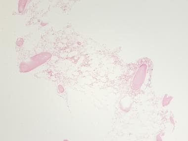 Aplastic anemia. Low-power view of hematoxylin-eosin–stained bone marrow showing hypocellularity, with increased adipose tissue and decreased hematopoietic cells in the marrow space.
Signs and symptoms
The clinical presentation of patients with aplastic anemia includes signs and symptoms related to the decrease in bone marrow production of hematopoietic cells. The onset is insidious, and the initial clinical manifestation is frequently related to anemia or bleeding, although fever or infections may be noted at presentation.
Signs and symptoms of aplastic anemia may include the following:
-
Pallor
-
Headache
-
Palpitations, dyspnea
-
Fatigue
-
Foot swelling
-
Gingival bleeding, petechial rashes
-
Overt and/or recurrent infections
-
Oropharyngeal ulcerations
A subset of patients with aplastic anemia present with jaundice or other evidence of hepatitis. [1, 2]
See Presentation for more detail.
Diagnosis
Testing
Laboratory testing for suspected aplastic anemia includes the following:
-
Complete blood count
-
Peripheral blood smears
-
Hemoglobin electrophoresis and blood-group testing
-
Biochemical profile
-
Serology for hepatitis and other viral entities
-
Autoimmune-disease evaluation for evidence of collagen-vascular disease
-
Fluorescence-activated cell sorter profiling
-
Fluorescent-labeled inactive toxin aerolysin testing
-
Diepoxybutane incubation
-
Histocompatibility testing
-
Kidney function studies
-
Liver function studies
-
Transaminase, bilirubin, and lactate dehydrogenase levels
Procedures
Bone marrow biopsy is performed in addition to aspiration to assess cellularity qualitatively and quantitatively. Bone marrow culture may be useful in diagnosing mycobacterial and viral infections; however, the yield is generally low.
See Workup for more detail.
Management
Severe or very severe aplastic anemia is a hematologic emergency, and care should be instituted promptly. Clinicians must stress the need for patient compliance with therapy. The specific medications administered depend on the choice of therapy and whether it is supportive care only, immunosuppressive therapy, or hematopoietic cell transplantation. [3]
Pharmacotherapy
The following medications are used in patients with aplastic anemia:
-
Immunosuppressive agents (eg, cyclosporine, methylprednisolone, equine antithymocyte globulin, rabbit antithymocyte globulin, cyclophosphamide, alemtuzumab)
-
Hematopoietic growth factors (eg, eltrombopag [4] , sargramostim, filgrastim)
-
Antimetabolite (purine) antineoplastic agents (eg, fludarabine)
-
Iron-chelating agents (eg, deferoxamine, deferasirox)
Nonpharmacotherapy
Nonpharmacologic management of aplastic anemia includes the following:
-
Supportive care
-
Blood transfusions with blood products that have undergone leukocyte reduction and irradiation
-
Hematopoietic cell transplantation
See Treatment and Medication for more detail.
The British Society for Standards in Haematology has issued guidelines on diagnosis and management of aplastic anemia in adults [5] and pediatric patients. [6] The Pediatric Haemato-Oncology Italian Association has issued guidelines on diagnosis and management of acquired aplastic anemia in childhood. [7]
For more information, see the following Medscape articles:
For patient education information, see What Is Aplastic Anemia?.
Background
Paul Ehrlich introduced the concept of aplastic anemia in 1888 when he reported the case of a pregnant woman who died of bone marrow failure. However, it was not until 1904 that Anatole Chauffard named this disorder aplastic anemia.
Etiology
The theoretical basis for marrow failure includes primary defects in or damage to the stem cell or the marrow microenvironment. [8, 9, 10, 11, 12] This condition is characterized by both qualitative loss in functions as well as quantitative loss in stem cell numbers. The distinction between acquired and inherited disease may present a clinical challenge, but more than 80% of cases are acquired. External insults (eg, infections, radiation, drugs) may disrupt stem cell homeostasis in marrow environment, leading to altered growth. Clinical and laboratory observations suggest that acquired aplastic anemia is an autoimmune disease.
On morphologic evaluation, the hematopoietic elements in the bone marrow are less than 25%, and they are largely replaced with fat cells. Flow cytometry shows that the CD34 cell population, which contains the stem cells and the early committed progenitors, is substantially reduced. [10] Fas-mediated apoptosis of CD34+ progenitor cells causes stem cell depletion. [13, 14]
Data from in vitro colony-culture assays suggest profound functional loss of the hematopoietic progenitors, so much so that they are unresponsive even to high levels of hematopoietic growth factors.
Previously, it had been hypothesized that aplastic anemia may be due to a defect at various levels, such as an intrinsic defect of hematopoietic cells; external injury to hematopoietic cells; and defective stroma, which is critical for normal proliferation and functioning of hematopoietic cells. Theoretically, all of these mechanisms could be responsible for aplastic anemia. This theory was the basis of many in vitro stem cell culture experiments using a crossover design in which stem cells from patients with aplastic anemia were cultured with normal stroma and vice versa. The conclusions from these studies led to the understanding that stem cell defect is the central mechanism in the majority of patients with aplastic anemia. [15, 16]
In patients with severe aplastic anemia, stromal cells have normal function, including growth factor production. Adequate stromal function is implicit in the success of hematopoietic cell transplantation (HCT) in aplastic anemia, because the stromal elements are almost entirely (frequently) of host origin.
The role of an immune dysfunction was suggested in 1970, when autologous recovery was documented in a patient with aplastic anemia who failed to engraft after HCT. Mathe proposed that the immunosuppressive regimen used for conditioning promoted the return of normal marrow function. Since then, numerous studies have shown that, in approximately 70% of patients with acquired aplastic anemia, immunosuppressive therapy improves marrow function. [11, 17, 18, 19, 20]
Immunity is genetically regulated (by immune response genes), and it is also influenced by environment (eg, nutrition, aging, previous exposure). [21, 22] Although the inciting antigens that breach immune tolerance with subsequent autoimmunity are unknown, human leukocyte antigen (HLA)-DR2 is overrepresented among European and United States patients with aplastic anemia, and its presence is predictive of a better response to cyclosporine.
Suppression of hematopoiesis is likely mediated by an expanded population of CD8+ HLA-DR+, cytotoxic T lymphocytes (CTLs) that are frequently detectable in the blood and bone marrow of patients with aplastic anemia. These cells produce inhibitory cytokines, such as gamma-interferon and tumor necrosis factor, which can suppress progenitor cell growth. Polymorphisms in these cytokine genes that are associated with an increased immune response are more prevalent in patients with aplastic anemia. These cytokines suppress hematopoiesis by affecting the mitotic cycle and cell killing by inducing Fas-mediated apoptosis.
In addition, such cytokines induce nitric oxide synthase and nitric oxide production by marrow cells, which contributes to immune-mediated cytotoxicity and the elimination of hematopoietic cells. Hirano et al reported that CD8+ cytotoxic T cells raised against kinectin-derived peptides suppress colony-forming units (CFUs) in an HLA class I–restricted fashion, findings that suggest kinectin may be a candidate autoantigen in the pathophysiology of aplastic anemia. [23]
Constitutive expression of Tbet, a transcriptional regulator that is critical to type 1 T helper cell (Th1) polarization, occurs in a majority of aplastic anemia patients. [17] Perforin is a cytolytic protein expressed mainly in activated cytotoxic lymphocytes and natural-killer cells. Mutations in the perforin gene are responsible for some cases of familial hemophagocytosis [24] ; mutations in SAP, a gene encoding for a small modulator protein that inhibits undefined-interferon production, underlie X-linked lymphoproliferation, a fatal illness associated with an aberrant immune response to herpesviruses and aplastic anemia. Perforin and SAP protein levels are markedly diminished in some cases of acquired aplastic anemia.
The transcription factors FOXP3 and NFAT1 have key roles in regulatory T-cell (Treg) development and function, and Tregs play a role in autoimmunity. Tregs are decreased at presentation in almost all patients with aplastic anemia; FOXP3 protein and messenger RNA levels also are significantly lower in patients with this condition, and NFAT1 protein levels are decreased or absent. [25]
Variations in telomere length in peripheral blood cells, especially neutrophils, have been reported in severe aplastic anemia, but the clinical significance of this finding is uncertain. A faster reduction in telomere length in aplastic anemia leads to decreased expression of cell cycle checkpoint genes such as CDK2/6 and MYC, and a high number of mutations is observed in the telomere reverse transcriptase (TERT) gene. [26, 27]
However, although telomere length was unrelated to response, it has been associated with the risk of relapse, clonal evolution, and overall survival in patients with severe aplastic anemia. [28] Studies in murine models also suggest a role of thrombopoietin (TPO) and associated signalling pathway during normal hematopoiesis. Raised levels of TPO have been observed in aplastic anemia, most likely due to compensatory response to diminished stem cells function. [29, 30] Moreover, a decrease in TPO levels is seen after immunosuppressive therapy. [31]
Congenital or inherited causes
Congenital or inherited causes of aplastic anemia are responsible for at least 25% of cases in children and for perhaps up to 10% of adults. [32] Patients may have dysmorphic features or physical stigmata, but marrow failure may be the initial presenting feature. Several loci have been identified that are associated not only with increased susceptibility to aplastic anemia but also with other physical findings.
Fanconi anemia
Fanconi anemia is characterized by the following:
-
Multiple congenital anomalies (60-75%): Short stature, abnormal skin pigmentation, malformations of the thumbs with or without dysplastic or absent radii, as well as microphthalmos and malformations of the heart, kidneys, intestines, and ears
-
Bone marrow failure: Thrombocytopenia, leukopenia, or aplastic anemia; most patients with Fanconi anemia have bone marrow failure by adulthood
-
Cancer: Hematologic malignancies are common with Fanconi anemia and myelodysplasia, with acute myeloid leukemia (AML) being the most common; solid tumors such as those in squamous cell head and neck cancer, female genital tumors, and liver tumors are also seen; Fanconi anemia is associated with increased chromosomal breakage and abnormal sister chromatid exchange in the presence of agents such as diepoxybutane or mitomycin C
-
Predominantly autosomal recessive inheritance pattern: Fifteen genes are now known to be causative for Fanconi anemia; only 1 of these— FANCB (X-linked recessive)—is not inherited in an autosomal recessive manner
-
Association between bone marrow progression and a complex pattern of recurrent chromosomal abnormalities: Some chromosomal abnormalities are commonly found in non–Fanconi anemia myelodysplastic syndrome (MDS) and secondary AML (eg, -7/7q or RUNX1 abnormalities), whereas others are specific for Fanconi anemia (eg, 1q+ and 3q+)
Dyskeratosis congenita
Dyskeratosis congenita is characterized by the diagnostic physical triad of dysplastic nails, lacy reticular pigmentation of the upper torso, and oral leukoplakia. However, patients may have dyskeratosis congenita without the triad. The following are also features of this condition:
-
Some signs of premature aging, such as early graying of the hair
-
Progressive bone marrow failure at any age, which can cause any combination of cytopenias, including aplastic anemia
-
Malignancy: Common; frequently MDS or AML; solid tumors such as head and neck cancer or genital cancers can also be seen
-
Pulmonary fibrosis
-
Autosomal dominant, autosomal recessive, and X-linked inheritance patterns; 6 genes are known to cause this disorder
Familial aplastic anemia
This is an isolated aplastic anemia. Mutations have been found in the TERC and TERT genes and are thought to confer a susceptibility to aplastic anemia. These genes encode proteins that are part of the telomerase apparatus that restores repeated regions in the telomere. [33]
Cartilage-hair hypoplasia
Cartilage-hair hypoplasia, which is caused by mutations in the RMRP gene, is inherited in an autosomal recessive manner. This condition is characterized by the following:
-
Short stature with short and bowed limbs
-
Sparse, lightly pigmented hair
-
Variably severe immune deficiency
-
Anemia during childhood
-
Hematopoietic malignancies, as well as malignancies of the skin, eyes, and liver
-
Gastrointestinal malformations and malabsorption
Pearson syndrome
Pearson syndrome causes sideroblastic anemia and exocrine pancreatic dysfunction. This condition results from mitochondrial DNA deletions.
Thrombocytopenia-absent radius syndrome
Thrombocytopenia-absent radius (TAR) syndrome is characterized by deletions located at chromosome 1q21.1 (which are typically about 200kb in size). Patients have bilateral absence of the radii with presence of the thumbs, as well as thrombocytopenia. Other congenital anomalies can also occur (eg, cardiac disease, skeletal anomalies, urogenital anomalies).
Shwachman-Diamond syndrome
Shwachman-Diamond syndrome is caused by mutations in the SBDS gene and is inherited in an autosomal recessive manner. This disease is characterized by dysfunction of the exocrine pancreas with malabsorption and growth failure, as well as cytopenias of single or multiple lineage. Patients with Shwachman-Diamond syndrome also have an increased risk of MDS and AML. [34]
Dubowitz syndrome
Dubowitz syndrome is characterized by intrauterine growth retardation, extremely short stature, and wizened facial appearance. Patients also have microcephaly and mild developmental delay. Dubowitz syndrome is also associated with eczema, immune deficiency, and aplastic anemia. Malignancy is more common with this disorder, particularly lymphoma and neuroblastoma. No single gene has been implicated as responsible for this syndrome. [35]
Diamond-Blackfan anemia
Diamond-Blackfan anemia (DBA) is characterized by a normochromic macrocytic anemia that can be isolated, or can be associated with growth retardation or congenital malformation in the upper limbs, heart, and genitourinary systems. In a small minority of patients, DBA can progress to aplastic anemia. Nine genes have been found to be causative for DBA, and they are inherited in an autosomal dominant manner. [36] Approximately 50% of cases are inherited from a parent, and about 50% result from de novo mutations.
Acquired causes
Acquired causes of aplastic anemia (80%) include the following:
-
Idiopathic factors
-
Infections, with pathogens such as hepatitis viruses, [37] Epstein-Barr virus (EBV), human immunodeficiency virus (HIV), parvovirus, and mycobacteria
-
Exposure to ionizing radiation
-
Exposure to toxic chemicals, such as benzene or pesticides [38]
-
Transfusional graft versus host disease (GVHD)
-
Orthotopic liver transplantation for fulminant hepatitis
-
Pregnancy
-
Eosinophilic fasciitis
-
Anorexia
-
Severe nutritional deficiencies (vitamin B12, folate)
-
Paroxysmal nocturnal hemoglobinuria (PNH)
-
MDS
-
Acute lymphoblastic leukemia (ALL)(rarely)
Drugs and elements (eg, chloramphenicol, gold) may cause aplasia of the marrow. The immune mechanism does not account for the marrow failure in idiosyncratic drug reactions. In such cases, direct toxicity may occur, perhaps due to genetically determined differences in metabolic detoxification pathways. For example, the null phenotype of certain glutathione transferases is overrepresented among patients with aplastic anemia.
PNH is caused by an acquired genetic defect affecting the PIGA gene and limited to the stem cell compartment. Mutations in the PIGA gene render cells of hematopoietic origin sensitive to increased complement lysis. Approximately one third of patients with aplastic anemia have evidence of PNH at presentation, as detected by means of flow cytometry. [39] Furthermore, patients whose disease responds after immunosuppressive therapy may recover with clonal hematopoiesis and PNH.
Very rarely, aplastic anemia has been reported following vaccination. Case reports describe aplastic anemia—de novo or relapse—following vaccination against hepatitis B, influenza, varicella, and COVID-19. [40, 41, 42]
Epidemiology
United States statistics
No accurate prospective data are available regarding the incidence of aplastic anemia in the United States. Findings from several retrospective studies usually overlap those from Europe and suggest that the incidence is 0.6-6.1 cases per million population; this rate is largely based on data from reviews of death registries.
International statistics
The annual incidence of aplastic anemia in Europe, as detailed in large, formal epidemiologic studies, is 2 cases per million population. [43] Aplastic anemia is thought to be more common in Asia than in the West. The incidence was accurately determined to be 4 cases per million population in Bangkok, [44] but based on prospective studies, it may actually be closer to 6 cases per million population in the rural areas of Thailand. This increased incidence may be related to environmental factors, such as increased exposure to toxic chemicals, rather than to genetic factors, because this increase is not observed in people of Asian ancestry who are living in the United States.
Race-, sex-, and age-related demographics
Although no racial predisposition for aplastic anemia is reported in the United States, the prevalence is increased in the Far East. The male-to-female ratio for acquired aplastic anemia is approximately 1:1, although a male preponderance has been reported in studies from Asia. [45]
Although aplastic anemia occurs in all age groups, a small peak in the incidence is observed in childhood because of the inclusion of inherited marrow-failure syndromes. A second peak is observed in people aged 20-25 years.
Prognosis
The outcome of patients with aplastic anemia has substantially improved because of improved supportive care. The natural history of aplastic anemia suggests that a small number of patients may spontaneously recover with supportive care [46] ; however, observation and/or supportive care alone is rarely indicated. Specific therapies, including hematopoietic cell transplantation (HCT) and immunosuppressive regiimens, can be curative, particularly in cases with an identical HLA family donor, where overall survival probability is as high as 90%. [47]
The estimated 10-year survival rate for the typical patient receiving immunosuppression is 68%, compared with 73% for HCT. [48] However, there is a significantly improved outcome for HCT over time, for matched sibling and alternative donors, and with younger age. [48] In cases of immunosuppression, relapse and late clonal disease are risks.
In a single-institution analysis of 183 patients who received immunosuppressive treatments for severe aplastic anemia, the telomere length of peripheral blood leukocytes was unrelated to treatment response. In a multivariate analysis, however, telomere length inversely correlated with risk of relapse, clonal evolution, and overall survival. [28] In a study of 330 patients with aplastic anemia (235 acquired, 85 Fanconi anemia, and 10 Diamond-Blackfan anemia) who underwent HCT from unrelated donors found that longer telomere length in donor leukocytes was associated with increased 5-year survival, but patient leukocyte telomere length was not associated with survival. [49]
In a Japanese study of 64 children with aplastic anemia, multivariate analysis showed that the following were significant variables for poor response to immunosuppressive therapy [50] :
-
Telomere length shorter than -1.0 standard deviations (hazard ratio [HR]: 22.0; 95% confidence interval [CI]: 4.19-115; P< 0.001)
-
Platelet count at diagnosis less than 25 × 10 9/L (HR: 13.9; 95%CI: 2.00-96.1; P = 0.008),
-
Interval from diagnosis to immunosuppressive therapy longer than 25 days (HR: 4.81; 95%CI: 1.15-20.1; P = 0.031)
Mortality/morbidity
Infection and bleeding are major causes of morbidity and mortality from aplastic anemia. Patients who undergo HCT have additional issues related to acute and chronic toxicity from the conditioning regimen and graft versus host disease (GVHD), as well as a potential for graft failure. [22, 51, 52, 53, 54, 55]
In approximately 25-30% of patients with aplastic anemia, the condition does not respond to immunosuppression. In cases with a treatment response, relapse and late-onset clonal disease, such as paroxysmal nocturnal hemoglobinuria (PNH), myelodysplastic syndrome (MDS), and leukemia, are risks—regardless of the treatment response or degree of response. [18, 56, 57, 58, 59]
Kulasekararaj and colleagues reported that the presence of somatic mutations (including ASXL1, DNMT3A, and BCOR) in patients with aplastic anemia for more than 6 months was associated with 40% risk of transformation to MDS. Nearly a fifth of patients with aplastic anemia have mutations in genes typically seen in myeloid malignancies that predicted for later transformation to MDS. [60]
In a Japanese study of 427 patients (16-72 years old) with aplastic anemia who underwent unrelated-donor bone marrow transplantation, outcome was significantly inferior in patients whose donors were 40 years of age or older than in those with younger donors. In the older donor group, overall survival was significantly inferior (adjusted hazard ratio, 1.64), the incidence of fatal infection was significantly higher (13.7% vs. 7.5%), and primary engraftment failure and acute GVHD occurred significantly more often (9.7% vs. 5.0% and 27.1% vs. 19.7%, respectively). [61]
An Australian population-based cohort study of adults receiving allogeneic HCT reported an elevated secondary cancer risk in several patient groups, including those transplanted for severe aplastic anemia. Overall, in patients alive 2 years after transplantation (n=1463), the cumulative incidence of late mortality was 22.2% at 10 years and the risk of death relative to the matched general population was 13.8. [62]
A study of 663 patients with severe aplastic anemia, 95 of whom developed clonal evolution, found that pre-treatment age > 48 years and absolute neutrophil count > 0.87 × 10⁹/L were strong predictors of high-risk evolution (defined as presence of an overt myeloid neoplasm, or any chromosome 7 abnormality or complex karyotype). Splicing factors and RUNX1 somatic variants were detected exclusively at high-risk evolution; RUNX1, splicing factors, and ASXL1 somatic mutations detected at 6 months after immunosuppressive therapy predicted high-risk evolution. [63]
Five-year overall survival (OS) was 37% in patients with high-risk clonal evolution, versus 94% in those with low-risk. OS was improved in high-risk patients who underwent HCT. [63]
-
Oral leukoplakia in dyskeratosis congenita.
-
Aplastic anemia. Low-power view of hematoxylin-eosin–stained bone marrow showing hypocellularity, with increased adipose tissue and decreased hematopoietic cells in the marrow space.


