Continually Updated Clinical Reference
 
 
  All Sources     eMedicine     Medscape     Drug Reference     MEDLINE
 
eMedicine - Cavernous Sinus Thrombosis : Article by

Quick Find
Authors & Editors
Introduction
Clinical
Differentials
Workup
Treatment
Medication
Follow-up
Miscellaneous
Multimedia
References

Related Articles
Cellulitis

Epidural and Subdural Infections

Epidural Hematoma

Glaucoma, Acute Angle-Closure

Orbital Infections

Periorbital Infections

Sinusitis

Subarachnoid Hemorrhage

Subdural Hematoma


Patient Education
Click here for patient education.


Author: Rahul Sharma, MD, MBA, Instructor in Medicine, Weill Medical College of Cornell University; Consulting Staff, Department of Emergency Medicine, New York Presbyterian Hospital-Weill Cornell Medical Center

Rahul Sharma is a member of the following medical societies: American College of Emergency Physicians

Coauthor(s): Edward Bessman, MD, Chairman, Department of Emergency Medicine, John Hopkins Bayview Medical Center; Assistant Professor, Department of Emergency Medicine, Johns Hopkins University

Editors: David FM Brown, MD, Assistant Professor, Department of Medicine, Division of Emergency Medicine, Harvard Medical School; Associate-Chief, Attending Physician, Department of Emergency Medicine, Massachusetts General Hospital; Francisco Talavera, PharmD, PhD, Senior Pharmacy Editor, eMedicine; J Stephen Huff, MD, Associate Professor of Emergency Medicine and Neurology, Department of Emergency Medicine, University of Virginia Health Sciences Center; John D Halamka, MD, MS, Associate Professor of Medicine, Harvard Medical School, Beth Israel Deaconess Medical Center; Chief Information Officer, CareGroup Healthcare System and Harvard Medical School; Attending Physician, Division of Emergency Medicine, Beth Israel Deaconess Medical Center; Barry E Brenner, MD, PhD, FACEP, Program Director, Professor, Department of Emergency Medicine, Professor, Internal Medicine, University Hospitals, Case Western Reserve School of Medicine

Author and Editor Disclosure

Synonyms and related keywords: cavernous sinus thrombosis, CST, thrombosis of the cavernous intracranial sinus, cavernous sinus syndrome, cavernous sinus, cavernous sinuses, parasellar lesions, tumors, carotid artery aneurysms, carotid-cavernous fistulas, C-C fistulas, cavernous sinus thrombosis

Background

Cavernous sinus thrombosis (CST) was initially described by Bright in 1831 as a complication of epidural and subdural infections. CST is usually a late complication of an infection of the central face or paranasal sinuses. Other causes include bacteremia, trauma, and infections of the ear or maxillary teeth. CST is generally a fulminant process with high rates of morbidity and mortality. Fortunately, the incidence of CST has been decreased greatly with the advent of effective antimicrobial agents.

Pathophysiology

The cavernous sinuses are irregularly shaped, trabeculated cavities located at the base of the skull. The cavernous sinuses are the most centrally located of the dural sinuses and lie on either side of the sella turcica. These sinuses are just lateral and superior to the sphenoid sinus and are immediately posterior to the optic chiasm (see Image 1). Each cavernous sinus is formed between layers of the dura mater, and multiple connections exist between the 2 sinuses.

The cavernous sinuses receive venous blood from the facial veins (via the superior and inferior ophthalmic veins) as well as the sphenoid and middle cerebral veins. They, in turn, empty into the inferior petrosal sinuses, then into the internal jugular veins and the sigmoid sinuses via the superior petrosal sinuses. This complex web of veins contains no valves; blood can flow in any direction depending on the prevailing pressure gradients. Since the cavernous sinuses receive blood via this distribution, infections of the face including the nose, tonsils, and orbits can spread easily by this route.

The internal carotid artery with its surrounding sympathetic plexus passes through the cavernous sinus. The third, fourth, and sixth cranial nerves are attached to the lateral wall of the sinus. The ophthalmic and maxillary divisions of the fifth cranial nerve are embedded in the wall (see Image 1).

This intimate juxtaposition of veins, arteries, nerves, meninges, and paranasal sinuses accounts for the characteristic etiology and presentation of CST.

Staphylococcus aureus accounts for approximately 70% of all infections. Streptococcus pneumoniae, gram-negative bacilli, and anaerobes can also be seen. Fungi are a less common pathogen and may include Aspergillus and Rhizopus species.

Frequency

United States

Occurrence of CST has always been low, with only a few hundred case reports in the medical literature. The majority of these date from before the modern antibiotic era. One review of the English-language literature found only 88 cases from 1940-1988.

Mortality/Morbidity

Prior to the advent of effective antimicrobial agents, the mortality rate from CST was effectively 100%. Typically, death is due to sepsis or central nervous system (CNS) infection. With aggressive management, the mortality rate is now less than 30%. Morbidity, however, remains high, and complete recovery is rare. Roughly one sixth of patients are left with some degree of visual impairment, and one half have cranial nerve deficits.

Race

No predilection

Sex

No predilection

Age

All ages are affected, with a mean of 22 years.


History

The early signs and symptoms of cavernous sinus thrombosis (CST) may not be specific. A patient who presents with headache and any cranial nerve findings should be potentially evaluated for CST. The most common signs of CST are related to the anatomical structures affected within the cavernous sinus (see Image 1).

  • Patients generally have sinusitis or a midface infection (most commonly a furuncle) for 5-10 days. In as many as 25% of cases in which a furuncle is the precipitant, it will have been manipulated in some fashion (eg, squeezing, surgical incision).
  • Headache is the most common presentation symptom and usually precedes fevers, periorbital edema, and cranial nerve signs. The headache is usually sharp, increases progressively, and is usually localized to the regions innervated by the ophthalmic and maxillary branches of the fifth cranial nerve.
  • In some patients, periorbital findings do not develop early on, and the clinical picture is subtle.
  • As the infection tracts posteriorly, patients complain of orbital pain and fullness accompanied by periorbital edema and visual disturbances.
  • Without effective therapy, signs appear in the contralateral eye by spreading through the communicating veins to the contralateral cavernous sinus. Eye swelling begins as a unilateral process and spreads to the other eye within 24-48 hours via the intercavernous sinuses. This is pathognomonic for CST.
  • The patient rapidly develops mental status changes including confusion, drowsiness, and coma from CNS involvement and/or sepsis. Death follows shortly thereafter.

Physical

Other than the findings associated with the primary infection, the following signs are typical:

  • Periorbital edema may be the earliest physical finding.
    • Chemosis results from occlusion of the ophthalmic veins.
    • Lateral gaze palsy (isolated cranial nerve VI) is usually seen first since CN VI lies freely within the sinus in contrast to CN III and IV, which lie within the lateral walls of the sinus.
    • Ptosis, mydriasis, and eye muscle weakness from cranial nerve III dysfunction
  • Manifestations of increased retrobulbar pressure follow.
  • Signs of increased intraocular pressure (IOP) may be observed.
    • Pupillary responses are sluggish.
    • Decreased visual acuity is common owing to increased IOP and traction on the optic nerve and central retinal artery.
  • Hypoesthesia or hyperesthesia in dermatomes supplied by the V1 and V2 branches of the fifth cranial nerve
  • Appearance of signs and symptoms in the contralateral eye is diagnostic of CST, although the process may remain confined to one eye.
  • Meningeal signs, including nuchal rigidity and Kernig and Brudzinski signs, may be noted.
  • Systemic signs indicative of sepsis are late findings. They include chills, fever, shock, delirium, and coma.

Causes

  • Most cases of septic CST are due to an acute infection in an otherwise healthy individual. However, patients with chronic sinusitis or diabetes mellitus may be at a slightly higher risk.
  • The causative agent is generally Staphylococcus aureus, although streptococci, pneumococci, and fungi may be implicated in rare cases.


Cellulitis
Epidural and Subdural Infections
Epidural Hematoma
Glaucoma, Acute Angle-Closure
Orbital Infections
Periorbital Infections
Sinusitis
Subarachnoid Hemorrhage
Subdural Hematoma

Other Problems to be Considered

Carotid artery aneurysm or fistula
Exophthalmic goiter
Orbital neoplasm


Lab Studies

  • Cavernous sinus thrombosis (CST) is a clinical diagnosis and lab studies are seldom specific. Most patients exhibit a polymorphonuclear leukocytosis, often marked with a shift toward immature forms. Examination of the cerebrospinal fluid is consistent with either a parameningeal inflammation or frank meningitis. Blood culture results generally are positive for the offending organism.

Imaging Studies

  • Historically, a number of techniques have been used to image CST, including plain sinus radiography, carotid angiography, and orbital venography. In current practice, computed tomography (CT) scan or magnetic resonance imaging (MRI) with contrast is the modality of choice to confirm the diagnosis of CST and to differentiate it from alternatives such as orbital cellulitis, which may have a similar clinical presentation.
  • MRI with MR venogram (MRV) is the preferred imaging choice as the MRV will show the absence of venous flow in the affected cavernous sinus. 
  • On noncontrast CT, thrombosis of the cavernous sinus can be appreciated as increased density. The introduction of intravenous contrast can reveal filling defects within the cavernous sinus as well as thickening of the superior ophthalmic vein. Nevertheless, CT scan findings may be subtle, and a negative CT scan cannot rule out CST reliably when the clinical suspicion is high.
  • Carotid angiography can demonstrate narrowing or obstruction of the intercavernous segment of the carotid artery. MRI and CT scan can also show this narrowing and/or obstruction of the carotid artery.

Procedures

  • Lumbar puncture may be helpful in distinguishing CST from more localized processes (eg, sinusitis, orbital cellulitis). Lumbar puncture reveals inflammatory cells in approximately 75% of cases.


Emergency Department Care

  • The mainstay of therapy for cavernous sinus thrombosis is early and aggressive antibiotic administration. Although S aureus is the usual cause, broad-spectrum coverage for gram-positive, gram-negative, and anaerobic organisms should be instituted pending the outcome of cultures.
  • Empiric antibiotic therapy should include a penicillinase-resistant penicillin plus a third- or fourth-generation cephalosporin. If dental infection or other anaerobic infection is suspected, an anaerobic coverage should also be added.
  • IV antibiotics are recommended for a minimum of 3-4 weeks.
  • Controversy exists on the use of anticoagulation for cavernous sinus thrombosis. Because of the rarity of this syndrome, no prospective trials have been performed on the use of anticoagulation for CST. Some retrospective studies have shown a decrease in mortality and clot propagation by anticoagulation. Therefore, anticoagulation with heparin should be considered since the goal is to prevent further thrombosis and to reduce the incidence of septic emboli. Heparin is contraindicated in the presence of intracerebral hemorrhage or other bleeding diathesis.
  • Corticosteroids may help to reduce inflammation and edema and should be considered as an adjunctive therapy. They should be instituted after antibiotic coverage. When the course of CST leads to pituitary insufficiency, however, corticosteroids definitely are indicated to prevent adrenal crisis. Dexamethasone or hydrocortisone should be considered.
  • Surgery on the cavernous sinus is technically difficult and has never been shown to be helpful. The primary source of infection should be drained, if feasible (eg, sphenoid sinusitis, facial abscess). It is important to recognize the infected sphenoid sinus early and to prevent spread of the infection to the cavernous sinus.

Consultations

If drainage is indicated, make arrangements for intensive care and request the appropriate surgical consultation.


Antibiotic therapy ideally is started after appropriate cultures but should not be delayed if difficulties exist in obtaining specimens. Antibiotics selected should be broad-spectrum, particularly active against S aureus, and capable of achieving high levels in the cerebrospinal fluid. With the recent increased prevalence of community-acquired MRSA, the emergency physician should consider additional coverage with intravenous antibiotics, such as vancomycin, if MRSA infection is suspected. 

Drug Category: Antibiotic, Miscellaneous

Empiric broad-spectrum coverage for gram-positive, gram-negative, and anaerobic organisms is necessary. Therapy must be comprehensive and should cover all likely pathogens in the context of the clinical setting.

In cases of suspected MRSA infection, vancomycin should be added for additional coverage.

Drug NameOxacillin (Bactocill)
DescriptionA bactericidal antibiotic that inhibits cell wall synthesis. Used in treatment of infections caused by penicillinase-producing staphylococci. May be used to initiate therapy when staphylococcal infection is suspected.
Adult Dose2 g IV q4h
Pediatric Dose50-100 mg/kg/d PO divided q6h
150-200 mg/kg/d IV/IM divided q6h; not to exceed 12 g/d
ContraindicationsDocumented hypersensitivity
InteractionsDecreases effects of contraceptives and tetracycline; disulfiram and probenecid may increase levels; large IV doses may increase effects of anticoagulants
PregnancyB - Fetal risk not confirmed in studies in humans but has been shown in some studies in animals
PrecautionsCaution in impaired renal function

Drug NameCeftriaxone (Rocephin)
DescriptionAlternate antimicrobial choice. Third-generation cephalosporin that has broad gram-negative spectrum, lower efficacy against gram-positive organisms, and higher efficacy against resistant organisms than earlier generation cephalosporins. By binding to 1 or more penicillin-binding proteins, arrests bacterial cell wall synthesis and inhibits bacterial growth.
Adult Dose2 g IV q12h
Pediatric Dose75 mg/kg/d IV divided bid
ContraindicationsDocumented hypersensitivity
InteractionsProbenecid may increase levels; ethacrynic acid, furosemide, and aminoglycosides may increase nephrotoxicity
PregnancyB - Fetal risk not confirmed in studies in humans but has been shown in some studies in animals
PrecautionsAdjust dose in renal impairment; caution in breastfeeding women and allergy to penicillin

Drug NameMetronidazole (Flagyl)
DescriptionAdditional anaerobic coverage. Imidazole ring-based antibiotic active against various anaerobic bacteria and protozoa. Usually employed in combination with other antimicrobial agents (except when used for Clostridium difficile enterocolitis, in which monotherapy appropriate).
Adult Dose500 mg IV q6h
Pediatric DoseNot established
ContraindicationsDocumented hypersensitivity, first trimester of pregnancy
InteractionsMay increase toxicity of anticoagulants, lithium, and phenytoin; cimetidine may increase toxicity; disulfiram reaction may occur with orally ingested ethanol
PregnancyB - Fetal risk not confirmed in studies in humans but has been shown in some studies in animals
PrecautionsAdjust dose in hepatic disease; monitor for seizures and development of peripheral neuropathy

Drug NameChloramphenicol (Chloromycetin)
DescriptionBinds to 50S bacterial-ribosomal subunits and inhibits bacterial growth by inhibiting protein synthesis. Effective against gram-negative and gram-positive bacteria.
Adult Dose1.5 g IV q6h
Pediatric Dose50-75 mg/kg/d PO/IV divided q6h
ContraindicationsDocumented hypersensitivity
InteractionsBarbiturates may decrease serum levels, while chloramphenicol may increase barbiturate levels, causing toxicity; manifestations of hypoglycemia may occur with sulfonylureas; rifampin may reduce serum levels, presumably through hepatic enzyme induction; may increase effects of anticoagulants; may increase serum hydantoin levels, possibly resulting in toxicity; hydantoin may increase or decrease chloramphenicol levels
PregnancyC - Fetal risk revealed in studies in animals but not established or not studied in humans; may use if benefits outweigh risk to fetus
PrecautionsUse only for indicated infections, or as prophylaxis for bacterial infections; serious and fatal blood dyscrasias (eg, aplastic anemia, hypoplastic anemia, thrombocytopenia, granulocytopenia) can occur; evaluate baseline blood studies and repeat approximately every 2 d while in therapy; discontinue upon appearance of reticulocytopenia, leukopenia, thrombocytopenia, anemia, or findings attributable to chloramphenicol; adjust dose in liver or kidney dysfunction; caution in pregnancy at term or during labor because of potential toxic effects on fetus (gray syndrome)

Drug NameVancomycin
DescriptionIndicated for patients who cannot receive or have failed to respond to penicillins and cephalosporins or have infections with resistant staphylococci. For abdominal penetrating injuries, it is combined with an agent active against enteric flora and/or anaerobes.
To avoid toxicity, current recommendation is to assay vancomycin trough levels after third dose drawn 0.5 h prior to next dosing. Use creatinine clearance to adjust dose in patients diagnosed with renal impairment.
Used in conjunction with gentamicin for prophylaxis in penicillin allergic patients undergoing gastrointestinal or genitourinary procedures.
Adult Dose1 g or 15 mg/kg IV q12h
Pediatric Dose30-40 mg/kg/d IV in 2 doses
ContraindicationsDocumented hypersensitivity
InteractionsErythema, histaminelike flushing, and anaphylactic reactions may occur when administered with anesthetic agents; taken concurrently with aminoglycosides, risk of nephrotoxicity may increase above that with aminoglycoside monotherapy; effects in neuromuscular blockade may be enhanced when coadministered with nondepolarizing muscle relaxants
PregnancyC - Fetal risk revealed in studies in animals but not established or not studied in humans; may use if benefits outweigh risk to fetus
PrecautionsCaution in renal failure, neutropenia; red man syndrome is caused by too rapid IV infusion (dose given over a few min) but rarely happens when dose given IV over 2 h administration or as PO or IP administration; red man syndrome is not an allergic reaction

Drug Category: Anticoagulants

Unfractionated IV heparin and fractionated low-molecular-weight SC heparins are the 2 options in anticoagulation therapy.

Drug NameHeparin
DescriptionAugments activity of antithrombin III and prevents conversion of fibrinogen to fibrin. Does not actively lyse thrombus but able to inhibit further thrombogenesis. Prevents reaccumulation of clot after spontaneous fibrinolysis. Various dosing nomograms available.
Adult Dose80 U/kg IV bolus; then 18 U/kg/h IV infusion; titrate to desired aPTT
Pediatric DoseAdminister as in adults
ContraindicationsDocumented hypersensitivity; subacute bacterial endocarditis; intracerebral hemorrhage, bleeding diathesis, or other active bleeding; history of heparin-induced thrombocytopenia
InteractionsDigoxin, nicotine, tetracycline, and antihistamines may decrease effects; NSAIDs, aspirin, dextran, dipyridamole, and hydroxychloroquine may increase toxicity
PregnancyC - Fetal risk revealed in studies in animals but not established or not studied in humans; may use if benefits outweigh risk to fetus
PrecautionsIn neonates, preservative-Free heparin is recommended to avoid possible toxicity (gasping syndrome) by benzyl alcohol, which is used as preservative; caution in severe hypotension and shock; monitor for bleeding in peptic ulcer disease, menstruation, increased capillary permeability, and when giving IM injections

Drug Category: Corticosteroids

These agents have anti-inflammatory properties and cause profound and varied metabolic effects. They modify the body's immune response to diverse stimuli. When the course of CST leads to pituitary insufficiency, corticosteroids definitely are indicated to prevent adrenal crisis.

Drug NameHydrocortisone (Solu-Cortef, Westcort)
DescriptionDOC due to its mineralocorticoid activity and glucocorticoid effects. Decreases inflammation by suppressing migration of polymorphonuclear leukocytes and reversing increased capillary permeability. Useful in management of inflammation caused by an immune response.
Adult Dose100 mg IV q6h
Pediatric DoseInfants and young children: 1-2 mg/kg IV as bolus followed by 25-150 mg/d divided q6-8h
ContraindicationsDocumented hypersensitivity; viral, fungal, or tubercular skin infections
InteractionsClearance may decrease with estrogens; may increase digitalis toxicity secondary to hypokalemia
PregnancyB - Fetal risk not confirmed in studies in humans but has been shown in some studies in animals
PrecautionsAbrupt discontinuation of glucocorticoids may cause adrenal crisis; caution in hyperthyroidism, osteoporosis, peptic ulcer, cirrhosis, nonspecific ulcerative colitis, diabetes, and myasthenia gravis


Further Inpatient Care

  • Admission to intensive care unit is indicated.

Deterrence/Prevention

  • Patients should be educated that furuncles or abscesses (pimples) in the central portion of the face should not be manipulated without prior antibiotic coverage.

Complications

  • Meningitis
  • Septic emboli
  • Blindness
  • Cranial nerve palsies
  • Sepsis and shock

Prognosis

  • Mortality rate is as high as 30%; the majority of survivors suffer permanent sequelae.


Medical/Legal Pitfalls

  • Failure to consider CST in the differential diagnosis of sinusitis, facial, or periorbital cellulitis


Media file 1:  
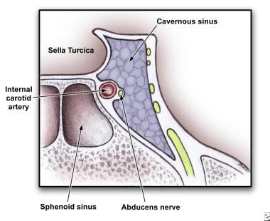
Anatomy of cross section of cavernous sinus showing close proximity to cranial nerves and sphenoid sinus.
Click to see larger pictureClick to see detailView Full Size Image
Media type:  Image


  • Bhatia K, Jones NS. Septic cavernous sinus thrombosis secondary to sinusitis: are anticoagulants indicated? A review of the literature. J Laryngol Otol. Sep 2002;116(9):667-76. [Medline].
  • Canhao P, Ferro JM, Lindgren AG. Causes and predictors of death in cerebral venous thrombosis. Stroke. Aug 2005;36(8):1720-5. [Medline].
  • Cannon ML, Antonio BL, McCloskey JJ, et al. Cavernous sinus thrombosis complicating sinusitis. Pediatr Crit Care Med. Jan 2004;5(1):86-8. [Medline].
  • DiNubile MJ. Septic thrombosis of the cavernous sinuses. Arch Neurol. May 1988;45(5):567-72. [Medline].
  • Duong DK, Leo MM, Mitchell EL. Neuro-ophthalmology. Emerg Med Clin North Am. Feb 2008;26(1):137-80, vii. [Medline].
  • Ferro JM, Canhao P, Bousser MG. Cerebral vein and dural sinus thrombosis in elderly patients. Stroke. Sep 2005;36(9):1927-32. [Medline].
  • Goodwin WJ. Orbital complications of ethmoiditis. Otolaryngol Clin North Am. Feb 1985;18(1):139-47. [Medline].
  • Heckmann JG, Tomandl B. Cavernous sinus thrombosis. Lancet. Dec 13 2003;362(9400):1958. [Medline].
  • Karlin RJ, Robinson WA. Septic cavernous sinus thrombosis. Ann Emerg Med. Jun 1984;13(6):449-55. [Medline].
  • Lessner A, Stern GA. Preseptal and orbital cellulitis. Infect Dis Clin North Am. Dec 1992;6(4):933-52. [Medline].
  • Levine SR, Twyman RE, Gilman S. The role of anticoagulation in cavernous sinus thrombosis. Neurology. Apr 1988;38(4):517-22. [Medline].
  • Peters KS. Secondary headache and head pain emergencies. Prim Care. Jun 2004;31(2):381-93, vii. [Medline].
  • Schnipper D, Spiegel JH. Management of intracranial complications of sinus surgery. Otolaryngol Clin North Am. Apr 2004;37(2):453-72, ix. [Medline].
  • Southwick FS, Richardson EP, Swartz MN. Septic thrombosis of the dural venous sinuses. Medicine (Baltimore). Mar 1986;65(2):82-106. [Medline].
  • Watkins LM, Pasternack MS, Banks M. Bilateral cavernous sinus thromboses and intraorbital abscesses secondary to Streptococcus milleri. Ophthalmology. Mar 2003;110(3):569-74. [Medline].
  • Yanofsky NN. The acute painful eye. Emerg Med Clin North Am. Feb 1988;6(1):21-42. [Medline].
  • Zimmer J, Bhatt J, et al. Is it all in his head?. The Internet Journal of Pediatrics and Neonatology. 2006;6.

Cavernous Sinus Thrombosis excerpt

Article Last Updated: Jul 18, 2008