Overview
Ticks are excellent vectors for disease transmission; consequently, tick-borne diseases are common. [1] They are second only to mosquitoes as vectors of human disease, both infectious and toxic. More than 800 species of these obligate blood-sucking creatures inhabit the planet. [2] The image below depicts the Ixodes scapularis and the Dermacentor variabilis.
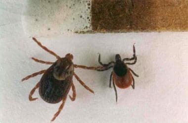 This photo shows the relative sizes of the adult forms of Ixodes scapularis (right) and Dermacentor variabilis (left). These ticks are shown next to a common match for scale. I scapularis also is referred to as Ixodes dammini. Photo by Darlyne Murawski; reproduced with permission.
See Lyme Disease and 4 Emerging Tick-Borne Illnesses, a Critical Images slideshow, to help identify and treat several tick-borne conditions.
Also, see the Bug Bites You Need to Know This Summer slideshow for helpful images and information on various bug bites.
From the perspective of disease transmission to humans, the essential characteristic of ticks is their need to ingest a blood meal to transform to their next stage of development. Not picky in their eating habits, they take their requisite blood meal from all classes of vertebrates (eg, mammals, reptiles, and birds), with the exception of fish.
Ticks feed by perching in low vegetation and waiting (questing) for a susceptible host on which they can attach themselves and feed. Once on a host, the tick inserts its hypostome, a central piercing element with hooks, into the host’s skin. Some ticks secrete a cementing material to fasten themselves to the host.
In addition, Ixodes ticks secrete anticoagulant, immunosuppressive, and anti-inflammatory substances into the area of the tick bite. These substances presumably help the tick to obtain a blood meal without the host’s noticing. These same substances also help any freeloading pathogens to establish a foothold in the host. [3]
Ticks can carry and transmit a remarkable array of pathogens, including bacteria, spirochetes, rickettsiae, protozoa, viruses, nematodes, and toxins. A single tick bite can transmit multiple pathogens, a phenomenon that has led to atypical presentations of some classic tick-borne diseases. In the United States, ticks are the most common vectors of vector-borne diseases. [4]
North American tick-borne diseases
In North America, the following diseases are caused by tick bites:
-
Tick paralysis
-
Mammalian meat allergy (alpha-gal syndrome)
-
Powassan virus disease
-
Bourbon virus disease
Tick paralysis
Tick paralysis is a neurotoxidrome that occurs in individuals with an actively attached tick. It is characterized by vague symptoms of malaise followed by worsening neurologic deficits. Findings may include bulbar symptoms such as diplopia, dysphagia, and dysarthria, as well as ataxia, generalized weakness, and areflexia. Treatment involves finding and removing the tick, which typically allows for complete neurological recovery in a matter of days.
More than 40 species can cause paralysis in both animals and humans, including Dermacentor variabilis, the most common species responsible for tick paralysis in the southeastern United States. Tick paralysis is most common from March through July, when ticks are most active, and should be considered in patients where Guillain–Barré syndrome and/or botulism are being diagnosed. [6]
Mammalian meat allergy (alpha-gal syndrome)
Over the past decade, the lone star tick, a medium-sized, reddish-brown tick common to the southeastern United States, has been implicated as the likely cause of severe mammalian meat allergies (alpha-gal syndrome [AGS]), which have been increasing in incidence. This allergy has been found in North America, Europe, Asia, Central America, and Africa. The lone star tick is believed to produce a sugar from its gut called galactose-alpha-1,3-galactose (“Alpha-Gal”), which is injected into the host during a tick bite. The sugar is also found in red meat (eg, beef, pork, venison, rabbit) and some dairy products. [7]
During 2010-2018, more than 34,000 suspected cases of AGS were identified in the United States. Places where the lone star tick is found are most affected. Cases predominantly occur in counties within the southern, midwestern, and mid-Atlantic regions. [21]
The primary management of mammalian meat allergy is avoidance of mammalian meat. The allergy can cause symptoms ranging from urticaria to angioedema and anaphylaxis. Delayed anaphylactic shock has also been reported to occur 4-6 hours after consumption of red meat. These symptoms are treated in the usual manner. Oral antihistamines are recommended for patients who remain symptomatic or have a potential high risk for exposure. Oral cromolyn may be considered for mast cell stabilization for patients with significant or persistent gastrointestinal symptoms, despite dietary avoidance. [22]
Powassan virus disease
Powassan (POW) virus may also be transmitted to humans by infected ticks, with approximately 60 US cases reported in the last decade, mostly in the Northwest United States and Great Lakes region.
Signs and symptoms of POW infection may include headache, fever, weakness, vomiting, seizures, confusion, and memory loss. Long-term neurologic problems have been reported. POW virus infection has no specific treatment, but infected persons often require hospitalization for respiratory support, intravenous fluids, or medications to reduce brain swelling. [8]
Bourbon virus disease
In June 2014, a newly discovered virus (Bourbon virus, named after the Kansas county in which it was found to have infected a farmer, ultimately leading to his death) is presumed to have been transmitted via tick bite, although this has not been proven. The patient’s symptoms, including fever, low red and white blood cell counts, elevated liver enzyme levels, and loss of appetite, suggested a tick-borne illness such as ehrlichiosis or the Heartland virus, but test results were negative. A laboratory with the Centers for Disease Control and Prevention (CDC) in Colorado finally determined that the virus was one that had never been encountered in the Western hemisphere.
The Bourbon virus belongs to the orthomyxovirus family and possesses a genome similar to that of viruses in Eastern Europe, Africa, and Asia. [9] Tests are planned to determine if prior undiagnosed but similar cases may have been caused by Bourbon virus.
European tick-borne diseases
In Europe, the list is similar to that in North America, but the following diseases should be considered as well [10] :
-
Boutonneuse fever (caused by a less virulent spotted fever rickettsial organism, Rickettsia conorii)
Most tick bites do not result in transmission of infection; in the case of Lyme disease, for example, only about 2-3% of all persons bitten by Ixodes scapularis ticks in endemic areas develop Lyme disease.
Secondary infections and allergic reactions to proteins in tick saliva are also possible. In fact, one study suggests that repeated tick bites may actually protect against Lyme disease, possibly because of hypersensitivity developed in response to previous bites of uninfected ticks. [13]
For patient education resources, see the Bites and Stings Center, as well as Ticks.
Biology and Life Cycle of Ticks
A basic familiarity with the biology of ticks is important for understanding their roles in the various tick-borne diseases and the ways in which these diseases might be prevented. [14] Ticks are arthropods of the class Arachnida, which includes spiders, scorpions, and mites. Of the 3 families of ticks, only hard ticks (family Ixodidae) and soft ticks (family Argasidae) are of medical importance. The principal difference between the 2 groups is the hard plate, or scutum, that hard ticks possess and soft ticks do not.
The life cycles of hard and soft ticks differ. Most hard ticks undergo a 2-year life cycle in which they begin as 6-legged larvae. Amblyomma, Dermacentor, and Ixodes are the 3 genera of hard ticks that transmit diseases to humans in the United States (see the images below). [15] These ticks generally feed for many days, a fact that has some bearing on the treatment of tick bites. Specific tick-borne illnesses are discussed in greater detail elsewhere, in the articles devoted to individual tick-borne diseases.
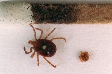 This photo is of an adult female, Amblyomma americanum, and a nymphal form of the same species (shown next to a common match for scale). This tick is the vector for monocytic ehrlichiosis and a Lymelike disease that occurs in the midcentral and southern United States. The agent for this latter disease has not yet been isolated. Photo by Darlyne Murawski; reproduced with permission.
This photo shows the relative sizes of the adult forms of Ixodes scapularis (right) and Dermacentor variabilis (left). These ticks are shown next to a common match for scale. I scapularis also is referred to as Ixodes dammini. Photo by Darlyne Murawski; reproduced with permission.
This photo is of an adult female, Amblyomma americanum, and a nymphal form of the same species (shown next to a common match for scale). This tick is the vector for monocytic ehrlichiosis and a Lymelike disease that occurs in the midcentral and southern United States. The agent for this latter disease has not yet been isolated. Photo by Darlyne Murawski; reproduced with permission.
This photo shows the relative sizes of the adult forms of Ixodes scapularis (right) and Dermacentor variabilis (left). These ticks are shown next to a common match for scale. I scapularis also is referred to as Ixodes dammini. Photo by Darlyne Murawski; reproduced with permission.
The following representative cycle is that of I scapularis (also known as Ixodes dammini) in the northeastern United States. The larvae hatch from eggs in summer and begin seeking hosts in August; at this stage, they have only 6 legs and are the size of the period at the end of this sentence. If the larvae do not find a host from which to obtain a blood meal, they die. The preferred host is the white-footed mouse, Peromyscus leucopus. Larvae that successfully feed then fall off the host and live in the soil and decaying vegetation over the winter.
The next spring, most often in May and June, the larvae molt into 8-legged nymphs (see the image below). These nymphs are quite small and seek their blood meal from a small vertebrate. Humans may be infected as accidental hosts at this point in the cycle. The nymphs then either die (if they fail to find a blood meal) or live in the soil and eventually molt into adults in the fall.
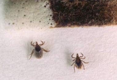 This photo shows 2 nymphal Ixodes scapularis ticks. The one on the right is unfed. Note the clear linear brown markings that are the midgut diverticula. The nymph on the left has been feeding on a mouse for 48 hours. Note that its body is larger compared with the scutum and that the midgut diverticula are less distinct. Photo by Darlyne Murawski; reproduced with permission.
The 8-legged adult ticks are somewhat larger and therefore seek a larger host. The white-tailed deer, Odocoileus virginianus, is the preferred host for adult ticks, which mate on deer over the winter. Because the deer plays a key role in tick mating, the increase in the deer population in many parts of the country is an important factor in the epidemiology of some tick-borne diseases (eg, Lyme disease). The adult female lays several thousand eggs and then dies. Eggs that survive the winter hatch into larvae the next season, and the 2-year cycle begins anew.
Soft ticks have no hard shell (scutum). In the United States, only ticks of the genus Ornithodoros (see the image below) transmit human disease—namely, relapsing fever. The biology of soft ticks differs from that of hard ticks in that meals last for only short periods (< 1 hour) and disease can be transmitted in less than 1 minute.
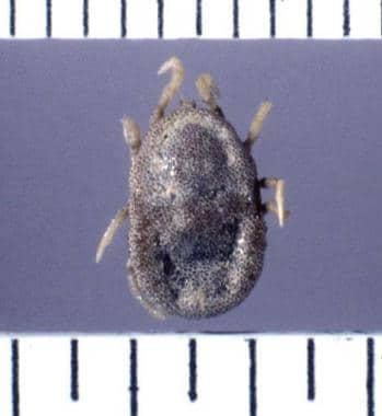 This is an example of a soft-bodied tick of the genus Ornithodoros. These ticks transmit various Borrelia species that cause relapsing fever. Photo courtesy of Julie Rawlings, MPH, Texas Department of Health.
Guidelines on the Diagnosis and Management of Tickborne Rickettsial Diseases
Guidelines on the diagnosis and management of tickborne rickettsial diseases by the Centers for Disease Control and Prevention are discussed below. [16]
The absence of known tick attachment should never dissuade a health care provider from considering the diagnosis of tickborne rickettsial disease. If the presentation supports the diagnosis, treatment should not be delayed pending results of confirmatory testing, even in the absence of definite tick exposure. [3]
Clustering of certain tickborne rickettsial diseases is well recognized and has been reported among family members, pet dogs, coworkers, military personnel, and other groups.
Dogs and humans are susceptible to infection with many of the same tickborne rickettsial pathogens, including Rickettsia rickettsii, Ehrlichia chaffeensis, Ehrlichia ewingii, and Anaplasma phagocytophilum; in some instances, pet dogs might serve as sentinels for tickborne rickettsial disease in humans.
Rocky Mountain spotted fever
Rocky Mountain spotted fever (RMSF) is the most severe rickettsial illness in the United States. Delay in recognition and treatment is the most important factor associated with risk of death from RMSF.
The classic triad of fever, rash, and reported tick bite is rarely present when patients with RMSF first seek care.
Rash is present in most patients during the course of RMSF, although it can appear late or be atypical, localized, faint, evanescent, and difficult to recognize in persons with darker pigmented skin.
Other tickborne diseases
Rickettsia parkeri rickettsiosis is typically milder than RMSF, and the first manifestation in nearly all patients is an inoculation eschar.
Symptoms of Ehrlichia chaffeensis ehrlichiosis typically include fever, headache, malaise, myalgia, and gastrointestinal symptoms. With E chaffeensis ehrlichiosis, rash is present in approximately one third of patients but is more common among children than among adults.
Neurologic manifestations are reported in approximately 20% of patients with E chaffeensis ehrlichiosis.
Increased severity of E chaffeensis ehrlichiosis has been associated with increased age (≥60 years) and immunosuppression.
Leukopenia, thrombocytopenia, and elevated hepatic transaminase levels are characteristic laboratory findings in the first week of E chaffeensis ehrlichiosis.
E chaffeensis has a predilection for mononuclear phagocytic cells, and morulae might be observed in monocytes of the blood, CSF, or bone marrow phagocytes.
Ehrlichia ewingii ehrlichiosis has similar clinical features as E chaffeensis ehrlichiosis; however, rash and gastrointestinal symptoms are less common. E ewingii has a predilection for granulocytes, and morulae might be observed in granulocytes of the blood, CSF, or bone marrow.
The case-fatality rate for E chaffeensis ehrlichiosis is approximately 3%; no deaths from E ewingii or E muris-like (EML) agent ehrlichiosis have been reported.
Anaplasma phagocytophilum has a predilection for granulocytes, and blood smear or bone marrow examination might reveal morulae within these cells.
The tick vector that transmits A phagocytophilum also transmits other pathogens, and coinfections with Borrelia burgdorferi or Babesia microti have been described.
Diagnosis
The reference standard for diagnosis of tickborne rickettsial diseases is the indirect immunofluorescence antibody (IFA) assay using paired serum samples obtained soon after illness onset and 2-4 weeks later. Demonstration of at least a 4-fold rise in antibody titer is considered confirmatory evidence of acute infection.
Patients usually do not have diagnostic serum antibody titers during the first week of illness, and a negative IFA assay or enzyme-linked immunosorbent assay (ELISA) result during this period does not exclude the diagnosis of tickborne rickettsial diseases.
For ehrlichiosis and anaplasmosis, diagnosis during the acute stage can be made using polymerase chain reaction (PCR) amplification of DNA extracted from whole blood.
PCR assay of whole blood is less sensitive for diagnosis of RMSF than it is for ehrlichiosis or anaplasmosis; however, sensitivity increases in patients with severe disease.
For spotted fever group (SFG) rickettsioses, immunostaining of skin rash or eschar biopsy specimens or a PCR assay using DNA extracted from these specimens can help provide a pathogen-specific diagnosis.
Immunostaining of autopsy specimens can be particularly useful for diagnosing fatal tickborne rickettsial infections.
Blood-smear or buffy-coat preparation microscopy might reveal the presence of morulae in infected leukocytes, which is highly suggestive of anaplasmosis or ehrlichiosis. Blood-smear microscopy is not useful for RMSF, other SFG rickettsioses, or EML agent ehrlichiosis.
Rickettsiae cannot be isolated with standard blood culture techniques because they are obligate intracellular pathogens; specialized cell culture methods are required. Because of limitations in availability and facilities, culture is not often used as a routine confirmatory diagnostic method for tickborne rickettsial diseases.
Treatment
Doxycycline is the drug of choice for treatment of all tickborne rickettsial diseases in children and adults; empiric therapy should be initiated promptly in patients with a clinical presentation suggestive of a rickettsial disease.
Tickborne rickettsial diseases respond rapidly to doxycycline, and fever persisting for more than 48 hours after initiation of therapy should prompt consideration of an alternative or additional diagnosis, including the possibility of coinfection.
Doxycycline is recommended by the American Academy of Pediatrics and CDC as the treatment of choice for patients of all ages, including children younger than 8 years, with a suspected tickborne rickettsial disease.
Delay in treatment of tickborne rickettsial diseases can lead to severe disease and death.
In persons with severe doxycycline allergy or who are pregnant, chloramphenicol may be an alternative treatment for RMSF; however, persons treated with chloramphenicol have a greater risk of death than those treated with doxycycline.
Chloramphenicol is not an acceptable alternative for the treatment of ehrlichiosis or anaplasmosis.
For mild cases of anaplasmosis, rifampin might be an alternative to doxycycline for patients with a severe drug allergy or who are pregnant.
Data on the risks of doxycycline use during pregnancy suggest that treatment at the recommended dose and duration for tickborne rickettsial diseases is unlikely to pose a substantial teratogenic risk; however, data are insufficient to state that no risk exists.
Prophylactic use of doxycycline after a tick bite is not recommended for the prevention of tickborne rickettsial diseases.
Treatment of asymptomatic persons seropositive for tickborne rickettsial disease is not recommended, regardless of past treatment status, because antibodies can persist for months to years after infection.
Antibacterial treatment should never be delayed while awaiting laboratory confirmation of rickettsial illness, nor should treatment be discontinued solely based on a negative test result with an acute-phase specimen.
Prevention of Tick-Borne Diseases
Prevention strategies for tick-borne diseases can be divided into 3 general categories: environmental, personal, and prophylactic (after a tick bite has occurred). [17, 18] Environmental strategies (eg, control of the population of deer and other vectors and tick control measures) are beyond the scope of this article.
CDC guidelines on the prevention of tickborne diseases are as follows: [16]
-
Use tick repellents containing DEET, IR3535, picaridin (1-piperidinecaboxylic acid, 2-[2-hydroxyethyl], 1-methlypropyl ester), or other EPA-registered products when outdoors. Follow package label instructions for application.
-
Wear protective clothing, including long-sleeved shirts, pants, socks, and closed-toe shoes.
-
Permethrin-treated or impregnated clothing can significantly reduce the number of tick bites when working outdoors.
-
Protect pets from tick bites by regularly applying veterinarian-approved ectoparasite control products, such as monthly topical acaricide products, acaricidal tick collars, oral acaricidal products, and acaricidal shampoos.
Personal strategies
Personal strategies for preventing tick-borne disease include the following:
-
Avoiding grassy areas with shrubs that attract ticks
-
Wearing white or light-colored clothing so that attached ticks can be easily noticed and removed
-
Tucking pant legs into socks
-
Walking in the center of paths to avoid vegetation on which ticks lie in wait of a host
-
Applying lotion containing diethyltoluamide to the skin (avoiding the face and hands) – Diethyltoluamide concentrations of about 30% are recommended; neurotoxicity (eg, seizures) is reported in children, and some authorities recommend avoiding repeated use in children
-
Applying permethrin to clothing
-
Performing daily tick checks and removing ticks as soon as they are detected
Tick removal is best accomplished by grabbing the tick as close to the skin as possible with a very fine forceps and pulling it with gentle upward traction. The bite site should be thoroughly disinfected with alcohol or another skin antiseptic solution. The use of household tweezers is discouraged, as they compress the tick and its feeding chamber within the host's skin and release allergen into the host's vascular bed. Use of gasoline, petroleum, and other organic solvents to suffocate ticks, as well as burning the tick with a match, should be avoided.
Often, the complete mouthparts do not come out with the rest of the tick. Leaving these in does not increase the risk of disease transmission, but they may cause a local infection or foreign body reaction.
Another method of tick removal that has been suggested is to inject a wheal of lidocaine with epinephrine intradermally beneath the tick. By blanching the area and thereby removing the blood, the ticks may be induced to crawl out of its own accord. This method seems intuitively feasible, but it has not been tested in any large clinical trials.
Prophylactic strategies
Prophylactic measures include the use of vaccines, which are available for some tick-borne diseases and are discussed in the individual articles bearing on those diseases. In addition, certain tick-borne diseases—specifically, Lyme disease and relapsing fever [19] —can be prevented by administering antibiotics (see Lyme disease and Relapsing Fever).
A vaccine (TicoVac) is available for prevention tick-borne encephalitis caused by Flaviviridae, including European or Western tick-borne encephalitis virus (transmitted by Ixodes ricinus), Siberian tick borne encephalitis virus (transmitted by I persulcatus), and Far-Eastern tick borne encephalitis virus, formerly known as Russian spring summer encephalitis virus (transmitted by I persulcatus).
-
This photo shows the relative sizes of the adult forms of Ixodes scapularis (right) and Dermacentor variabilis (left). These ticks are shown next to a common match for scale. I scapularis also is referred to as Ixodes dammini. Photo by Darlyne Murawski; reproduced with permission.
-
This photo is of an adult female, Amblyomma americanum, and a nymphal form of the same species (shown next to a common match for scale). This tick is the vector for monocytic ehrlichiosis and a Lymelike disease that occurs in the midcentral and southern United States. The agent for this latter disease has not yet been isolated. Photo by Darlyne Murawski; reproduced with permission.
-
This photo shows 2 nymphal Ixodes scapularis ticks. The one on the right is unfed. Note the clear linear brown markings that are the midgut diverticula. The nymph on the left has been feeding on a mouse for 48 hours. Note that its body is larger compared with the scutum and that the midgut diverticula are less distinct. Photo by Darlyne Murawski; reproduced with permission.
-
This is an example of a soft-bodied tick of the genus Ornithodoros. These ticks transmit various Borrelia species that cause relapsing fever. Photo courtesy of Julie Rawlings, MPH, Texas Department of Health.