Overview
Laryngotracheobronchitis (ie, croup) is a viral infection of the upper respiratory tract that causes varying degrees of airway obstruction but that, with aggressive emergent management, only infrequently requires hospital admission. [1, 2] Although the disease is most often self-limited, it occasionally is severe and can in rare cases be fatal. A barking cough, stridor, and fever are characteristic symptoms; laryngotracheobronchitis is the most common cause of stridor in children. [3, 4, 5] (See the image below.)
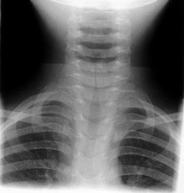 Child with croup. Note the steeple or pencil sign of the proximal trachea evident on this anteroposterior film. Courtesy of Dr. Kelly Marshall, CHOA at Scottish Rite.
Emergency Care
Prehospital care
Avoid actions that may agitate the child with laryngotracheobronchitis and lead to worsened respiratory distress. Transport the child in a parent's lap and give oxygen as tolerated, usually via a blow-by technique.
Emergency department care
Goals of emergency department (ED) care are to reduce respiratory distress, monitor for worsening condition, and consider or evaluate for other etiologies of stridor. Evidence-based guidelines have been established for the management of laryngotracheobronchitis. [6]
Make the child as comfortable as possible, and avoid agitating the patient with unnecessary procedures and examinations. Humidified air or mist therapy may be used, but both have unproven efficacy. [7] A randomized controlled trial by Siebert et al showed that exposure to cold outdoor air for 30 minutes reduced the severity of mild to moderate croup symptoms in children. [8]
Provide oxygen (humidified) to all hypoxic patients. A Cochrane review that evaluated the safety and efficacy of heliox for the treatment of mild to moderate croup did not conclusively show that heliox was superior to other methods of oxygen administration. [9]
L-epinephrine (1:1000) is as effective as racemic epinephrine. Nebulized epinephrine has proven to significantly reduce symptoms of laryngotracheobronchitis within 30 minutes of administration. (Epinephrine therapy does not indicate the need for admission.) [10, 11]
Rebound stridor after epinephrine therapy has been described in patients with laryngotracheobronchitis, but it appears to be less of a problem if corticosteroid therapy is initiated early in the ED course.
Dexamethasone has been shown to reduce symptoms in patients with moderate to severe laryngotracheobronchitis (0.6 mg/kg IM, not to exceed 10 mg). [12, 13] The intravenous formulation of dexamethasone may be administered orally as it is readily bioavailable from the GI tract. [14] Some authorities recommend a repeat dose of dexamethasone in 6 hours. [15, 16] Prednisolone (2 mg/kg/dose/day for a total of 3 days) may be an alternative if dexamethasone is not available. [17, 18]
Nebulized budesonide (2 mg) has been shown in several studies to be equivalent to oral dexamethasone. Inhaled dexamethasone is also used, when budesonide is unavailable. [19]
Always consider other causes of stridor, such as foreign bodies, bacterial tracheitis, and epiglottitis. Be sure to observe patients for an adequate period before ED discharge and to document satisfactory pulse oximetry.
Consultation with an otorhinolaryngologist and anesthesia prior to rapid sequence induction (RSI) may be necessary if the patient is exhibiting rapid deterioration that might suggest an alternative diagnosis.
-
Child with croup. Note the steeple or pencil sign of the proximal trachea evident on this anteroposterior film. Courtesy of Dr. Kelly Marshall, CHOA at Scottish Rite.