Practice Essentials
Simple rib fractures are the most common injury sustained following blunt chest trauma, accounting for more than half of thoracic injuries from nonpenetrating trauma. Approximately 10% of all patients admitted after blunt chest trauma have one or more rib fractures. [1] These fractures are rarely life-threatening in themselves but can be an external marker of more severe visceral injury inside the abdomen and the chest. [2, 3, 4]
There are 12 pairs of ribs in the thoracic region: the first 7 attach anteriorly to the sternum and posteriorly to the spinal column; 8 through 10 attach similarly but connect to the costal cartilage of the sternum anteriorly; and 11 and 12 are considered floating because they only attach posteriorly, not anteriorly. [4]
First-rib fractures are considered indicators for increased morbidity and mortality in major trauma. [5, 6, 7, 8] According to a study by Sammy et al based on information in the UK Trauma Audit and Research Network, first-rib fracture was a significant predictor of injury severity (Injury Severity Score >15) and polytrauma. [5]
In a study by Murphy et al, patients with a fracture of the first or second rib had higher mortality than patients with other rib fractures (7.4% vs 4.1%), as well as increased great vessel injury (2.8% vs 0.6%). [6]
Peek et al performed a retrospective study of 564,798 patients with one or more rib fractures, using the National Trauma Data Bank, and 44.9% had polytrauma and 49.5% were admitted to the ICU, with a quarter of the ICU patients requiring invasive mechanical ventilatory support. Overall mortality was 5.6%. Motor vehicle accidents were associated with 51.6% of injuries, and blunt chest injury accounted for 95% of rib fractures. [3]
In a study by Abdelwahed and Martinez of 215 patients with rib fractures, prolonged length of stay in the ICU (>7 days) was associated with an increased Injury Severity Score (median score, 24), greater number of rib fractures (median, 6), higher rate of lung contusion, and flail chest (35.3% of patients). [9]
(The image below depicts aortic injury, closely associated with a widening of greater than 8 cm measured at the widest points of the mediastinum.)
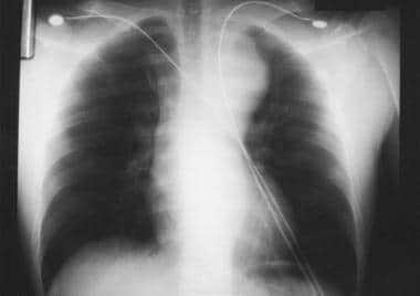 Aortic injury is closely associated with a widening of greater than 8 cm measured at the widest points of the mediastinum on an upright anteroposterior chest radiograph.
The most common mechanism of injury for rib fractures in elderly persons is a fall from height or from standing. In adults, motor vehicle accident (MVA) is the most common mechanism. Youths sustain rib fractures most often secondary to recreational and athletic activities, as well as by nonaccidental trauma. Rib fractures may also be pathologic. Cancers that metastasize to bone (eg, prostate, breast, renal) frequently become apparent in a rib. Ribs are relatively thin compared to major long bones and are more likely to fracture when invaded by a metastatic lesion. In a study, by Ochi et al, of Japanese patients with rheumatoid arthritis who were followed over a mean duration of 5.2 years, 13.5% reported incident fractures, with rib fractures being the most common fractures in men and vertebral fractures being the most common fractures in women, followed by rib fractures. [10]
Anteroposterior (AP) and lateral chest films are used routinely to assist in the diagnosis of rib fractures, yet sensitivity as low as 50% has been reported. Delayed or follow-up radiographs can be very helpful. Obtaining a rib radiograph series remains controversial, as the additional information rarely changes the clinical picture or alters treatment. Rib detail radiographs can be helpful in evaluation of the 1st and 2nd ribs and the 7th through 12th ribs. Chest CT scan (see the image below) is more sensitive than plain radiographs for detecting rib fractures. [11] Because first and second rib fractures are often associated with vascular injury, ED physicians should consider angiography for such patients, especially if symptoms and signs of neurovascular compromise are present. [7, 12, 13, 14, 15]
Pain control is fundamental to the management of rib fractures to decrease chest wall splinting and alveolar collapse in order to clear pulmonary secretions. Patients with minor rib injuries who are able to cough and clear secretions may be discharged with adequate analgesic medications. Intercostal nerve blocks provide pain relief without affecting respiratory function, although risks of this procedure include intravascular injection and pneumothorax. A small percentage of rib fractures do not heal even though a fibrous capsule may envelope the fracture. A nonunion may present months to years after injury and can cause discomfort with respiration because of movement of the fracture site. [16, 17, 18]
Pathophysiology
The chest wall protects underlying sensitive structures by surrounding internal organs with hard osseous structures, including the ribs, clavicles, sternum, and scapulae. An intact chest wall is necessary for normal respiration.
Rib fractures may compromise ventilation by a variety of mechanisms. Pain from rib fractures can cause respiratory splinting, resulting in atelectasis and pneumonia. Multiple contiguous rib fractures (ie, flail chest) interfere with normal costovertebral and diaphragmatic muscle excursion, potentially causing ventilatory insufficiency. Fragments of fractured ribs can also act as penetrating objects leading to the formation of a hemothorax or a pneumothorax. Ribs commonly fracture at the point of impact or at the posterior angle (structurally their weakest area). Ribs 4 through 9 are the ones most commonly injured.
The thinnest and weakest portion of the first rib is at the groove for the subclavian artery. [19] The mechanism of first-rib injury in motor vehicle accidents seems to be a violent contraction of the scalene muscles brought on by the sudden forward movement of the head and neck. [20]
A single blow may cause rib fractures in multiple places. Traumatic fractures most often occur at the site of impact or the posterolateral bend, where the rib is weakest.
Due to the greater pliability of children's ribs, greater force is required to produce a fracture.
Etiology
Causes of rib fracture include the following:
Epidemiology
The incidence of rib fractures is dramatically underreported. More than 2 million blunt mechanisms of injury occur annually just as motor vehicle collisions, with reported incidence of chest injury between 67 and 70% of those. [21] The prevalence of rib fractures is linked to the prevalence of the underlying cause of the trauma. Rib fractures are more common in countries with higher incidence of MVAs.
Because children have more elastic ribs, they are less likely than adults to sustain fractures following blunt chest trauma. Elderly individuals are more likely to have associated injuries and complications. Children present more frequently with trauma to the underlying chest and abdominal organs without the associated rib fractures commonly seen in adults. Classically, this made rib fractures in children an ominous sign of potential high-force injury. Bruising near the fracture site is uncommon with pediatric rib fractures, seen in only 9.1% of pediatric rib fractures in one study. [26]
Older persons are more prone to rib fractures than younger adults and, therefore, the pulmonary sequelae such as atelectasis, pneumonia, and respiratory arrest. [22] The presence of cardiopulmonary disease also significantly increases morbidity and mortality rates in patients older than 65 years. The clinical benefits of a rib scoring system has been tested at one site for hospitalized older adults. [28]
Child abuse
According to a report by the American College of Radiology, rib fractures may be the only abnormality in up to 30% of abused children. [29] Consider child abuse in children who lack a significant mechanism for multiple rib fractures or have fractures in different stages of healing. Children younger than 2 years with rib fractures have a prevalence of child abuse as high as 83%.
In a retrospective study, by Brennan et al, of 67 children younger than 5 years diagnosed with rib fractures, additional injuries causing concern for child abuse were identified in 40 (60%), and 58% were deemed likely or definite abuse. Posterior rib fractures, multiple rib fractures, and presence of rib fractures of multiple ages were all associated with the presence of additional injuries and classification as definite or likely abuse. Children who were in motor vehicle crashes, were hospitalized after birth, or had previously diagnosed metabolic bone disease were excluded from the study. [30]
Kriss et al reviewed the medical records of 78 abused children aged zero to 18 months old with rib fractures and found a total of 360 rib fractures in 273 ribs. Sixty-three (81%) of the children had multiple rib fractures. Other skeletal injuries were present in 54% of the children. [31]
Mortality/morbidity
Rib fractures are not usually dangerous in and of themselves. Patients may develop pneumonia from splinting. Morbidity correlates with the degree of injury to underlying structures.
In one study of patients with rib fractures, the mortality rate reached 12%; of these, 94% had associated injuries and 32% had a hemothorax or a pneumothorax. [32] More than half of all patients required either operative or ICU management. Average blood loss per fractured rib is reportedly 100-150 mL.
In one retrospective study of 99 elderly patients, 16% of patients (95% confidence interval [CI], 9.5-24.9%) developed adverse events, including 2 deaths. [33] Adverse events were defined as acute respiratory distress syndrome (ARDS), pneumonia, unanticipated intubation, transfer to ICU for hypoxemia, or death. Risk factors associated with these adverse events were age 85 years or older, initial systolic blood pressure less than 90 mm Hg, hemothorax, pneumothorax, 3 or more unilateral rib fractures, or pulmonary contusion. These risk factors predicted adverse events with 100% sensitivity (95% CI, 79.4-100%) and 38.6% specificity (95% CI, 28.1-49.9%), and they may identify variables that might aid in identifying patients at high risk for serious adverse events if validated in a larger prospective study.
A study of rib fractures in patients younger than 21 years found that mortality increased nearly linearly for increasing numbers of pediatric rib fractures. Odds of mortality increased with each additional rib fractured in all pediatric age groups. Mortality doubled from 1.79% without rib fracture to 5.81% for 1 rib fracture and then nearly linearly increased to 8.23% for 7 fractures. Ventilator days also increased with increasing number of rib fractures. [34]
Rib fractures are the most common injury in elderly blunt chest trauma patients, and each additional rib fracture increases the odds of dying by 19% and of developing pneumonia by 27%. [35, 36]
Position of the fractured rib in the thorax helps identify potential injury to specific underlying organs. Fracture of the lower ribs is usually associated with injury to abdominal organs rather than to lung parenchyma. Fracture of the left lower ribs is associated with splenic injuries, and fracture of the right lower ribs is associated with liver injuries. Fracture of the floating ribs (ribs 11, 12) is often associated with renal injuries.
First-rib fractures have often been described as having a high association with serious or lethal spinal or vascular injuries. [37, 5] They are the rarest of all rib fractures [20] and were once thought to be a harbinger of severe trauma, [38] since the first rib is very well protected by the shoulder, lower neck musculature, and clavicle.
First-rib fractures were thought to require a much higher impact force to fracture than other ribs, but that theory is now in question. Until further studies are done, fractures of the first rib should raise suspicion of significant chest trauma. The presence of a first-rib injury requires a multidisciplinary approach. CT of the spine and chest allows for an early diagnosis. Appropriate treatment and observation in the intensive care unit may prevent further morbidity and/or mortality. [37]
While first-rib fractures have a high association with spinal fractures and are associated with multisystem injuries, the occurrence of first-rib fractures is not always associated with increased morbidity and mortality. [37] Mortality rates as high as 36% have been reported with fractures of the first rib, which are associated with injury to the lung, ascending aorta, subclavian artery, and brachial plexus. [7] Other complications associated with first-rib fractures include delayed subclavian vessel thrombosis, aortic aneurysm, tracheobronchial fistula, thoracic outlet syndrome, [39] , and Horner syndrome. [40]
The association of lower rib fractures with pelvic fractures has been associated with a higher incidence of solid-organ injury. [41]
Prognosis
Isolated rib fractures in younger patients have a good prognosis. Older patients have a higher incidence of significant pulmonary complications. In one study, 16% of patients 65 years and older with isolated blunt chest trauma had some delayed adverse event, defined as pneumonia, ARDS, unanticipated intubation, need to transfer patient to ICU for hypoxemia, and death from pulmonary sequelae. [33]
Return to work or sport depends on the activity involved and the level of pain. Heavy labor and intensive training for athletes with stress fractures are not recommended for the first 3 weeks. When pain is not present at rest, the patient can begin to increase his or her activity level but this should be gradually done. Most rib fractures heal within 6 weeks. Many patients are able to resume daily activities much sooner.
Virtually all nonpathologic rib fractures heal well with conservative management. Some patients are able to return to work within a few days, depending on their occupation.
-
Aortic injury is closely associated with a widening of greater than 8 cm measured at the widest points of the mediastinum on an upright anteroposterior chest radiograph.
-
Anteroposterior (AP) radiograph of an elderly female patient with severe left chest wall pain after a minor fall. This image demonstrates a left lateral rib fracture (arrow) that is not seen on the standard AP chest radiograph.
-
Axial computed tomography image of the chest in a patient with both anterior and posterior thoracic injuries. A fracture of the sternum (white arrow) and a posterior left rib fracture (yellow arrow) are present.
-
Right rib radiograph in a 48-year-old male who presented with severe right posterior chest wall pain following a fall. This image demonstrates 2 fractures of the right chest wall (white arrows).


