Practice Essentials
Injuries of the hand can cause significant functional impairment, diminished quality of life, and delayed return to work. [1] Hand injuries are commonly seen in the emergency department, and emergency physicians should be able to identify and provide emergent management of digital dislocations. Complications can occur if the diagnosis is missed or delayed, or if the joint is incompletely reduced or improperly splinted. [2, 3, 4, 5, 6, 7]
Management
Every emergency physician should have a firm understanding of acute management of simple dislocations of the digits. Historical, physical, and radiographic findings often guide management of the dislocation. When the dislocation is complicated, consult with and/or refer to a hand surgeon. Generally, reduced dislocations without evidence of instability and with near-normal range of motion can be treated with brief immobilization and subsequent referral.
For patients with a hand dislocation who are being transported by emergency medical services (EMS), immobilization of the deformed joint, protection of injured soft tissue, and pain management are the mainstays of prehospital treatment.
Providing rapid pain relief, obtaining imaging studies, and performing expeditious reduction (closed or open) must be the triad that drives the ED protocol for treating patients with isolated digital or hand dislocation.
Patients treated and discharged from the ED should have oral analgesia prescribed as part of their outpatient care.
Patients with dislocations that are not reducible in the ED should discuss their disposition with a hand specialist. If one is not immediately available, the patient may need to be transferred to a facility that can provide a higher level of care.
Patients with open joints or other significant hand injuries usually require admission. Complex and open dislocations should be evaluated by a hand surgeon for open reduction. Individuals with fracture-related dislocation should be further evaluated by a hand surgeon. Patients should be referred to a hand specialist following ED treatment of hand dislocation.
Few studies have tracked hand injury patients past the acute care period. Postdischarge tracking of hand injury patients may reveal time points when most patients require assistance, which can help direct interventions to reduce post–hand injury sequelae. Primary care providers should perform a comprehensive assessment of patients prior to their discharge. Patients should undergo regular follow-ups to reduce comorbidities and improve outcomes. [8]
Epidemiology
In the United States, approximately 30% of about 10,000 annual blast injuries involve the hand, causing a broad spectrum of injury severity. Typically, the first web space is most severely affected. Blast injury to the hand often results in severe deficits, frequently affecting thumb functionality and irreversibly altering occupational capabilities. [9]
The severity of carpal injuries resulting from fireworks is highly variable but is likely to follow predictable patterns due to the position of the hand and the location of the firework prior to explosion. Surgical reconstruction can be challenging, but adequate outcomes with a functional hand can be achieved through a systematic approach. [10]
Carpometacarpal fracture-dislocation of the hand is a relatively uncommon injury. This injury is difficult to diagnose because of gross swelling of the hand. Diagnosis of this unusual form of injury requires a high index of suspicion, vigilant examination, and use of high-quality radiography. Intensive postoperative physiotherapy is vital for achieving a good outcome. [11]
Emergency Department Treatment
Providing rapid pain relief, obtaining imaging studies, and performing expeditious reduction (closed or open) must be the triad that drives the ED protocol for treating patients with isolated digital or hand dislocation.
Triage/initial evaluation and treatment
Many patients with an isolated digital or hand dislocation can be expeditiously treated from the time they arrive until initial physician contact if protocols allow the nursing staff to provide analgesia and to order the appropriate radiologic study.
Pain management can consist of oral medication, intramuscular injection, intravenous injection, or regional anesthesia, as advisable, according to severity of the injury and the patient's medical history, allergic reaction profile, and expressed comfort level.
Oral medication can be problematic if the patient eventually requires open reduction. Pain management protocols should take this into account.
Imaging studies of the affected digit or hand can easily be part of an ED protocol. For example, radiographs of the isolated digit can be obtained if the triage or evaluating nurse assesses the patient to have a deformity at the distal or proximal finger joints.
Hand dislocation
Once hand dislocation has been identified and pain control initiated, relocation is the next step in patient care. Most hand dislocations are easily reduced by the emergency physician. Some dislocations may not be reducible by closed means because of the interposition of the volar plate or associated ligaments or tendons in the joint. If several attempts at reduction are not successful, consultation and open reduction internal fixation (ORIF) are often indicated. [12, 13] Stability should be thoroughly assessed following a successful reduction.
Digital dislocation
Distal interphalangeal joints of the fingers
The distal interphalangeal (DIP) joint of the finger is a highly vulnerable area. Dislocations in this area are uncommon because of strong joint support provided by skin and periarticular structures. The strong support network is unyielding, however, and if force of appropriate intensity is applied, the skin may tear, leading to an open dislocation.
(A DIP dislocation is shown below.)
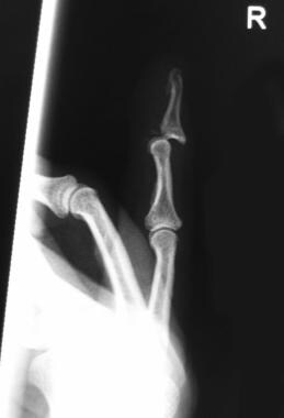 Dorsal distal interphalangeal (DIP) joint finger dislocation (lateral view). Note small fracture fragments.
Reduce the dislocation with longitudinal traction and hyperextension, with firm dorsal pressure at the base of the distal phalanx. Open reduction is rarely needed with this type of dislocation.
After the dislocation has been reduced, assess the stability of the joint to rule out evidence of tendon injury. Immobilize the joint with a dorsal splint in flexion if volar dislocation has occurred without tendon injury, and in extension if the dislocation is dorsal and without tendon injury.
Proximal interphalangeal joints of the fingers
Volar dislocation of the proximal interphalangeal (PIP) joint of a finger is relatively uncommon. When a volar dislocation occurs, the proximal phalanx can rupture through the transverse retinacular fibers between the lateral band and the central tendon. The lateral bands may become interposed, making closed reduction difficult.
If the volar plate is ruptured and the extensor mechanism avulsed, a Boutonnière deformity may result. Open reductions usually are performed for these injuries. Occasionally, closed reduction may be performed. If the joint remains stable, immobilize the digit briefly in a slightly flexed position.
Dorsal dislocations are reduced by applying longitudinal traction and mild hyperextension with dorsal pressure to the proximal aspect of the middle phalanx.
Immobilization of a simple dislocation without instability should be brief. If the patient continues to perform activities that may put the digit at risk for subsequent dislocation, the digit should be protected with buddy taping and/or splinting during the activity. [14, 15]
Metacarpophalangeal joints of the fingers
Dislocation of a metacarpophalangeal (MCP) joint of the fingers most often involves the index or small finger. Dislocations here are relatively uncommon because of the strength of periarticular structures. Dislocation may be simple or complex. A complex dislocation nearly always needs open reduction because of an interposed volar plate.
Closed reduction may be accomplished by using traction along the axis of the hyperextended phalanx and firmly pushing the base of the dislocated phalanx toward the MCP joint.
Assess the stability of the joint after reduction, and follow the procedure with immobilization. Some controversy exists regarding the length of time this takes and the position of immobilization to be applied. Some recommend early range of motion if no evidence of postreduction instability is observed.
Interphalangeal joint of the thumb
Reduction of this joint is usually accomplished via closed means. This particular dislocation may present with associated rupture of the flexor pollicis longus. Following evaluation and reduction, immobilize the involved joint with a thumb spica splint. The period of joint immobilization should be brief to avoid joint stiffening.
Metacarpophalangeal joint of the thumb
Anterior dislocation of the MCP joint of the thumb is classified as simple or complex. The appropriate method of reduction of a dislocation depends on the type of dislocation.
For a simple dislocation, the clinician should avoid pure traction, as this can convert a simple dislocation into a complex one.
Reduction is achieved by pushing the phalanx into the MCP joint rather than by pulling it into place. If 1-2 attempts at reduction are unsuccessful, an open reduction must be performed. More aggressive and repeated attempts at reduction may lead to fracture. An interposed volar plate or intrinsic muscle may be the reason for failed attempts at closed reduction.
After the dislocation has been reduced, immobilize the joint with a thumb spica splint. The duration of immobilization varies, but clinicians should avoid extended immobilization and should minimize immobilization of unaffected areas.
Instability of the thumb is an indication for referring the patient to a hand specialist.
(A thumb MCP dislocation is shown in the image below.)
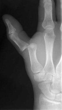 Thumb metacarpophalangeal (MCP) joint dislocation. Image courtesy of David T. Schwartz, MD.
Post-emergency care
Patients treated and discharged from the ED should have oral analgesia prescribed as part of their outpatient care.
Patients with dislocations that are not reducible in the ED should discuss their disposition with a hand specialist. If one is not immediately available, the patient may need to be transferred to a facility that can provide a higher level of care.
Patients with open joints or other significant hand injuries usually require admission. Complex and open dislocations should be evaluated by a hand surgeon for open reduction. Individuals with fracture-related dislocation should be further evaluated by a hand surgeon. Patients should be referred to a hand specialist following emergency treatment for hand dislocation.
-
Dorsal distal interphalangeal (DIP) joint finger dislocation (lateral view). Note small fracture fragments.
-
Thumb metacarpophalangeal (MCP) joint dislocation. Image courtesy of David T. Schwartz, MD.


