Practice Essentials
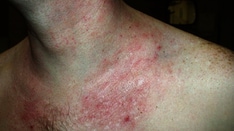
Individuals with allergic contact dermatitis (see the image below) may have persistent or relapsing dermatitis, particularly if the material(s) to which they are allergic is not identified or if they practice inappropriate skin care. The longer an individual has severe dermatitis, the longer, it is believed, that the dermatitis will take to resolve once the cause is identified.
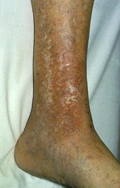 Chronic stasis dermatitis with allergic contact dermatitis to quaternium-15, a preservative in moisturizer. Allergic contact dermatitis produces areas of erythema in areas of atrophie blanche and varicose veins.
See 5 Body Modifications and Piercing: Dermatologic Risks and Adverse Reactions, a Critical Images slideshow, to help recognize various body modifications and the related potential complications.
Signs and symptoms
Acute allergic contact dermatitis is characterized by pruritic papules and vesicles on an erythematous base. Lichenified pruritic plaques may indicate a chronic form of the condition.
Individuals with allergic contact dermatitis typically develop the condition within a few days of exposure, in areas that were exposed directly to the allergen. Certain allergens (eg, neomycin), however, penetrate intact skin poorly; in such cases, the onset of dermatitis may be delayed for up to a week following exposure.
Individuals may develop widespread dermatitis from topical medications applied to leg ulcers or from cross-reacting systemic medications administered intravenously.
Intraoral metal contact allergy may result in mucositis that mimics lichen planus, which has an association with intraoral squamous cell carcinoma.
See Clinical Presentation for more detail.
Diagnosis
Diagnostic studies for allergic contact dermatitis include the following:
-
Potassium hydroxide preparation and/or fungal culture: To exclude tinea; these tests are often indicated for dermatitis of the hands and feet
-
Patch testing: To identify external chemicals to which the person is allergic
-
Repeat open application test (ROAT): To determine whether a reaction is significant in individuals who develop weak or 1+ positive reactions to a chemical
-
Dimethylgloxime test: To determine whether a metallic object contains enough nickel to provoke allergic dermatitis
-
Skin biopsy: May help to exclude other disorders, particularly tinea, psoriasis, and cutaneous lymphoma
See Workup for more detail.
Management
The definitive treatment for allergic contact dermatitis is the identification and removal of any potential causal agents; otherwise, the patient is at increased risk for chronic or recurrent dermatitis. Treatments also include the following:
-
Corticosteroids: Topical corticosteroids are the mainstay of treatment, although acute, severe allergic contact dermatitis, such as from poison ivy, often needs to be treated with a 2-week course of systemic corticosteroids
-
Topical immunomodulators (TIMs): Approved for atopic dermatitis, but they are also prescribed for cases of allergic contact dermatitis when they offer safety advantages over topical corticosteroids
-
Phototherapy: Administered to individuals with chronic allergic contact dermatitis that is not controlled well by topical corticosteroids; these patients may benefit from treatment with a combination of psoralen (a photosensitizer) and ultraviolet-A (PUVA)
-
Immunosuppressive agents: Chronic immunosuppressive agents are, in rare instances, used to treat recalcitrant cases of severe, chronic, widespread allergic contact dermatitis or severe hand dermatitis that prevents a patient from working or performing daily activities
-
Disulfiram: Occasionally, an individual who is highly allergic to nickel and has severe vesicular hand dermatitis will benefit from treatment with disulfiram (Antabuse); the drug has a chelating effect
See Treatment and Medication for more detail.
Background
Allergic contact dermatitis (ACD) is a delayed type of induced sensitivity (allergy) resulting from cutaneous contact with a specific allergen to which the patient has developed a specific sensitivity. This allergic reaction causes inflammation of the skin manifested by varying degrees of erythema, edema, and vesiculation.
The term contact dermatitis sometimes is used incorrectly as a synonym for allergic contact dermatitis. Contact dermatitis is inflammation of the skin induced by chemicals that directly damage the skin (see Irritant Contact Dermatitis) and by specific sensitivity in the case of allergic contact dermatitis.
Jadassohn first described allergic contact dermatitis in 1895. He developed the patch test to identify the chemicals to which the patient was allergic. Sulzberger popularized patch testing in the United States in the 1930s. The Finn chamber method for patch testing was designed in the 1970s; these chambers consist of small metal cups, typically attached to strips of tape, filled with allergens dispersed in either petrolatum or water. The thin-layer rapid use epicutaneous (TRUE) test for patch testing became available in the United States in the 1990s.
The importance of specific substances as causes of allergic contact dermatitis varies with the prevalence of that substance in the environment. Mercury compounds once were significant causes of allergic contact dermatitis but rarely are used as topical medications and, currently, are uncommon as a cause of allergic contact dermatitis. Ethylenediamine, which was present in the original Mycolog cream, declined as a primary cause of allergic contact dermatitis once Mycolog cream was reformulated to no longer contain this allergen.
A detailed history, both before and after patch testing, is crucial in evaluating individuals with allergic contact dermatitis. Before patch testing, the history identifies potential causes of allergic contact dermatitis and the materials to which individuals are exposed that should be included in patch testing. After patch testing, the history determines the clinical significance of the findings. (See Clinical.)
Topical corticosteroids are the mainstay of treatment, while a variety of symptomatic treatments can provide short-term relief of pruritus. However, the definitive treatment of allergic contact dermatitis is the identification and removal of any potential causal agents; otherwise, the patient is at increased risk for chronic or recurrent dermatitis. (See Treatment.)
Go to Irritant Contact Dermatitis, Pediatric Contact Dermatitis, and Protein Contact Dermatitis for complete information on these topics.
Pathophysiology
Approximately 3000 chemicals are well documented as specific causes of allergic contact dermatitis.
Compounds must be less than 500 d for efficient penetration through the stratum corneum barrier, which is the water-impermeable outer layer of the skin. Small organic molecules that are chemically reactive (chemical sensitizers) bind with self-proteins to generate immunogenic neoantigens through a process termed haptenization. Although haptens can penetrate through intact skin, patients with certain disease states that impair barrier function (eg, leg ulcers, perianal dermatitis) have an increased risk of sensitization to topically applied medications and their vehicle components.
Many patients with atopic dermatitis or allergic contact dermatitis to nickel harbor a defective form of the filaggrin gene. [1] Filaggrin helps aggregate cytoskeletal proteins that form the cornified cell envelope. In its absence, the barrier is defective.
Prehaptens are chemicals that are not activated by host proteins, but instead require chemical transformation by oxidative derivatization by ambient or air oxidation to form hydroperoxide. Examples include certain fragrance materials and dyes used in hair coloring, such as para-phenylenediamine.
Haptens activate Toll-like receptors (TLRs) and activate innate immunity. The importance of hapten-mediated activation of innate immunity is highlighted by the clinical observation that the irritancy of chemicals (ie, the ability of these chemicals to cause grossly visible skin inflammation upon primary exposure) correlates with their ability to act as contact sensitizers and to induce acute contact dermatitis.
Haptens or haptenated self-proteins are recognized by innate immune mechanisms in the skin, and this leads to the elaboration of a number of proinflammatory mediators, including interleukin (IL)–1β. As a result, skin-resident dendritic cells (DCs) become activated. There are several populations of DCs. Langerhans cells are the only DC subtype in the epidermis. Like all skin-resident DCs, Langerhans cells efficiently acquire antigen in the periphery and migrate to regional lymph nodes where they present antigen to naïve and memory T cells. These DCs, which may have been directly haptenated or could have acquired haptenated proteins from their surroundings, migrate to skin-draining lymph nodes where they present peptides from haptenated proteins to activate memory and naïve T cells.
In the final step, hapten-induced inflammation recruits activated effector T cells back to the initial site of antigen encounter in the skin. The effector T cells release proinflammatory cytokines, such as interferon-γ, and promote the killing of haptenated cells, resulting in the development of the classic inflammatory rash seen in allergic contact dermatitis.
Keratinocytes are crucial for the development of allergic contact dermatitis. They constitute the vast majority of cells in the epidermis and form the anatomic barrier of the skin. Keratinocytes express most TLRs, and this allows them to respond to TLR4-triggering haptens, such as nickel. Keratinocytes are also a source of IL-10, an immunosuppressive cytokine that limits the extent of contact hypersensitivity
The initial sensitization typically takes 10-14 days from initial exposure to a strong contact allergen such as poison ivy. Some individuals develop specific sensitivity to allergens following years of chronic low-grade exposure; for example, sensitivity to chromate in cement can eventually develop in individuals with chronic irritant contact dermatitis resulting from the alkaline nature of cement. Once an individual is sensitized to a chemical, allergic contact dermatitis develops within hours to several days of exposure.
CD4+ CCR10+ memory T cells persist in the dermis after clinical resolution of allergic contact dermatitis.
Etiology
Approximately 25 chemicals appear to be responsible for as many as one half of all cases of allergic contact dermatitis. These include nickel, preservatives, dyes, and fragrances.
Poison ivy
Poison ivy (Toxicodendron radicans) is the classic example of acute allergic contact dermatitis in North America. Allergic contact dermatitis from poison ivy is characterized by linear streaks of acute dermatitis that develop where plant parts have been in direct contact with the skin.
Nickel
Nickel is the leading cause of allergic contact dermatitis in the world. The incidence of nickel allergic contact dermatitis in North America is increasing; in contrast, new regulations in Europe have resulted in a decreasing prevalence of nickel allergy in young and middle-aged women. [2, 3]
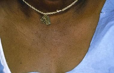
Allergic contact dermatitis to nickel typically is manifested by dermatitis at the sites where earrings or necklaces (see the image below) containing nickel are worn or where metal objects (including the keypads of some cell phones [4] ) containing nickel are in contact with the skin.
Nickel may be considered a possible occupational allergen. Workers in whom nickel may be an occupational allergen primarily include hairdressers, retail clerks, caterers, domestic cleaners, and metalworkers. Individuals allergic to nickel occasionally may develop vesicles on the sides of the fingers (dyshidrotic hand eczema or pompholyx) from nickel in the diet.
Rubber gloves [5]
Allergy to 1 or more chemicals in rubber gloves is suggested in any individual with chronic hand dermatitis who wears them, unless patch testing demonstrates otherwise. Allergic contact dermatitis to chemicals in rubber gloves typically occurs maximally on the dorsal aspects of the hand. Usually, a cutoff of dermatitis occurs on the forearms where skin is no longer in contact with the gloves. Individuals allergic to chemicals in rubber gloves may develop dermatitis from other exposures to the chemicals (eg, under elastic waistbands).
Hair dye and temporary tattoos
p-Phenylenediamine (PPD) is a frequent component of and sensitizer in permanent hair dye products and temporary henna tattoos [6] ; exposure in to it in hair dye products may cause acute dermatitis with severe facial edema. Severe local reactions from PPD may occur in black henna tattoos in adults and children. Epidemiologic data indicate that the median prevalence of positive patch test reactions to PPD among dermatitis patients is 4.3% (increasing) in Asia, 4% (plateau) in Europe, and 6.2% (decreasing) in North America. [7]
Textiles
Individuals allergic to dyes and permanent press and wash-and-wear chemicals added to textiles typically develop dermatitis on the trunk, which occurs maximally on the lateral sides of the trunk but spares the vault of the axillae. Primary lesions may be small follicular papules or may be extensive plaques.
Individuals in whom this allergic contact dermatitis is suspected should be tested with a series of textile chemicals, particularly if routine patch testing reveals no allergy to formaldehyde. New clothing is most likely to provoke allergic contact dermatitis, since most allergens decrease in concentration in clothing following repeated washings.
Preservatives
Preservative chemicals added to cosmetics, moisturizers, and topical medications are major causes of allergic contact dermatitis (see the image below). The risk of allergic contact dermatitis appears to be highest to quaternium-15, followed by allergic contact dermatitis to isothiazolinones. Methylisothiazolinone is used as an individual preservative and may be a significant allergen. [8] Kathon CG is methylchloroisothiazolinone in combination with methylisothiazolinone.
Although parabens are among the most widely used preservatives, they are not a frequent cause of allergic contact dermatitis.
 Severe allergic contact dermatitis resulting from preservatives in sunscreen. Patch testing was negative to the active ingredients in the sunscreen.
Schnuch et al estimated that preservatives found in leave-on topical products varied over 2 orders of magnitude in relative sensitization risk. [9]
Formaldehyde is a major cause of allergic contact dermatitis (see the image below). Certain preservative chemicals widely used in shampoos, lotions, other moisturizers, and cosmetics are termed formaldehyde releasers (ie, quaternium-15 [Dowicil 200], imidazolidinyl urea [Germall 115], and isothiazolinones [9] ). They are, in themselves, allergenic or may produce cross-sensitization to formaldehyde.
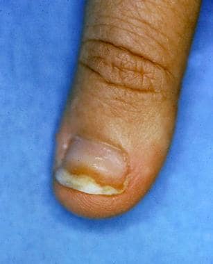 Onycholysis developing from allergic contact dermatitis to formaldehyde used to harden nails.
Individuals may develop allergy to fragrances. Fragrances are found not only in perfumes, colognes, aftershaves, deodorants, and soaps, but also in numerous other products, often as a mask to camouflage an unpleasant odor. Unscented products may contain fragrance chemicals used as a component of the product and not labeled as fragrance.
Individuals allergic to fragrances should use fragrance-free products. Unfortunately, the exact chemicals responsible for a fragrance in a product are not labeled. Four thousand different fragrance molecules are available to formulate perfumes. The fragrance industry is not required to release the names of ingredients used to compose a fragrance in the United States, even when individuals develop allergic contact dermatitis to fragrances found in topical medications.
Deodorants may be the most common cause of allergic contact dermatitis to fragrances because they are applied to occlude skin that is often abraded by shaving in women.
Massage and physical therapists and geriatric nurses are at higher risk of occupational allergic contact dermatitis to fragrances.
Corticosteroids
In the last decade, it has become clear that some individuals with chronic dermatitis develop allergy to topical corticosteroids. Most affected individuals can be treated with some topical corticosteroids, but an individual can be allergic to all topical and systemic corticosteroids. Budesonide and tixocortol pivalate are useful patch test corticosteroids for identifying individuals allergic to topical corticosteroids.
Neomycin
The risk of allergy to neomycin is related directly to the extent of its use in a population. The risk of allergy to neomycin is much higher when it is used to treat chronic stasis dermatitis and venous ulcers than when it is used as a topical antibiotic on cuts and abrasions in children. Assume that individuals allergic to neomycin are allergic to chemically related aminoglycoside antibiotics (eg, gentamicin, tobramycin). [12] Avoid these drugs both topically and systemically in individuals allergic to neomycin.
Benzocaine
Avoid topical use of benzocaine. Benzocaine is included in most standard patch test trays. Individuals allergic to benzocaine may safely use or be injected with lidocaine (Xylocaine), which does not cross-react with benzocaine.
Sunscreens
Many individuals complain of adverse reactions to sunscreens, but many of these individuals are not allergic to the sunscreen materials. They may be allergic to preservatives in these products or may have nonspecific cutaneous irritation from these products.
Photoallergy
Occasionally, individuals develop photoallergic contact dermatitis. Allergic contact dermatitis may be accentuated by ultraviolet (UV) light, or patients may develop an allergic reaction only when a chemical is present on the skin and when the skin is exposed sufficiently to ultraviolet light A (UV-A; 320-400 nm).
Acrylates and methacrylates [13, 14]
These agents are used in manufacturing, nail acrylics, and wound dressings, among other uses.
Epidemiology
United States statistics
The National Health and Nutrition Examination Survey (NHANES) estimated the prevalence of contact dermatitis to be 13.6 cases per 1000 population, using physical examinations by dermatologists of a selected sample of patients. NHANES underreported the prevalence compared with the physical examination findings.
The National Ambulatory Medical Care Survey conducted in 1995 estimated 8.4 million outpatient visits to American physicians for contact dermatitis. This was the second most frequent dermatologic diagnosis. Of office visits to dermatologists, 9% are for dermatitis. At a student health center dermatology clinic, 3.1% of patients presented for allergic contact dermatitis, and 2.3% presented for irritant contact dermatitis.
The TRUE test Web site can provide accurate basic information on common allergens. The Contact Allergen Management Program is provided as a service to the American Contact Dermatitis Society (ACDS) members and is particularly valuable for allergens found in topical skin care products. The Contact Allergen Management Program (CAMP) database contains more than 8100 known ingredients cataloged in more than 5500 commercial skincare products and is available as a Smartphone application.
International statistics
A Swedish study found that prevalence of allergic contact dermatitis of the hands was 2.7 cases per 1000 population. A Dutch study found that prevalence of allergic contact dermatitis of the hands was 12 cases per 1000 population.
Race, sex, and age-related demographics
No racial predilection exists for allergic contact dermatitis. Allergic contact dermatitis is more common in women than in men. This predominantly is a result of allergy to nickel, which is much more common in women than in men in most countries.
Allergic contact dermatitis may occur in neonates. In elderly individuals, the development of allergic contact dermatitis may be delayed somewhat, but the dermatitis may be more persistent once developed. Contact allergy to topical medicaments is more common in persons older than 70 years. [15]
Prognosis
The prognosis depends on how well the affected individual can avoid the offending allergen. [16]
Individuals with allergic contact dermatitis may have persistent or relapsing dermatitis, particularly if the material(s) to which they are allergic is not identified or if they continue to practice skin care that is no longer appropriate (ie, they continue to use harsh chemicals to wash their skin, they do not apply creams with ceramides or bland emollients to protect their skin).
The longer an individual has severe dermatitis, the longer it is believed it will take the dermatitis to resolve once the cause is identified.
Some individuals have persistent dermatitis following allergic contact dermatitis, which appears to be true especially in individuals allergic to chromates.
A particular problem is neurodermatitis (lichen simplex chronicus), in which individuals repeatedly rub or scratch an area initially affected by allergic contact dermatitis.
Mortality
Death from allergic contact dermatitis is rare in the United States. Allergic contact dermatitis to the weed wild feverfew caused deaths in India when the seeds contaminated wheat shipments to India. This plant then became widespread and a primary cause of severe airborne allergic contact dermatitis.
Patient Education
Patients have the best prognosis when they are able to remember the materials to which they are allergic and how to avoid further exposures. Provide patients with as much information as possible concerning the chemical to which they are allergic, including all known names of the chemical. Web sites, Smartphone applications, standard textbooks, and the TRUE test kit contain basic information about the chemicals.
Susceptible individuals need to read the list of ingredients before applying cosmetic products to their skin, since preservative chemicals are used widely in consumer, medical, and workplace products. The same chemical may have different names when used for consumer or industrial purposes.
Provide pamphlets with color pictures of poison ivy to individuals allergic to the plant. The American Academy of Dermatology also has pamphlets on allergic contact dermatitis and hand eczema.
For patient education information, see the Skin Conditions & Beauty Center, as well as Contact Dermatitis.
-
Chronic stasis dermatitis with allergic contact dermatitis to quaternium-15, a preservative in moisturizer. Allergic contact dermatitis produces areas of erythema in areas of atrophie blanche and varicose veins.
-
Erythema multiformelike reaction that developed acutely following hair dying.
-
Allergic contact dermatitis to nickel in a necklace.
-
Severe allergic contact dermatitis resulting from preservatives in sunscreen. Patch testing was negative to the active ingredients in the sunscreen.
-
Onycholysis developing from allergic contact dermatitis to formaldehyde used to harden nails.