Practice Essentials
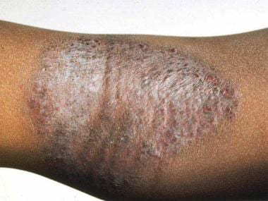
Atopic dermatitis (AD) is a chronic, pruritic inflammatory skin condition (see image below) that typically affects the face (cheeks), neck, arms, and legs but usually spares the groin and axillary regions. AD usually starts in early infancy, but also affects a substantial number of adults. AD is commonly associated with elevated levels of immunoglobulin E (IgE). That it is the first disease to present in a series of allergic diseases—including food allergy, asthma, and allergic rhinitis, in order—has given rise to the “atopic march” theory, which suggests that AD is part of a progression that may lead to subsequent allergic disease at other epithelial barrier surfaces. [1, 2]
See All About Allergies: Be Ready for Spring, a Critical Images slideshow, to help identify a variety of allergens and symptoms.
Signs and symptoms of atopic dermatitis
Incessant pruritus (itchiness) is the only symptom of AD. The disease typically has an intermittent course with flares and remissions occurring, often for unexplained reasons.
Primary physical findings include the following:
-
Xerosis (dry skin)
-
Lichenification (thickening of the skin and an increase in skin markings)
-
Eczematous lesions (skin inflammation)
The eczematous changes and its morphology are seen in different locations, depending on the age of the patient (ie, infant, child, or adult).
The following is a constellation of symptoms and features commonly seen in AD:
-
Pruritus
-
Early age of onset
-
Chronic and relapsing course
-
IgE reactivity
-
Peripheral eosinophilia
-
Staphylococcus aureus superinfection
-
Personal history of asthma or hay fever or a history of atopic diseases in a first-degree relative
See Clinical Presentation for more detail.
Diagnosis of atopic dermatitis
The following features should be considered in the diagnosis of AD in accordance with the American Academy of Dermatology (AAD) 2014 Guidelines [3] :
Essential features (must be present) are as follows:
-
Pruritus
-
Eczema (acute, subacute, chronic): (1) Typical morphology and age-specific patterns (facial/neck/extensor involvement in children, flexural involvement in any age group, sparing the groin and axillary regions); (2) chronic or relapsing history
Important features (supports the diagnosis) are as follows:
-
Early age of onset
-
Atopy: (1) Personal and/or family history; (2) IgE reactivity
-
Xerosis
Associated features (nonspecific but suggest the diagnosis of AD) are as follows:
-
Atypical vascular responses (eg, facial pallor, delayed blanch response)
-
Keratosis pilaris/pityriasis alba/hyperlinear palms/ichthyosis
-
Ocular/periorbital changes
-
Other regional findings (eg, perioral changes/periauricular lesions)
-
Perifollicular accentuation/lichenification/prurigo
Exclusionary conditions (conditions that should be excluded) are as follows:
-
Scabies
-
Seborrheic dermatitis
-
Contact dermatitis
-
Ichthyoses
-
Cutaneous T-cell lymphoma
-
Psoriasis
-
Photosensitivity dermatoses
-
Immune deficiency diseases
-
Erythroderma of other causes
Additional considerations in the diagnosis of AD are as follows:
-
No reliable biomarker exists for the diagnosis of AD
-
Laboratory testing is seldom necessary but a complete blood cell count can be useful to exclude immune deficiency; an IgE level can be helpful to confirm an atopic pattern; a swab of skin can be helpful to identify S aureus superinfection
-
Allergy and radioallergosorbent testing is of little value
-
Biopsy shows an acute, subacute, or chronic spongiotic dermatitis pattern that is nonspecific but can be helpful to rule out other conditions (eg, cutaneous T-cell lymphoma)
See Workup for more detail.
Management of atopic dermatitis
Agents typically used to treat AD include the following:
-
Moisturizers: Petrolatum, Aquaphor, or newer agents such as Atopiclair and Mimyx
-
Topical steroids (current mainstay of treatment; commonly used in conjunction with moisturizers): Hydrocortisone, triamcinolone, or betamethasone; ointment bases are generally preferred, particularly in dry environments
-
Broad immunomodulators: Tacrolimus and pimecrolimus (calcineurin inhibitors; generally considered second-line therapy)
-
Targeted biologic therapies
- Dupilumab (anti-IL-4Ra monoclonal antibody)
- Tralokinumab (anti-IL-13 monoclonal antibody)
See the list below:
-
Janus kinase (JAK) inhibitors
- Abrocitinib
- Upadacitinib
- Ruxolitinib topical
Other treatments that have been tried include the following:
-
Ultraviolet (UV)-A, UV-B, a combination of both, psoralen plus UV-A (PUVA), or UV-B1 (narrow-band UV-B) therapy
-
In severe disease, methotrexate, azathioprine, cyclosporine, and mycophenolate mofetil [4]
-
Probiotics
-
Antibiotics for clinical infection caused by S aureus or flares of disease [7]
-
Intranasal mupirocin ointment and diluted bleach (sodium hypochlorite) baths
Nonmedical measures that may be helpful include the following:
-
Using soft clothing (eg, cotton) next to the skin; wool products should be avoided
-
Maintaining mild temperatures, particularly at night
-
Using a humidifier (cool mist) in both winter and summer
-
Washing clothes in a mild detergent, with no bleach or fabric softener
-
Avoiding specific foods as appropriate if there is concomitant food allergy
See Treatment and Medication for more detail.
Background
Atopic dermatitis (AD) is a pruritic skin condition of unknown origin that usually starts in early infancy (an adult-onset variant is recognized); it is characterized by pruritus, eczematous lesions, xerosis (dry skin), and lichenification (thickening of the skin and an increase in skin markings).
AD may be associated with other atopic (immunoglobulin E [IgE]–associated) diseases (eg, acute allergic reaction to foods, asthma, urticaria, and allergic rhinitis). [8] AD has enormous morbidity, and the incidence and prevalence appear to be increasing. Further, AD is the first disease to present in a series of allergic diseases such as food allergy, asthma, and allergic rhinitis (in order), provoking the “atopic march” theory, which suggests that early or severe AD and cutaneous sensitization to environmental allergens may lead to subsequent allergic disease at other epithelial barrier surfaces (eg, gastrointestinal or respiratory tract). This hypothesis is supported by cross-sectional and longitudinal studies. [1]
Pathophysiology
Despite recent advances in the understanding of the genetics of atopic dermatitis (AD), the pathophysiology remains poorly defined. Two main hypotheses have been proposed regarding the development of inflammation that leads to AD. The first suggests a primary immune dysfunction resulting in IgE sensitization, allergic inflammation, and a secondary epithelial barrier disturbance. The second proposes a primary defect in the epithelial barrier leading to secondary immunologic dysregulation and resulting in inflammation.
In healthy individuals, balance exists between important subsets of T cells (eg, Th1, Th2, Th17, Th22). The primary immune dysfunction hypothesis invokes an imbalance in the T cell subsets, with Th2 cells predominating; this results in the production of type 2 cytokines such as interleukin (IL)–4, IL-5, and IL-13, causing an increase in IgE from plasma cells. Later, in persons with chronic AD, the Th1 cells have been shown to predominate. More recently, Th17 cells have been found to be elevated in patients with AD. [9] Although primarily considered a Th2 cell‒associated cytokine-mediated disease, the precise contributions of Th1 and Th17 cell responses remain to be fully defined.
In addition to the role of T and B cells in AD, other innate immune cells have also been implicated in the pathogenesis of AD, including eosinophils and mast cells. [10, 11] More recently, basophils and newly identified innate immune cells called group 2 innate lymphoid cells (ILC2s) have been shown to underlie the pathogenesis of AD. [12, 13, 14, 15, 16] Together, basophils and ILC2s are critical sources of the type 2 cytokines IL-4, IL-5, and IL-13. [12, 13] Further, these cells appear to be potently regulated by a family of epithelial cell‒derived cytokines directly released from damaged keratinocytes, including thymic stromal lymphopoietin (TSLP), IL-25, and IL-33. [17] Taken together, these studies highlight a new paradigm in which, in addition to classical adaptive Th2 cells, innate type 2 immune cells play critical roles in the etiology of AD through interactions with epidermal-derived cytokines.
In terms of AD-associated itch, Th2 cells are known to be significant sources of the itch-inducing cytokine or pruritogen IL-31. [18] Emerging clinical trials data indicate that blocking this pathway may be a key mechanism by which atopic itch can be treated clinically. Additionally, a 2017 study identified that neuronal, rather than immune, signaling of the type 2 cytokines IL-4 and IL-13 critically regulate AD-associated itch. [19] Indeed, the dual IL-4 and IL-13 blocker, dupilumab, has emerged as a highly effective treatment for AD, which received FDA approval in March of 2017. Thus, blocking cytokine-nerve interactions with targeted biologic therapies has emerged as a novel therapeutic strategy in AD.
The epidermal barrier dysfunction hypothesis suggests that AD patients develop AD as a result of skin barrier defects that allow for the entry of antigens, resulting in the production of inflammatory cytokines. Some authors question whether such antigens can also be absorbed from the gut (eg, from food) and/or the lungs (eg, from house dust mites). Xerosis and ichthyosis are known to be associated signs in many AD patients. Clinically, 37-50% of people with ichthyosis vulgaris have atopic disease and up to 37% of people with AD have clinical evidence of ichthyosis vulgaris. Mutations in the gene encoding filaggrin, a key epidermal barrier protein, cause ichthyosis vulgaris and are the strongest known genetic risk factors for the development of AD. [20, 21]
Further, filaggrin mutations are associated with early-onset AD and with airway disease in the setting of AD. [22] One mechanism by which filaggrin defects may influence inflammation is by the release of epithelial cell‒derived cytokines, including TSLP, IL-25, and IL-33, which are all known to be up-regulated in the context of AD. [23, 24, 25, 26] TSLP has been shown to be a potent promoter of basophil and ILC2 responses in the skin, while IL-25 and IL-33 preferentially elicit ILC2s. [12, 13, 16] Although filaggrin is strongly linked to AD, mutations are only found in 30% of European patients, begging the question of whether other genetic variants may also be responsible for some of the findings in the pathogenesis of AD. Indeed, genetic variants of TSLP have been shown to interact with mutations in filaggrin to influence AD disease persistence in patients. [27]
In AD, transepidermal water loss is increased. Whether the primary immune dysregulation causes secondary epithelial barrier breakdown or primary epithelial barrier breakdown causes secondary immune dysregulation that results in disease remains unknown. However, given the fact that filaggrin is critical for epithelial integrity, it is now thought that loss of filaggrin function leads to increased transepidermal penetration of environmental allergens, increasing inflammation and sensitivity and potentially leading to the atopic march. [28]
Etiology of Atopic Dermatitis
A family history of atopic dermatitis (AD) is common. The strongest known genetic risk factor for developing AD is the presence of a loss-of-function mutation in filaggrin. More recently, genome-wide association studies (GWAS) have identified susceptibility loci at 11q13.5 in European populations, at 5q22.1 and 1q21.3 in a Chinese Han population, and at 20q13.33 in both Chinese Han and German populations. A recent meta-analysis of GWAS studies in European populations identified SNPs rs479844 near OVOL1, rs2164983 near ACTL9, and rs2897442 in intron 8 of KIF3A. Many of these loci contain genes that encode proteins involved in epidermal proliferation and differentiation or inflammatory cytokines.
Infection
The skin of patients with AD is colonized by S aureus. Clinical infection with S aureus often causes a flare of AD, and S aureus has been proposed as a cause of AD by acting as a superantigen. Similarly, superinfection with herpes simplex virus can also lead to a flare of disease and a condition referred to as eczema herpeticum.
Hygiene
The hygiene hypothesis is touted as a cause for the increase in AD. This attributes the rise in AD to reduced exposure to various childhood infections and bacterial endotoxins. [31, 32]
Climate
AD flares occur in extremes of climate. Heat is poorly tolerated, as is extreme cold. A dry atmosphere increases xerosis. Sun exposure improves lesions, but sweating increases pruritus. These external factors act as irritants or allergens, ultimately setting up an inflammatory cascade.
Food antigens
The role of food antigens in the pathogenesis of AD is controversial, both in the prevention of AD and by the withdrawal of foods in persons with established disease. Because of the controversy regarding the role of food in AD, most physicians do not withdraw food from the diet. Nevertheless, acute food reactions (urticaria and anaphylaxis) are commonly encountered in children with AD.
Probiotics [33]
The role of probiotics in the diet of patients with AD remains controversial.
Aeroallergens
A role for aeroallergens and house dust mites has been proposed, but this awaits further corroboration.
Tobacco
A study by Lee et al suggested a correlation between early and/or current exposure to cigarette smoking and adult onset of AD. [34] The study also determined that exposure to tobacco smoke in childhood is linked to adult onset of AD.
Epidemiology of Atopic Dermatitis
Frequency
United States
The prevalence rate for atopic dermatitis (AD) is 10-12% in children and 0.9% in adults. More recent information examining physician visits for AD in the United States from 1997-2004 estimates a large increase in office visits for AD occurred. In addition, blacks and Asians visit more frequently for AD than whites. Note that this increase involves all disease under the umbrella of AD and it has not been possible to allocate which type has increased so rapidly. [35]
International
The prevalence rate of AD is rising, and AD affects 15-30% of children and 2-10% of adults. This figure estimates the prevalence in developed countries. In China and Iran, the prevalence rate is approximately 2-3%. The frequency is increased in patients who immigrate to developed countries from underdeveloped countries. [36]
Race
AD affects persons of all races. Immigrants from developing countries living in developed countries have a higher incidence of AD than the indigenous population, and the incidence is rapidly rising in developed countries.
Sex
The male-to-female ratio for AD is 1:1.4.
Age
In 85% of cases, AD occurs in the first year of life; in 95% of cases, it occurs before age 5 years. The incidence of AD is highest in early infancy and childhood. The disease may have periods of complete remission, particularly in adolescence, and may then recur in early adult life.
In the adult population, the rate of AD frequency is 3% or higher, but onset may be delayed until adulthood.
Prognosis
Most patients with this skin condition improve; this can occur at any age. While the frequency of atopic dermatitis (AD) is as high as 20% in childhood, [37] it is 0.9% in adults. One third of patients develop allergic rhinitis. One third of patients develop asthma.
In a longitudinal study of 7157 children and adolescents with AD from the Pediatric Eczema Elective Registry, researchers found that symptoms of mild to moderate AD are likely to persist into the teen years or beyond. [38, 39, 40]
Approximately two-thirds of the patients were followed for at least 2 years and the rest were followed for at least 5 years. From ages 2 to 26 years, more than 80% of patients reported having continued symptoms and/or use of topical medications to control symptoms. By age 20, approximately half of the patients had experienced at least one 6-month symptom- and medication-free period. Living in southern states, having a relative with an atopic illness, and exposure to pollen, wool, pets, cigarettes, fumes, some foods or drinks, and soaps/detergents were linked to persistent symptoms. [38, 39, 40]
Mortality/morbidity
Incessant itch and work loss in adult life is a great financial burden. A number of studies have reported that the financial burden to families and government is similar to that of asthma, arthritis, and diabetes mellitus. In children, the disease causes enormous psychological burden to families and loss of school days. Sleep disturbance is common in AD patients, owing to the incessant pruritus. Sleep disturbances can significantly impact quality of life. Mortality due to AD is unusual.
Kaposi varicelliform eruption (eczema herpeticum) is a well-recognized complication of AD. It usually occurs with a primary herpes simplex infection, but it may also be seen with recurrent infection. Vesicular lesions usually begin in areas of eczema and spread rapidly to involve all eczematous areas and healthy skin. Lesions may become secondarily infected. Timely treatment with acyclovir ensures a relative lack of severe morbidity or mortality.
Another cause of Kaposi varicelliform eruption is vaccination with vaccinia for the prevention of small pox, but because this is no longer mandatory, patients with AD do not develop the sequelae of eczema vaccinatum that has been seen in the past. It was usually contracted by the patient from the vaccination of themselves or their close relatives. This condition had a high mortality rate (up to 25%). In the current climate of threats of bioterrorism, vaccination may once again become necessary, and physicians should be aware of eczema vaccinatum in this setting.
Note that chickenpox vaccine does not carry the same risk as herpes simplex and vaccinia.
Bacterial infection with S aureus or Streptococcus pyogenes is not infrequent in the setting of AD. The skin of patients with AD is colonized by S aureus. Colonization does not imply clinical infection, and physicians should only treat patients with clinical infection. The emergence of methicillin-resistant S aureus (MRSA) may prove to be a problem in the future in these patients. Eczematous and bullous lesions on the palms and soles are often infected with beta-hemolytic group A Streptococcus.
Urticaria and acute anaphylactic reactions to food occur with increased frequency in patients with AD. The food groups most commonly implicated include peanuts, eggs, milk, soy, fish, and seafood. In studies in peanut-allergic children, the vast majority were atopic.
Latex and nickel allergy is more common in patients with AD than in the general population.
Of AD patients, 30% develop asthma and 35% have nasal allergies.
Patient Education
Frequently reinforce treatment and maintenance regimens with patients. Advise patients to contact the National Eczema Association for Science and Education at 4460 Redwood Hwy, Suite 16-D, San Rafael, CA 94903-1953.
Inform patients that treatment of this skin condition does not produce cure but good itch control can be achieved
Show videos to patients that show how to apply medication and that discuss the role of moisturization. A randomized controlled trial by Armstrong et al demonstrated improved patient education and clinical outcome in patients who watched a video on atopic dermatitis (AD) compared with those who received a pamphlet. [41] The study emphasized the importance of lifestyle changes and daily care in the successful treatment of AD.
For patient education resources, see Eczema (Atopic Dermatitis).
-
Atopic dermatitis. Typical atopic dermatitis on the face of an infant.
-
Atopic dermatitis. Flexural involvement in childhood atopic dermatitis.
-
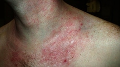
Atopic dermatitis. Dirty neck sign in chronic atopic dermatitis.
-
Atopic dermatitis. Irritation around mouth of an infant with atopic dermatitis.