Practice Essentials
Postinflammatory hyperpigmentation (PIH) is a frequently encountered problem and represents the sequelae of various cutaneous disorders as well as therapeutic interventions. [1] This acquired excess of pigment can be attributed to various preceding disease processes that affect the skin such as infections, allergic reactions, mechanical injuries, reactions to medications, phototoxic eruptions, trauma (eg, burns), and inflammatory diseases (eg, lichen planus, lupus erythematosus, atopic dermatitis).
Postinflammatory hyperpigmentation can also be seen following treatment with a number of electromagnetic devices such as ultrasound, radiofrequency, lasers, light-emitting diodes, and visible light, as well as secondary to microdermabrasion. [2] Typically, postinflammatory hyperpigmentation is most severe in patients whose basal cell layer of the epidermis is disrupted such as lichenoid dermatoses or lupus erythematosus.
Signs and symptoms
A diagnosis of postinflammatory hyperpigmentation should be considered if a history of a preceding pathologic process or injury to the affected area of hyperpigmentation is present.
The distribution of the hypermelanotic lesions depends on the location of the original inflammatory dermatosis. The color of the lesions ranges from light brown to black, with a lighter brown appearance if the pigment is within the epidermis (ie, epidermal melanosis) and a darker gray to bluish appearance if lesions contain dermal melanin (ie, dermal melanosis). Note the image below.
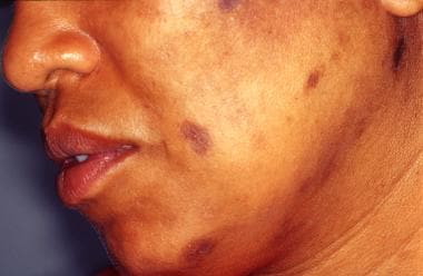 Photo of a 42-year-old African American woman with macules of postinflammatory hyperpigmentation on the left side of her face as a result of acne excoriée.
Diagnostics
A Wood lamp examination enables distinction of epidermal postinflammatory hyperpigmentation (PIH) from dermal PIH. Epidermal lesions tend to have accentuated borders under a Wood lamp examination, whereas those of dermal lesions appear poorly circumscribed and are not accentuated with a Wood lamp examination.
If a history of preceding inflammatory dermatosis is unclear or absent, skin biopsy is warranted to exclude other underlying causes of hyperpigmentation. Staining of the biopsy specimen with Fontana-Masson silver stain for melanin enables localization of the melanin in the epidermis and/or the dermis.
Epidermal PIH involves increased melanin pigment in the basal cell layer of the epidermis. Occasionally, giant melanosomes are evident in the epidermis.
Dermal PIH involves melanin pigment in the upper dermis, with pigment incontinence due to increased numbers of melanophages in the papillary dermis.
Management
Also see Surgical Care, Prevention, and Medication.
The treatment of postinflammatory hyperpigmentation (PIH) tends to be a difficult and prolonged process that often takes 6-12 months to achieve the desired results of depigmentation. Each of these treatment options potentially improves epidermal hypermelanosis, but none is proven effective for dermal hypermelanosis. Daily use of a broad-spectrum sunscreen (sun protection factor [SPF] 15 or greater) is an essential part of any therapeutic regimen.
A variety of topical treatments have been used to treat epidermal postinflammatory hyperpigmentation, with varying degrees of success. These agents include hydroquinone, tretinoin cream, corticosteroids, glycolic acid (GA), and azelaic acid. [3] Lightening of hyperpigmented areas may be achieved with one of the previously named topical agents; however, a combination of topical creams and gels, chemical peels, and sunscreens may be necessary for significant improvement. [4] They are only effective for epidermal hyperpigmentation. In one study, the combination of salicylic acid peel and topical tretinoin treatment was better than either treatment alone. [5] Another study showed combining glycolic acid peels with a modified Kligman formula of hydroquinone 2%, tretinoin 0.05%, and hydrocortisone 1% was of particular benefit in facial postinflammatory hyperpigmentation in dark-skinned Indians. [6]
Topical tretinoin 0.1% has been effective in treating postinflammatory hyperpigmentation. GA peels, in combination with tretinoin and hydroquinone, are an effective treatment of postinflammatory hyperpigmentation in dark-complexioned individuals. [7] All-trans retinoic acid aqueous gel 0.1-0.4% may be applied concomitantly with hydroquinone–lactic acid ointment for bleaching. [8, 9] After sufficient improvement of the hyperpigmentation is achieved, a corticosteroid may be applied topically with hydroquinone to promote healing. This combination of various topical therapeutic agents has been shown to be beneficial, especially on the face.
Topical azelaic acid, which has been approved for the treatment of acne vulgaris, is useful for PIH as well. [10] In acne patients who are prone to postinflammatory hyperpigmentation, azelaic acid may be a good treatment option. The efficacy of tazarotene 0.1% cream for the treatment of dyschromia associated with photoaging and for acne vulgaris may also be beneficial, particularly in people with dark skin tone. [11]
Early and efficacious treatment of acne in patients with dark-toned skin helps minimize pigmentary abnormalities. [12]
Other treatment modalities include use of trichloroacetic acid and gentle cryotherapy with liquid nitrogen. Each method must be used with extreme caution to avoid necrosis or blistering of the treated skin. These 2 methods of treatment should be avoided in dark-skinned patients because of the risk of permanent depigmentation and scarring.
Pigmented makeup creams have also been successfully used to camouflage hyperpigmented skin to a hue similar to that of the surrounding unaffected skin.
More options will be available in the future. Retinaldehyde (RAL) has shown depigmenting activity, while GA decreases the excess of pigment by a wounding and reepithelization process. A combination of RAL 0.1% and GA 6% (Diacneal) in the treatment of acne vulgaris and postinflammatory hyperpigmentation has been reported. [13] The peroxidase inhibitor methimazole, a noncytotoxic inhibitor of melanin production, is a possible agent for topical use in the years ahead. [14]
The efficacy and safety of a combined treatment regimen including serial GA peels, topical azelaic acid cream, and adapalene gel in the treatment of recalcitrant melasma was evaluated in 28 patients in a prospective, randomized, controlled trial lasting 20 weeks. [15] Those receiving chemical peels underwent serial GA peels in combination with topical azelaic acid 20% cream (twice daily) and adapalene 0.1% gel (4 times daily, applied at night). Combined treatment with serial GA peels, azelaic acid cream, and adapalene gel may be an effective and safe therapy for recalcitrant melasma. Choi et al report that Lepidium apetalum is a potential inhibitor of hyperpigmentation caused by UV radiation. [16]
Other studies suggest that decapeptide-12 was 17-fold more potent than hydroquinone at inhibiting tyrosinase in vitro. [17, 18] Decapeptide-12 was shown not to be cytotoxic to melanocytes, making it a safer alternative to hydroquinone. A pilot study by Hantash and Jimenez showed that twice a day treatment for 4 months with decapeptide-12 formulated in a topical emulsion also resulted in a 50% improvement in melasma in patients who had failed 6 months of Tri-Luma therapy. [18] The potential of decapeptide-12 as a therapeutic option for melasma was recently reviewed. [19]
Depigmenting agents abound in a variety of formations. Aloe vera leaf extract and its active ingredient aloin are considered potent skin depigmenting agents. [20] Delivery too can be important. Glabridin microsponge-loaded gel may be beneficial in treating hyperpigmentation. [21]
Patients should never be treated with monobenzyl ether of hydroquinone because of the risk of developing disfiguring depigmented patches of skin either at the application site or at distal cutaneous sites.
Topical application of epidermal growth factor reduces laser-induced PIH. [22]
Use of sunscreen for both ultraviolet and visible light protection is important to inhibit exacerbation of hyperpigmentation and to improve the appearance. [23]
Also see Skin Lightening and Depigmenting Agents.
Pathophysiology
Postinflammatory hyperpigmentation is caused by 1 of 2 mechanisms that result in either epidermal or dermal melanosis. The epidermal inflammatory response (ie, dermatitis) results in the release and subsequent oxidation of arachidonic acid to prostaglandins, leukotrienes, and other products. These products of inflammation alter the activity of both immune cells and melanocytes. When 35% trichloroacetic acid (TCA) solution was applied to skin, it was found that TCA-induced postinflammatory hyperpigmentation might serve as a good in vivo model for the study of acne-induced postinflammatory hyperpigmentation. [24]
Specifically, these inflammatory products stimulate epidermal melanocytes, causing them to increase the synthesis of melanin and subsequently to increase the transfer of pigment to surrounding keratinocytes. Such increased stimulation and transfer of melanin granules results in epidermal hypermelanosis.
On the contrary, dermal melanosis occurs when inflammation disrupts the basal cell layer, causing melanin pigment to be released and subsequently trapped by macrophages in the papillary dermis, also known as pigmentary incontinence.
Fibroblast-derived melanogenic growth factors may be salient in mesenchymal-epithelial interactions modulating melanocyte function. [25]
Etiology
Postinflammatory hyperpigmentation can occur with various disease processes that affect the skin. These processes include allergic reactions, infections, trauma, and phototoxic eruptions. Fractional laser photothermolysis occasionally induces postinflammatory hyperpigmentation. [26, 27]
Common inflammatory diseases that result in postinflammatory hyperpigmentation include acne excoriée, lichen planus, systemic lupus erythematosus, chronic dermatitis, and cutaneous T-cell lymphoma, especially erythrodermic variants.
Furthermore, lesions of postinflammatory hyperpigmentation can darken with exposure to UV light and various chemicals and medications, such as tetracycline, bleomycin, doxorubicin, 5-fluorouracil, busulfan, arsenicals, silver, gold, antimalarial drugs, hormones, and clofazimine. Hyperpigmentation after foamed bleomycin sclerotherapy for vascular malformations has been delineated. [28]
Epidemiology
Frequency
United States
Postinflammatory hyperpigmentation is a universal response of the skin, but it is more common in individuals with darker skin (Fitzpatrick skin types III to VI). Postinflammatory hyperpigmentation can be caused by any inflammatory process of the skin; however, it is more apparent in photo-induced dermatoses and more severe in lichenoid dermatoses.
International
Internationally, postinflammatory hyperpigmentation is a common inflammatory response of the skin, developing more commonly in darker skin. Despite their lighter skin color, certain Asians (from Pacific rim countries such as Japan, Taiwan, China) are more susceptible to developing PIH following one of the inciting factors listed above.
Race
Although postinflammatory hyperpigmentation occurs in whites, it is more common in darker pigmented individuals including African Americans or Asians.
Sex
Postinflammatory hyperpigmentation occurs with equal incidence in males and females; it has no sexual predilection.
Age
Postinflammatory hyperpigmentation can occur in persons of any age.
Prognosis
Morbidity associated with postinflammatory hyperpigmentation is related to the underlying inflammatory process that causes postinflammatory hyperpigmentation. If the hyperpigmentation is located in cosmetically sensitive regions, a significant amount of emotional distress may result.
Postinflammatory hyperpigmentation tends to fade with time and therapy, as previously discussed. Remnants of epidermal hyperpigmentation may persist for indefinite periods, typically 6-12 months, after the initial inflammatory process resolves. Dermal postinflammatory hyperpigmentation may even persist for years.
Patient Education
Educate patients about the cause of postinflammatory hyperpigmentation, prolonged therapy, and persistence of hyperpigmented lesions.
-
Photo of a 42-year-old African American woman with macules of postinflammatory hyperpigmentation on the left side of her face as a result of acne excoriée.


