Practice Essentials
Lymphangiomas are uncommon, hamartomatous, congenital malformations of the lymphatic system that involve the skin and subcutaneous tissues. The classification of lymphangiomas lacks a standard clear definition and universal application, in part because of the nature of lymphangiomas, which represent a clinicopathologic continuum. The classification most frequently used divides these lesions into 2 major groups based on the depth and the size of these abnormal lymph vessels. The superficial vesicles are called lymphangioma circumscriptum. The more deep-seated group includes cavernous lymphangioma and cystic hygroma. Many categorize cystic hygroma as a variant of cavernous lymphangioma. Note the image below.
Also see Oral Lymphangiomas and Cervicofacial Lymphangiomas.
Signs and symptoms
Lymphangiomas may affect a number of sites, including the scrotum. Multilocular lymphangiomatosis of the scrotum is unusual. [1] The site of a lymphangioma may determine its clinical signs and symptoms. For example, a lacrimal gland lymphangioma may result in acute proptosis and inferonasal displacement of an eye. [2]
Note the images below.
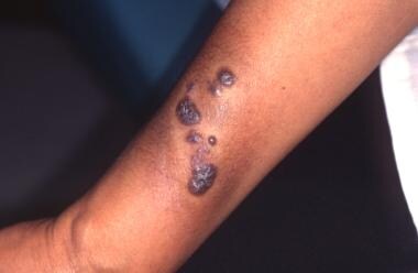 A 44-year-old woman with plaque on her forearm since birth that clinically appeared to be typical lymphangioma circumscriptum. Histologically, however, it had features of both hemangioma and lymphangioma.
Lymphangioma circumscriptum
Lymphangioma circumscriptum involves small clusters of vesicles measuring about 2-4 mm. These clear vesicles can vary from pink to red to black secondary to hemorrhage. [3] Although it may appear localized to the dermis, this neoplasm frequently extends deeply and laterally. The lesions can have a warty appearance on their surface; as a result, they are often confused with warts. Shah et al report a lymphangioma presenting on the penis. [4]
Cavernous lymphangioma
Typically, cavernous lymphangiomas appear as subcutaneous nodules with a rubbery consistency. They may have large dimensions. The overlying skin has no lesions or changes. The area of involvement varies, ranging from lesions smaller than 1 cm in diameter to larger lesions that involve an entire limb. Rarely, nasolacrimal lymphomas may be seen, with one first evident with unilateral blood tearing. [5]
Cystic hygroma
Cystic hygromas are usually larger than cavernous lymphangiomas, and they more commonly occur in the neck and parotid area. Often, deep cavernous lymphangiomas are not evident on superficial examination, but cystic hygromas are detected with ease because of their size and location. These large cystic lesions are soft and translucent. Congenital and acquired lymphangiomas of the vulva are rare. [6, 7] Diffuse lymphangiomas may go unnoticed, evident as asymptomatic, erythematous flat, indurated, or atrophic plaques with or without swelling with or without any surface changes.
Fetal lymphangioma
Fetal lymphangioma may involve fetal skin and occasionally mucosa and subcutaneous tissue, typically on neck or axilla. [8] The fetal chest wall cystic lymphangioma is rare. [9]
Diagnostics
The diagnosis of lymphangiomas is based mainly on the clinical history and findings from physical examination and conventional light microscopy.
Also see Histologic Findings.
MRI can help define the degree of involvement and the entire anatomy of the lymphangioma lesion. MRI can help prevent unnecessary extensive, incomplete surgical resection, because of the association with a high recurrence rate.
The scrotum is not a usual site, but a painless scrotal swelling was documented as a lymphangioma by typical sonography and MRI findings, followed by excision and pathologic confirmation. [10]
Immunohistochemical study is useful in differentiating lymphangiomas from hemangiomas in difficult cases. Test results with factor VIII – related antigen are positive for hemangiomas but negative or weakly positive in the endothelium of lymphangiomas. Immunohistochemical studies for laminin show the typical multilayered basal lamina of normal blood vessels and the discontinuous basal lamina in lymphangiomas.
Dermoscopic findings may aid in the diagnosis of cutaneous lymphangioma circumscriptum. [11, 12, 13] Nodules filled with clear fluid show light brown lacunas surrounded by paler septa. Those tinged with blood may have focal reddish areas inside the lagoons, pink diffuse coloration, and/or reddish to violaceous lacunar structures. In those lacunae that contained blood, it accumulated in the lowest part. [14] Thus, they are characterized by a lacunar pattern and with a marked hematic content may be indistinguishable from a hemangioma.
Management
The preferred treatment for lymphangiomas is complete surgical excision. [15] On the basis of the Whimster hypothesis, the large subcutaneous cisterns should be removed to prevent the lesion from resurfacing.
Also see Treatment.
Background
Lymphangiomas can occur anywhere in the skin and the mucous membranes. The most common sites are the head and the neck, followed by the proximal extremities, the buttocks, and the trunk. However, they sometimes can be found in the intestines, the pancreas, and the mesentery. Deeper cystic lesions usually occur in areas of loose and areolar tissue, typically the neck, the axilla, and the groin. Their skin involvement ranges from small, well-demarcated areas to large, diffuse regions with unclear borders.
Lymphangioma circumscriptum, the common form of cutaneous lymphangioma, is characterized by persistent, multiple clusters of translucent vesicles that usually contain clear lymph fluid (often compared with frog spawn). [3] These vesicles represent superficial saccular dilations from underlying lymphatic vessels that occupy the papilla and push upward against the overlying epidermis. Each skin lesion may range from a minute vesicle to a small bulla-sized lesion. These vesicles can be clear or vary from pink to dark red because of serosanguineous fluid and hemorrhage. These vesicles often are associated with verrucous changes, which give them a warty appearance.
In the case of lymphangioma circumscriptum, the underlying lesions constitute abnormal dilated lymph vessels involving the upper part of the dermis. The sites of predilection are the proximal extremities, trunk, axilla, and oral cavity, especially the tongue. [16] Involvement in other areas, such as the scrotum, is not uncommon. [17] Lymphangioma circumscriptum has a high recurrence rate after excision because of its deep component (see Pathophysiology).
Cavernous lymphangioma are also uncommon and usually arise during infancy. The most common sites are the head and neck areas and, less frequently, the extremities. These lesions are seated deep in the dermis, forming a painless swelling or thickening of the skin, mucous membranes, and subcutaneous tissue. Unlike lymphangioma circumscriptum, the overlying skin usually is uninvolved. Occasionally, patients report pain when the involved area is pressed. The affected area may be 1 cm, it may be as large as several centimeters in diameter, or it may involve an entire extremity. Upon examination and palpation, lipomas or cysts can be mistaken for these lesions. Lymphangioma circumscriptum can occur in conjunction with cavernous lymphangioma and cystic hygroma.
Some authors categorize cystic hygroma or cystic lymphangioma as an independent entity. Many authors agree that cystic hygroma is a form of cavernous lymphangioma in which the degree of involvement and character is determined by its location. These congenital lesions are deeply seated in areas of areolar or loose connective tissue. They appear early in life as large soft-tissue masses, usually on the axilla, neck, or groin. These lesions are soft, vary in size and shape, and tend to grow extensively if not surgically excised. Typical lesions are multilocular cysts filled with clear or yellow lymph fluid. Usually, cystic hygroma is diagnosed clinically with its large size, location, and translucence.
Terminology for these lesions can be confusing. [18] Some lymphangiectasias have sometimes been called acquired lymphangiomas, secondary lymphangiomas and acquired lymphangioma circumscriptum.
Pathophysiology
In 1976, Whimster [19] studied the pathogenesis of lymphangioma circumscriptum. According to Whimster, the basic pathologic process is the collection of lymphatic cisterns in the deep subcutaneous plane. These cisterns are separated from the normal network of lymph vessels, but they communicate with the superficial lymph vesicles through vertical, dilated lymph channels.
Whimster postulated that these cisterns might arise from a primitive lymph sac that fails to connect with the rest of the lymphatic system during its embryonic development. A thick coat of muscle fibers that cause rhythmic contractions line these sequestered primitive sacs. Rhythmic contractions increase the intramural pressure, causing dilated channels to protrude from the walls of the cisterns toward the skin. He suggested that the vesicles seen in lymphangioma circumscriptum are outpouchings of these dilated projecting vessels.
Whimster's observations are supported by those of lymphangiographic and radiographic studies. These studies revealed that large multilobulated cisterns extend deep in the dermis and laterally beyond the obvious clinical lesions. These deep lymphangiomas show no evidence of communication with the adjacent normal lymphatics. The cause for the failure of these primitive lymph sacs to connect to the rest of the lymphatic system is not known.
Some lymphangiomas may represent vascular malformations during embryonic development rather than as true neoplasms. [20] Vascular endothelial growth factor (VEGF)–C and VEGF receptor-3 are active in the formation of lymphangiomas. Based upon their expression, superficial lymphangiomas more likely result from peripheral lymphatic dilatation than from a growth factor.
Fetal lymphangioma is presumably a result of failure in lymphatic drainage. [8]
Etiology
The reason that these embryonic lymph sacs remain disconnected from the rest of the lymphatic system is not known. Fetal lymphangioma is presumably a result of failure in lymphatic drainage, documented on routine prenatal ultrasonographic studies. [8] Vulva lymphangioma circumscriptum may be secondary to pelvic lymphatic obstruction, linked in some patients to rectal adenocarcinoma, cervical carcinoma, or endometrial carcinoma. [7, 21]
Epidemiology
Frequency
Lymphangiomas are rare. They account for 4% of all vascular tumors and approximately 25% of all benign vascular tumors in children.
Its incidence in India has been estimated at 1.2-2.8 cases per 1000 newborns. [17]
Race, sex, and age
No racial predominance is reported for lymphangiomas.
Equal sex incidences are reported for lymphangiomas in most studies. Some groups have reported that lymphangioma circumscriptum is more common in females than in males, while others report a 3:1 male-to-female ratio.
Lymphangioma can become evident at any age, but the greatest incidence occurs at birth or early in life. About 50% of lymphangiomas are seen at birth, and most lymphangiomas are evident by the time the patient is aged 5 years. It may be documented in fetuses too. [8]
Prognosis
Lymphangiomas are benign hamartomatous malformations instead of true neoplasms. The prognosis for lymphangiomas is excellent.
Rarely do cutaneous lymphangiomas interfere with the well-being of patients. Patients are expected to live a full healthy life, and they usually seek medical intervention because of cosmetic reason.
Lymphangiomas represent hamartomatous malformations with no risk of malignant transformation. In the case of cystic hygroma, total surgical excision is appropriate to prevent complications such as respiratory compromise, aspiration, and infections in critical areas, such as the neck. Lymphangiomas have a strong tendency for local recurrence unless they are completely excised. Recurrent episodes of cellulitis and minor bleeding are not uncommon.
Patient Education
Patients should receive reassurance. Lymphangiomas represent benign lymphatic malformations and not premalignant lesions. Patients should be aware of the risk of recurrence.
-
A 44-year-old woman with plaque on her forearm since birth that clinically appeared to be typical lymphangioma circumscriptum. Histologically, however, it had features of both hemangioma and lymphangioma.
-
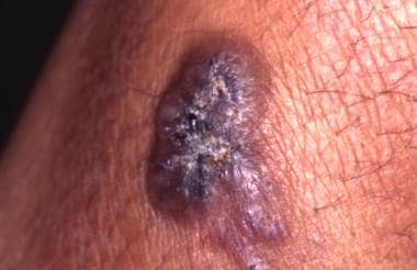
Close-up demonstrating the clinical morphology to better advantage.
-
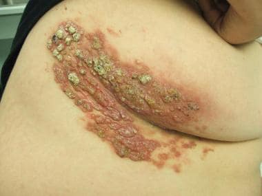
A 16-year-old obese boy with large unilateral verrucous lymphangioma.


