| Patient Education |
|
Click here for patient education.
|
|
You are in: eMedicine Specialties >
Dermatology > PEDIATRIC DISEASES
Proteus Syndrome
Article Last Updated: Feb 15, 2007
AUTHOR AND EDITOR INFORMATION
Section 1 of 11
Author: Matthew J Mahlberg, MD, Resident Physician, Department of Dermatology, New York University School of Medicine
Matthew J Mahlberg is a member of the following medical societies: American Medical Association and Phi Beta Kappa
Coauthor(s):
William D James, MD, Paul R Gross Professor of Dermatology, University of Pennsylvania School of Medicine; Vice-Chair, Program Director, Department of Dermatology, University of Pennsylvania Health System
Editors: Albert C Yan, MD, Section Chief, Assistant Professor, Department of Pediatrics, Section of Dermatology, Children's Hospital of Philadelphia and University of Pennsylvania; David F Butler, MD, Professor of Dermatology, Texas A&M University College of Medicine; Director, Division of Dermatology, Scott and White Clinic; Director Dermatology Residency Training Program, Scott and White Clinic; Jeffrey J Miller, MD, Associate Professor, Department of Dermatology, Penn State University, Milton S Hershey Medical Center; Catherine Quirk, MD, Clinical Assistant Professor, Department of Dermatology, Brown University; Dirk M Elston, MD, Director, Department of Dermatology, Geisinger Medical Center
Author and Editor Disclosure
Synonyms and related keywords:
PS, hamartomatous disorder, multifarious mesodermal malformation, plurifocal overgrowth, partial gigantism, regional gigantism
Background
Proteus syndrome (PS) is a sporadically occurring hamartomatous disorder associated with irregular overgrowth of multiple body tissues and cell lineages. Most malformations in patients with PS have a mesodermal origin. Characteristic plurifocal overgrowths (partial or regional gigantism) can involve any structure of the body. Although some evidence of this syndrome was published in the medical literature as early as 1907, the modern medical description of the disease is attributed to Cohen and Hayden, who identified the syndrome in 1979.1 In 1983, to stress the polymorphic nature of the clinical manifestations of PS, Wiedermann coined the term Proteus syndrome after the Greek god Proteus (meaning polymorphous), the "Old Man of the Sea" who could change his shape at will to avoid capture.2 Perhaps the most famous case of PS is that of Joseph Merrick, the "Elephant Man" described by Sir Frederick Treves in 1884, who was made famous by a stage play and movie of the same name. Although first thought to have neurofibromatosis, Merrick is now believed to have had PS. Preserved castings of his soles show cerebriform cutaneous hyperplasia, a nearly pathognomonic finding in persons with PS.3
Pathophysiology
PS is a rare, sporadic disease with patchy or mosaic manifestations. The etiology of PS remains unknown; however, the predominant hypothesis is that PS is caused by a postzygotic mosaic alteration in a gene that is lethal in the nonmosaic state.4 Chromosomal aberrations have not been demonstrated with routine cytogenetic studies, but the karyotype in 2 patients with PS showed structural abnormalities of chromosome 16 and chromosome 1 in a mosaic distribution, further supporting the hypothesis of somatic mutation.5 Several authors initially reported a common PTEN gene mutation in their patients; however, these patients were later determined to have a Proteuslike syndrome.6 The concept of a somatic mutation that involves tissue growth factors or their receptors can explain several aspects of PS, such as the mosaic distribution of lesions, its sporadic occurrence, the unaffected offspring from affected individuals, and the existence of discordant twins.7 Despite the evidence implicating a somatic mutation, no causal gene mutations are yet known for PS.
Mortality/Morbidity
Patients with PS have difficulty ambulating because of toe macrodactyly, scoliosis, and joint instability, with frequent hip dislocations, expansive subcutaneous tumors, and compression neuropathies due to intraneural hamartomas. Some patients may have persistent atelectasis, pneumonia, or symptoms of pulmonary insufficiency. Mental retardation is present in approximately 30% of patients. Premature death has been reported in 20% of PS patients, most often related to deep venous thrombosis leading to pulmonary embolus, postoperative complications, or pneumonia.8
Race
PS has no predilection for any particular race.
Sex
The male-to-female ratio is 1.9:1 (n = 96).8
Age
At least some of the abnormalities associated with PS are present at birth or they appear in the first years of life. They usually progress until puberty.9 Notably, the cerebriform nevi typically do not manifest until age 2 years, on average, often delaying the correct diagnosis of PS.10
History
Patients present with the characteristic abnormalities of PS, which may include the following:
- Asymmetric hypertrophy of the face is apparent.
- Asymmetric hypertrophy of part or all of one or both limbs, particularly the digits, is noted.
- Asymmetric hypertrophy of the trunk is also typical.
- Patients have heterogeneous skin lesions, which may include palmoplantar cerebriform hyperplasia, soft subcutaneous tumors, epidermal nevi, vascular hamartomas, and hyperpigmented or hypopigmented areas.
- Many patients have normal intelligence, but mental retardation and seizures are reported. Mental retardation is found in approximately 30% of PS patients and is more common in patients with typical facial phenotype.8
Physical
The intrinsic variability of the clinical features may result in overdiagnosis. Diagnostic criteria and guidelines for differential diagnosis and patient evaluation have been established. See the PS diagnostic criteria at the end of this section.11 Only those manifestations that are believed to have diagnostic value are considered because of the tremendous variability in the manifestations of PS among and within patients. Physical findings can be classified as cutaneous or noncutaneous.
- Cutaneous manifestations
- Virtually all patients with PS have at least one type of cutaneous lesion, of which several different types have been reported. Skin lesions can generally be classified into 2 groups, those that are congenital or neonatal in onset and are stable (group 1) and those that are postnatal and progressive (group 2).10 In some patients, the presenting symptom may be one or more of the many types of heterogeneous skin lesions that characterize PS. These include cerebriform connective-tissue nevi, epidermal nevi, vascular hamartomas, lipomas, and hyperpigmented or hypopigmented areas. Epidermal nevi and vascular malformations are considered group 1 lesions, while lipomas and cerebriform connective-tissue nevi generally occur later and are considered group 2.10
- Group 1 cutaneous lesions are characterized as follows:
- Epidermal nevi are a common finding in patients with PS. These manifest at birth as tan-to-brown, flat-topped, hyperkeratotic or verrucous papules, which run in a linear or whorled pattern along the lines of Blaschko.12, 13 See Media File 1. Epidermal nevi are found asymmetrically, scattered around the body. Histologic findings are acantholysis and hyperkeratosis in a clinical lesion consistent with an epidermal nevus.12
- Vascular malformations are another common cutaneous finding usually present at birth. These anomalies can be of venous, capillary, or lymphatic origin and include nevus flammeus, angiokeratomas, cavernous hemangiomas, superficial and deep lymphangiomas, and varicosity of the superficial veins. These lesions are developmental abnormalities and grow proportionately through a patient's lifetime without regression.7 In some patients, these vascular malformations have been noted to expand beyond proportionate growth.14 Prominence of the veins may be enhanced in areas of regional lipodystrophy.13
- Group 2 cutaneous lesions are characterized as follows:
- Cerebriform connective-tissue nevi are one of the most common and characteristic features of PS, often sufficient for diagnosis when present, although their presence is not required. These nevi have a relatively delayed onset, making the diagnosis difficult in neonates and infants. Cerebriform nevi occur as well-demarcated, skin-colored plaques with a cerebriform or rugated appearance (see Media File 2). Most often, they occur on the palms or soles but have also been noted on the forearm, on the trunk, and inferior to the nasal ala.12 Lesions on the sole typically cause the most morbidity because they cause difficulty walking, are prone to ulceration and infection, and are malodorous.12 Histologically, the lesions consist of an irregular proliferation of highly collagenized fibrous tissue.9, 15
- PS patients have adipose tissue abnormalities, resulting in lipomas and areas of lipohypoplasia. Lipomas occur as hamartomatous masses consisting of subcutaneous tissue or a variable combination of adipose, lymphatic, and hemangiomatous components (eg, lipolymphohemangiomas) and are the second most frequent type of skin findings that characterize PS. The soft subcutaneous tumors can affect any area of the body but are most common on the head, abdomen, groin, or legs. They appear as soft, skin-colored nodules or tumors. Patchy areas of lipohypoplasia or dermal hypoplasia are also observed in some patients.12 Lipohypoplasia occurs as regions of skin with minimal fat, while dermal hypoplasia appears as depressed, red plaques in areas with prominent veins.
- Cutaneous manifestations also include striking overgrowth anomalies, including asymmetric hypertrophy of the face, part or all of one or both limbs (particularly digits), the trunk, or any combination of these; often, hemihypertrophy of one side of the body results.16 See Media Files 3-5. These abnormalities can also be the first to attract the parents' attention and cause considerable anxiety. Café au lait spots and areas of hypopigmentation or hyperpigmentation with a linear or whorled arrangement are also hallmark skin findings in persons with PS.12 Hypertrichosis and nail abnormalities are also seen.12
- Noncutaneous manifestations
- The most characteristic noncutaneous findings in patients with PS involve skeletal overgrowth and include corporal hemihypertrophy, partial gigantism of hands and/or feet, and skeletal anomalies such as long-bone overgrowth and scoliosis.13 Overgrowth of the hands, feet, or both was universal in one series of 24 patients with PS. Arm or leg overgrowth is nearly always present.12
- Craniofacial abnormalities are common and progressive and include cranial hemihyperplasia, hyperostosis of the skull or external auditory canal, craniosynostosis, and unilateral condylar hyperplasia.7 Hyperostosis and unilateral condylar hyperplasia are common, while cranial hemihyperplasia and craniosynostosis are rare. A facial phenotype with dolichocephaly, down-slanting palpebral fissures, ptosis, anteverted nares, and open mouth has been associated with mental retardation and seizures.17
- Specific tumors have been noted to occur with increased frequency in patients with PS. In addition to being found subcutaneously, lipomas infiltrate muscle and internal organs, including the heart, pancreas, spinal cord, and pharynx.18 Other tumors noted to occur in several patients include ovarian cystadenomas, testicular tumors, and parotid adenomas. Other benign neoplasms, such as bronchial hamartomas and multiple meningiomas, have been reported.
- Cystic lung disease is found in approximately 10% of patients with PS and can lead to significant morbidity; with rapid progression, it can be fatal.19 Renal cysts have also been described.8
- Visceral overgrowth is noted, most often of the spleen or thymus.
- Internal vascular malformations of the gastrointestinal tract, spleen, kidneys, and testicles have been reported. One report of intravesicular venous lesions was associated with life-threatening hematuria.20
- Many ocular manifestations are reported in patients with PS. The most commonly recorded include strabismus, nystagmus, and epibulbar tumors. Other ocular defects include a high degree of myopia, retinal pigmentary abnormalities, retinal detachment, cataracts, posterior segment hamartomas, retinal coloboma, heterochromia irides, and glaucoma.21, 8
- Additional features include asymmetric myopathy, congenital heart defects, malocclusion, hypodontia, and hypoplastic enamel.8 Immunodeficiency has been reported in one patient with PS.22
- PS diagnostic criteria: Diagnosis requires the presence of all general criteria and various specific criteria, including the presence of the category A criterion, 2 category B criteria, or 3 category C criteria.11
- General criteria are as follows:
- Mosaic distribution of lesions
- Sporadic occurrence
- Progressive course
- Specific criteria are as follows:
- Category A - Cerebriform connective-tissue nevus
- Category B - Epidermal nevus; asymmetric, disproportionate overgrowth (limbs, skull, external auditory meatus, vertebrae, and/or viscera); and specific tumors that occur in the second decade (ie, ovarian cystadenoma, parotid monomorphic adenoma)
- Category C - Dysregulated adipose tissue (lipomas, regional lipohypoplasia), vascular malformations (capillary, venous, and/or lymphatic), lung cysts, and facial phenotype (as described above)
Causes
The etiology of PS is unknown, but a Proteuslike syndrome has been associated with PTEN mutations.
- The syndrome occurs sporadically in healthy families.
- Routine cytogenetic studies have not consistently revealed chromosomal aberrations.
- The concept of a somatic mutation that affects the production or regulation of tissue growth factors or their receptors could explain the sporadic occurrence, the random distribution of overgrowth, and the wide range of findings within the phenotype.
Klippel-Trenaunay-Weber Syndrome
Maffucci Syndrome
Neurofibromatosis
Other Problems to be Considered
Carefully distinguish PS from other overgrowth disorders, particularly Klippel-Trenaunay syndrome, hemihyperplasia/lipomatosis syndrome, and neurofibromatosis type 1.23 Several other syndromes can also be considered in the differential diagnosis10; these are discussed below. Vascular syndromes The classic definition of Klippel-Trenaunay syndrome is the triad of vascular stain, soft tissue and/or bony hypertrophy, and venous varicosities. It often involves vascular malformation involving the lower or upper limbs, but usually only 1 limb is affected. The vascular (ie, capillary, lymphatic, and venous) malformations always exist in combination. Overgrowth is present at birth; commonly, this is more severe than that in PS. Moreover, Klippel-Trenaunay syndrome lacks subcutaneous tumors, palmar and/or plantar cerebriform hyperplasia, and cranial exostoses, which are usually found in PS patients.24 The delayed onset of cerebriform hyperplasia often leads to the misdiagnosis of PS as Klippel-Trenaunay syndrome in neonates and infants.10 Parkes-Weber syndrome is a vascular malformation involving the upper and lower limbs. It is characterized by a diffuse capillary blushing, warmth, and underlying arteriovenous shunts. Maffucci syndrome is characterized by hemangiomas and enchondromas. Syndromes with pigmentation and lipomatosis Neurofibromatosis type 1 involves café au lait macules, axillary freckling, Lisch nodules, and neurofibromas, rather than epidermal nevi and hamartomas. Bannayan-Riley-Ruvalcaba syndrome, which is characterized by macrocephaly, lipomas, capillary malformations, and polyposis of the colon and rectum, does not cause asymmetric growth, cranial exostoses, epidermal nevi, or palmar or plantar changes. Patients with hemihyperplasia lipomatosis syndrome lack the progressive overgrowth seen in patients with PS. Encephalocraniocutaneous lipomatosis has been considered a circumscribed form of PS, although some authors believe that PS and encephalocraniocutaneous lipomatosis are different entities.25
Imaging Studies
- Imaging studies are useful in helping to establish the diagnosis of PS and in tracking the progression of the disease.
- Radiography of the skull, vertebral column, long bones, and pelvis: The most characteristic forms of overgrowth are macrodactyly, clinodactyly, asymmetrical hypertrophy of a limb, vertebral body abnormalities, and hyperostosis.18
- Comparative radiographic study of the hands and feet: The most characteristic noncutaneous features are progressive asymmetric macrodactyly, hemihypertrophy of any part of the skeleton, scoliosis and spinal canal stenosis, macrocephaly, and exostoses, especially of the skull.
- Intracranial MRI: This is an essential initial examination to evaluate for malformations of the CNS that may be associated with mental retardation or seizures. Such findings may include multiple meningiomas, polymicrogyria, and periventricular heterotopias.
- Abdominal MRI: Even in the absence of symptoms, abdominal MRI is important to exclude intra-abdominal lipomas. If present, these can behave aggressively, invading adjacent structures.
- High-resolution chest CT scanning: This examination may be useful in evaluating pulmonary cystic malformations.
Other Tests
- Skin biopsy: Skin biopsy should be considered to confirm the clinical diagnosis of a cerebriform connective-tissue nevus. See PS diagnostic criteria, category A, in Physical.
- Karyotype analysis: Chromosome analysis may reveal a translocation or deletion that may suggest a candidate gene.
- Electroencephalography: Although most patients appear to be of normal intelligence, mental retardation may occur. Seizures have also been reported.
Procedures
- The ongoing evaluation of the patient with PS should include the following procedures11:
- Serial clinical photography
- Initial skeletal survey with targeted follow-up radiography
- MRI of all clinically affected areas
- MRI of the chest and abdomen in the absence of symptoms
- Consultation with a dermatologist, followed by biopsy if indicated
- Consultation with an orthopedic surgeon, followed by surgical treatment if indicated
- Ongoing treatment from a geneticist, pediatrician, or both
- Consultations with a neurologist and an ophthalmologist
- Referral to family support group
Histologic Findings
The histologic findings in PS are specific to the particular type of lesion. The histology of cerebriform connective-tissue nevi and epidermal nevi are discussed in History and Physical, along with a description of their clinical manifestations.
Medical Care
The aim of medical treatment is to minimize the physical and psychosocial consequences of PS; this requires a multidisciplinary approach.26
- Antithrombotic prophylaxis should be considered if the patient is undergoing a surgical procedure because the patient's vascular malformations predispose them to deep venous thromboses and fatal pulmonary emboli.27
- Cystic pulmonary lesions should be followed closely because they may progress to pneumonia, atelectasis, and, potentially, pulmonary insufficiency.
- Cerebriform connective-tissue nevi are of considerable concern for patients, especially when present on the plantar surfaces, making walking uncomfortable. Conservative medical treatment for cerebriform connective-tissue nevi includes (1) keeping the feet clean and dry, (2) regularly applying antibacterial lotion to reduce odor, (3) closely monitoring for ulceration and infection, and (4) using orthotic devices. Attempts at surgical excision have led to poor outcomes; therefore, surgery is not recommended.12
Surgical Care
- Preoperative evaluation should include assessment of the airway anatomy and pulmonary reserve because of the frequent presence of tonsillar hypertrophy and pulmonary cysts.28
- Plastic surgeons and orthopedic surgeons can correct some skeletal defects. Epiphysiodesis can help correct asymmetric epiphyseal growth, and reduction osteotomy can be used to shorten long bones. Spinal fusion for overgrown vertebrae may prevent the development of severe kyphoscoliosis and the risk of pulmonary compromise.7, 26
- Subcutaneous tumors should be removed in the early stages.29
- Gonadal deformities (ovarian or testicular) should be managed aggressively because of the high incidence of neoplastic transformation.28
Consultations
- Consult a dermatologist.
- Consult a plastic surgeon for the correction of defects.
- Consult an orthopedic surgeon for the correction of skeletal deformities.
- Ophthalmologists and neurologists may be helpful because many ocular manifestations and CNS anomalies associated with seizures or developmental delays are reported in patients with PS.
- Referral to psychologist or psychiatrist is important for patients and for their families. The disease creates an isolating social stigma associated with having a very rare, progressive, disfiguring condition. Symptoms of depression have been reported in 23% of parents of affected children.30
Complications
- Sequelae in patients with PS include ambulatory difficulty due to toe macrodactyly, scoliosis, and joint instability, with frequent hip dislocations.
- Dysregulated adipose growth can result in aggressive, infiltrating lipomas that can involve adjacent structures.
- Pulmonary complications are a frequent cause of morbidity and mortality in PS patients.
Prognosis
- The disease is progressive but somewhat variable in prognosis. With appropriate medical and surgical care, patients with PS may age normally.31 However, despite treatment efforts, PS may result in extreme musculoskeletal, cutaneous, and visceral deformities.16 Some patients (as many as 20%) die prematurely, usually from pulmonary embolus or pneumonia.
Medical/Legal Pitfalls
- Failure to correctly evaluate patients with PS may result in the nonidentification of neoplasia that requires surgical treatment or other disorders (eg, bone or neural diseases).
- Failure to evaluate and manage ocular involvement in patients with PS can result in visual impairment.
The authors and editors of eMedicine gratefully acknowledge the contributions of previous authors, Carlo Tolone, MD, Eleonora Ruocco, MD, and Antonio Vozza, MD, to the development and writing of this article.
| Media file 1:
Epidermal nevus of the trunk follows the Blaschko lines. |
 | View Full Size Image | |
Media type: Photo
|
| Media file 2:
Cerebriform connective-tissue nevus on the plantar surface. |
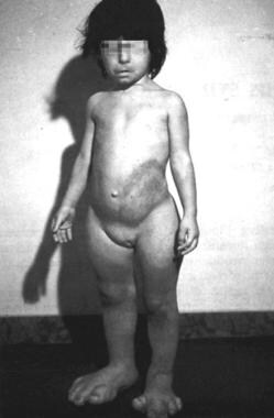
 | View Full Size Image | |
Media type: Photo
|
| Media file 4:
Proteus syndrome with hemihypertrophy of the limbs. |
 | View Full Size Image | |
Media type: Photo
|
| Media file 5:
Proteus syndrome with gigantism of the feet and macrodactyly. |
 | View Full Size Image | |
Media type: Photo
|
- Cohen MM Jr, Hayden PW. A newly recognized hamartomatous syndrome. Birth Defects Orig Artic Ser. 1979;15(5B):291-6. [Medline].
- Wiedemann HR, Burgio GR, Aldenhoff P, Kunze J, Kaufmann HJ, Schirg E. The proteus syndrome. Partial gigantism of the hands and/or feet, nevi, hemihypertrophy, subcutaneous tumors, macrocephaly or other skull anomalies and possible accelerated growth and visceral affections. Eur J Pediatr. Mar 1983;140(1):5-12. [Medline].
- Braun-Falco O, Plewig G, Wolff HH, et al. Dermatology. 2000. 2nd ed. Berlin: Springer; 849-50.
- Happle R. Lethal genes surviving by mosaicism: a possible explanation for sporadic birth defects involving the skin. J Am Acad Dermatol. Apr 1987;16(4):899-906. [Medline].
- Cardoso MT, de Carvalho TB, Casulari LA, Ferrari I. Proteus syndrome and somatic mosaicism of the chromosome 16. Panminerva Med. Dec 2003;45(4):267-71. [Medline].
- Happle R. The manifold faces of proteus syndrome. Arch Dermatol. Aug 2004;140(8):1001-2. [Medline].
- Cohen MM Jr. Proteus syndrome: an update. Am J Med Genet C Semin Med Genet. Aug 15 2005;137(1):38-52. [Medline].
- Turner JT, Cohen MM Jr, Biesecker LG. Reassessment of the Proteus syndrome literature: application of diagnostic criteria to published cases. Am J Med Genet A. Oct 1 2004;130(2):111-22. [Medline].
- Nazzaro V, Cambiaghi S, Montagnani A, Brusasco A, Cerri A, Caputo R. Proteus syndrome. Ultrastructural study of linear verrucous and depigmented nevi. J Am Acad Dermatol. Aug 1991;25(2 Pt 2):377-83. [Medline].
- Twede JV, Turner JT, Biesecker LG, Darling TN. Evolution of skin lesions in Proteus syndrome. J Am Acad Dermatol. May 2005;52(5):834-8. [Medline].
- Biesecker LG, Happle R, Mulliken JB, Weksberg R, Graham JM Jr, Viljoen DL, et al. Proteus syndrome: diagnostic criteria, differential diagnosis, and patient evaluation. Am J Med Genet. Jun 11 1999;84(5):389-95. [Medline].
- Nguyen D, Turner JT, Olsen C, Biesecker LG, Darling TN. Cutaneous manifestations of proteus syndrome: correlations with general clinical severity. Arch Dermatol. Aug 2004;140(8):947-53. [Medline].
- del Rosario Barona-Mazuera M, Hidalgo-Galván LR, de la Luz Orozco-Covarrubias, Durán-McKinster C, Tamayo-Sánchez L, Ruiz-Maldonado R. Proteus syndrome: new findings in seven patients. Pediatr Dermatol. Jan-Feb 1997;14(1):1-5. [Medline].
- Cohen MM Jr. Proteus syndrome. In: Cohen MM Jr, Neri G, Weksberg R, eds. Overgrowth Syndromes. New York, NY: Oxford University Press; 2002:75-110.
- Samlaska CP, Levin SW, James WD, Benson PM, Walker JC, Perlik PC. Proteus syndrome. Arch Dermatol. Aug 1989;125(8):1109-14. [Medline].
- Atherton DJ. Naevi and other developmental defects. In: Champion RH, Burton JL, Burns DA, eds. Rook/Wilkinson/Ebling's Textbook of Dermatology. 6th ed. Oxford, England: Blackwell Science; 1998:519-616.
- Cohen MM Jr. Proteus syndrome: clinical evidence for somatic mosaicism and selective review. Am J Med Genet. Oct 1 1993;47(5):645-52. [Medline].
- Jamis-Dow CA, Turner J, Biesecker LG, Choyke PL. Radiologic manifestations of Proteus syndrome. Radiographics. Jul-Aug 2004;24(4):1051-68. [Medline].
- Newman B, Urbach AH, Orenstein D, Dickman PS. Proteus syndrome: emphasis on the pulmonary manifestations. Pediatr Radiol. 1994;24(3):189-93. [Medline].
- Franc-Guimond J, Houle AM, Barrieras D. The Proteus syndrome associated with life threatening hematuria. J Urol. Dec 2003;170(6 Pt 1):2418-9. [Medline].
- De Becker I, Gajda DJ, Gilbert-Barness E, Cohen MM Jr. Ocular manifestations in Proteus syndrome. Am J Med Genet. Jun 19 2000;92(5):350-2. [Medline].
- Hodge D, Misbah SA, Mueller RF, Glass EJ, Chetcuti PA. Proteus syndrome and immunodeficiency. Arch Dis Child. Mar 2000;82(3):234-5. [Medline].
- Biesecker LG, Peters KF, Darling TN, Choyke P, Hill S, Schimke N, et al. Clinical differentiation between Proteus syndrome and hemihyperplasia: description of a distinct form of hemihyperplasia. Am J Med Genet. Oct 2 1998;79(4):311-8. [Medline].
- Hagari Y, Aso M, Shimao S, Okano T, Kurimasa A, Takeshita K. Proteus syndrome: report of the first Japanese case with special reference to differentiation from Klippel-Trenaunay-Weber syndrome. J Dermatol. Aug 1992;19(8):477-80. [Medline].
- McCall S, Ramzy MI, Curé JK, Pai GS. Encephalocraniocutaneous lipomatosis and the Proteus syndrome: distinct entities with overlapping manifestations. Am J Med Genet. Jul 1 1992;43(4):662-8. [Medline].
- Biesecker LG. Proteus Syndrome. In: Cassidy SB, Allanson JE, eds. Management of Genetic Syndromes. 2nd ed. New York, NY: Wiley-Liss; 2005:437-44.
- Cohen MM Jr. Causes of premature death in Proteus syndrome. Am J Med Genet. Jun 1 2001;101(1):1-3. [Medline].
- Lublin M, Schwartzentruber DJ, Lukish J, Chester C, Biesecker LG, Newman KD. Principles for the surgical management of patients with Proteus syndrome and patients with overgrowth not meeting Proteus criteria. J Pediatr Surg. Jul 2002;37(7):1013-20. [Medline].
- Vaughn RY, Selinger AD, Howell CG, Parrish RA, Edgerton MT. Proteus syndrome: diagnosis and surgical management. J Pediatr Surg. Jan 1993;28(1):5-10. [Medline].
- Peters KF, Biesecker LG. An opportunity for genetic counseling intervention: Depression in parents of individuals with proteus syndrome. J Genet Couns. 2000;9:161-70.
- Choi ML, Wey PD, Borah GL. Pediatric peripheral neuropathy in proteus syndrome. Ann Plast Surg. May 1998;40(5):528-32. [Medline].
Proteus Syndrome excerpt Article Last Updated: Feb 15, 2007
|