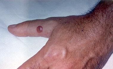
| Patient Education |
|
Click here for patient education.
|
|
You are in: eMedicine Specialties >
Dermatology > VIRAL INFECTIONS
Orf
Article Last Updated: Aug 18, 2008
AUTHOR AND EDITOR INFORMATION
Section 1 of 10
Author: Lana H Hawayek, MD, Assistant Professor of Dermatology, University of Cincinnati, Veterans Affairs Medical Center; Consulting Staff, University Dermatology Consultants
Lana H Hawayek is a member of the following medical societies: American Academy of Dermatology, American Society for Dermatologic Surgery, and Women's Dermatologic Society
Coauthor(s):
Nelly Rubeiz, MD, Consulting Staff, Department of Dermatology, American University of Beirut Medical Center; Associate Professor, Department of Dermatology, American University of Beirut, Lebanon
Editors: Shyam Verma, MBBS, DVD, FAAD, Adjunct Clinical Assistant Professor, Department of Dermatology, University of Virginia, State University of New York at Stonybrook, Penn State University; Richard P Vinson, MD, Assistant Clinical Professor, Department of Dermatology, Texas Tech University School of Medicine; Consulting Staff, Mountain View Dermatology, PA; Jeffrey J Miller, MD, Associate Professor, Department of Dermatology, Penn State University, Milton S Hershey Medical Center; Glen H Crawford, MD, Assistant Clinical Professor, Department of Dermatology, University of Pennsylvania School of Medicine; Chief, Division of Dermatology, The Pennsylvania Hospital; Dirk M Elston, MD, Director, Department of Dermatology, Geisinger Medical Center
Author and Editor Disclosure
Synonyms and related keywords:
orf, viral infection, zoonotic infection, animal-borne disease, ecthyma contagiosum, ecthyma infectiosum, infectious pustular dermatitis, contagious pustular dermatitis, sheep-pox, lip scab of sheep, sore mouth disease, scabby mouth disease
Background
Orf is a viral disease that is widespread in sheep and goats. Orf can be transmitted to humans by contact with an infected animal or contaminated fomites. Reindeer have also caused similar lesions in humans. Orf is frequently seen in farming communities1, 2 and meat handlers. Clinically, reddish weeping nodules of orf are seen on the dorsa of the hands and the fingers that heal spontaneously in approximately 35 days
A related Medscape CME article is Companion Animals and Human Health: Part II -- Zoonotic Diseases.
Pathophysiology
Orf is primarily a disease involving the skin, though the lymph nodes are occasionally involved.
Frequency
International
A higher frequency of orf reports has occurred from Europe and New Zealand compared with North America, but this is of unknown significance.
Mortality/Morbidity
The natural course of orf is spontaneous recovery in 3-6 weeks.
- Immunocompromised patients with orf can have progressive, destructive lesions requiring medical interventions such as antiviral therapy and surgical debridement. However, reports exist of immunosuppressed individuals with large, fungating lesions that have been refractory to treatment.
- Mortality from orf has not been reported.
Race
Orf has been reported exclusively in whites.
Sex
No sexual predilection is reported for orf.
Age
No age predilection is described for orf.
History
- Orf usually appears as a small papule on the dorsum of the index finger 1 week following contact with an infected animal or contaminated fomite.
- With orf, a low-grade fever may occur but usually subsides within 3-4 days.
Physical
Orf appears as a solitary lesion or as a few lesions on the fingers, the hands, or the forearms, and orf has even been reported on the face.3 The orf lesion starts as a small, firm, red-to-blue papule that grows to form a hemorrhagic, flat-topped pustule or bulla. The bulla may have a crust in its umbilicated center. The fully developed orf lesion is typically 2-3 cm in diameter, but it may reach 5 cm. It is often tender and may bleed easily.
- Regional adenitis and/or mild lymphangitis may be found.
- Mild fever and malaise may be associated with orf.
- Large, fungating orf lesions have been reported in patients who are immunosuppressed and in patients with atopic dermatitis.
- The orf infection goes through 6 clinical stages, each lasting about 1 week.
- Stage 1 (maculopapular) - A red elevated lesion
- Stage 2 (targetoid) - A bulla with an irislike configuration (nodule with a red center, a white middle ring, and a red periphery)
- Stage 3 (acute) - A weeping nodule
- Stage 4 (regenerative) - A firm nodule covered by a thin crust through which black dots are seen
- Stage 5 (papillomatous) - Small papillomas appearing over the surface
- Stage 6 (regressive) - A thick crust covering the resolving elevation
Causes
Orf is caused by infection with the orf virus that belongs to the Parapoxvirus genus, which also includes the milker's nodule virus.4 Parapoxvirus is a member of the family Poxviridae, which contains double-stranded DNA viruses known to be the largest viruses.
- The orf virus is a cylindrical virus measuring 260 X160 nm. Its surface tubules form a long crisscross design that is seen on negatively stained preparations by electron microscopy. This virus resists physical damage and persists through the winter months on hedges, feeding troughs, and barns.
- Orf is transmitted by direct contact inoculation. Humans acquire the infection from contact with infected animals, carcasses, or contaminated, nonliving material. Orf is very common among shepherds, veterinary surgeons, and farmers' wives who bottle-feed young lambs, as well as in butchers and meat porters from handling infected carcasses.
- No transmission of orf occurs to cattle, and no human-to-human transmission occurs.
Acute Febrile Neutrophilic Dermatosis
Erysipeloid
Milker's Nodules
Other Problems to be Considered
Furuncle Tularemia Anthrax5 Localized benign cutaneous tumors
Lab Studies
- No serologic tests are routinely performed for orf, but a detectable antibody response may be present. Serology cannot distinguish orf virus from other parapoxviruses such as paravaccinia (pseudocowpox) virus.
Other Tests
- Confirmation of the clinical diagnosis of orf is performed by electron microscopy with negative staining of the crust or a small biopsy. Electron microscopy has also been performed on fluid obtained from the orf lesion.6 It demonstrates classic ovoid cross-hatched virions but cannot be used to distinguish orf virus from other parapoxviruses such as paravaccinia (pseudocowpox) virus.
- Tissue culture with passage of the orf virus to sheep cell cultures is an option; however, the growth of the virus is slow and inconstant.
- Conventional histopathology of an orf skin biopsy may be of significant diagnostic value.
- Polymerase chain reaction (PCR) can definitively identify a parapoxvirus as orf virus.7 Two assays have been used by the US Centers for Disease Control and Prevention, standard PCR and real-time PCR.8 A 2006 report demonstrated the increased sensitivity of the newer real-time PCR technique (nearly 1000 times more sensitive than standard PCR).9 These assays are ideally performed on frozen tissue specimens, vesicle material, or scab debris from orf lesions.
Histologic Findings
The epidermis usually shows marked pseudoepitheliomatous hyperplasia. Necrosis of the epidermis with ulceration occurs in the center of the lesion. The orf viral infection causes intranuclear and intracytoplasmic inclusion bodies with vacuolization and disaggregation of keratinocytes. Pyknosis of individual keratinocytes occurs. A dense inflammatory infiltrate of plasma cells, macrophages, histiocytes, and lymphocytes is also observed in orf.
Medical Care
Although orf is a self-limited disease, symptomatic treatment with moist dressings, local antiseptics, and finger immobilization is helpful. Secondary bacterial infection from orf is not uncommon and must be treated with topical or systemic antibiotics. Several reports describe complicated orf cases of orf successfully treated with topical imiquimod, resulting in rapid regression of the lesions.10, 11, 12 Reports also describe promising results in treating orf lesions with cidofovir cream.13, 14
Surgical Care
For large exophytic orf lesions, dissection from the underlying dermis is performed.15 If an orf lesion is persistent, curettage and electrodesiccation may be curative. Shave excision is another procedure used to treat orf lesions. Cryotherapy (liquid nitrogen) has been reported to speed up the recovery process from orf treatment.16, 17 Radiation or amputation must be avoided.
Deterrence/Prevention
- The best preventive measure in animals is orf vaccination every 6-8 months. Veterinary vaccines for orf virus use nonattenuated, live virus preparations. Recently vaccinated animals pose an increased risk to humans. Once an animal is infected, it should be put in isolation.
- Humans infected with orf do not need to be isolated because no human-to-human transmission occurs.
- Patients who are immunosuppressed or persons with compromised skin barriers (eg, trauma, skin disease) must avoid contact with infected animals because they have an increased susceptibility for contracting the orf infection.
Complications
- Toxic erythema
- Erythema multiforme18, 19
- Growth of the skin lesion to a giant size
- Widespread papulovesicular eruption of the skin and the mucosae
- Unusual localization of the infection (ocular or perianal)
- Bullous pemphigoid20
- Orf-induced immunobullous disease (reported in 2008)21
Prognosis
- The prognosis of orf is excellent. The orf lesions usually heal completely with no scarring in about 35 days.
Patient Education
- Most patients infected with orf are farmers or people who handle animals; therefore, they are usually familiar with the disease. Those who are unacquainted with this condition must be reassured. They must also be informed that recurrences may occur and result in lesions that are less pronounced than the primary infection.
Special Concerns
- Pregnancy and fetal development have not been reported to be adversely affected by this viral infection.22
| Media file 4:
The regenerative stage of orf with a central crust. |
 | View Full Size Image | |
Media type: Photo
|
| Media file 5:
Orf lesion (arrow) associated with erythema multiforme. |
 | View Full Size Image | |
Media type: Photo
|
- Buchan J. Characteristics of orf in a farming community in mid-Wales. BMJ. Jul 27 1996;313(7051):203-4. [Medline].
- Paiba GA, Thomas DR, Morgan KL, Bennett M, Salmon RL, Chalmers R, et al. Orf (contagious pustular dermatitis) in farmworkers: prevalence and risk factors in three areas of England. Vet Rec. Jul 3 1999;145(1):7-11. [Medline].
- Bodnar MG, Miller OF 3rd, Tyler WB. Facial orf. J Am Acad Dermatol. May 1999;40(5 Pt 2):815-7. [Medline].
- Groves RW, Wilson-Jones E, MacDonald DM. Human orf and milkers' nodule: a clinicopathologic study. J Am Acad Dermatol. Oct 1991;25(4):706-11. [Medline].
- Centers for Disease Control and Prevention. Human orf mimicking cutaneous anthrax---California. MMWR. 1973;22:108.
- Sanchez RL, Hebert A, Lucia H, Swedo J. Orf. A case report with histologic, electron microscopic, and immunoperoxidase studies. Arch Pathol Lab Med. Feb 1985;109(2):166-70. [Medline].
- Torfason EG, Gunadóttir S. Polymerase chain reaction for laboratory diagnosis of orf virus infections. J Clin Virol. Feb 2002;24(1-2):79-84. [Medline].
- Olson VA, Laue T, Laker MT, Babkin IV, Drosten C, Shchelkunov SN, et al. Real-time PCR system for detection of orthopoxviruses and simultaneous identification of smallpox virus. J Clin Microbiol. May 2004;42(5):1940-6. [Medline].
- Gallina L, Dal Pozzo F, Mc Innes CJ, Cardeti G, Guercio A, Battilani M, et al. A real time PCR assay for the detection and quantification of orf virus. J Virol Methods. Jun 2006;134(1-2):140-5. [Medline].
- Erbagci Z, Erbagci I, Almila Tuncel A. Rapid improvement of human orf (ecthyma contagiosum) with topical imiquimod cream: report of four complicated cases. J Dermatolog Treat. 2005;16(5-6):353-6. [Medline].
- Lederman ER, Green GM, DeGroot HE, Dahl P, Goldman E, Greer PW, et al. Progressive ORF virus infection in a patient with lymphoma: successful treatment using imiquimod. Clin Infect Dis. Jun 1 2007;44(11):e100-3. [Medline].
- Ara M, Zaballos P, Sánchez M, Querol I, Zubiri ML, Simal E, et al. Giant and recurrent orf virus infection in a renal transplant recipient treated with imiquimod. J Am Acad Dermatol. Feb 2008;58(2 Suppl):S39-40. [Medline].
- Geerinck K, Lukito G, Snoeck R, De Vos R, De Clercq E, Vanrenterghem Y, et al. A case of human orf in an immunocompromised patient treated successfully with cidofovir cream. J Med Virol. Aug 2001;64(4):543-9. [Medline].
- De Clercq E. Clinical potential of the acyclic nucleoside phosphonates cidofovir, adefovir, and tenofovir in treatment of DNA virus and retrovirus infections. Clin Microbiol Rev. Oct 2003;16(4):569-96. [Medline].
- Shelley WB, Shelley ED. Surgical treatment of farmyard pox. Orf, milker's nodules, bovine papular stomatitis pox. Cutis. Feb 1983;31(2):191-2. [Medline].
- Degraeve C, De Coninck A, Senneseael J, Roseeuw D. Recurrent contagious ecthyma (Orf) in an immunocompromised host successfully treated with cryotherapy. Dermatology. 1999;198(2):162-3. [Medline].
- Ocampo Candiani J, González Soto R, Welsh Lozano O. Orf nodule: treatment with cryosurgery. J Am Acad Dermatol. Aug 1993;29(2 Pt 1):256-7. [Medline].
- Ferrando MF, Léauté-Labrèze C, Fleury H, Taďeb A. Orf and erythema multiforme in a child. Pediatr Dermatol. Mar-Apr 1997;14(2):154-5. [Medline].
- Mourtada I, Le Tourneur M, Chevrant-Breton J, Le Gall F. [Human orf and erythema multiforme]. Ann Dermatol Venereol. Apr 2000;127(4):397-9. [Medline].
- Murphy JK, Ralfs IG. Bullous pemphigoid complicating human orf. Br J Dermatol. May 1996;134(5):929-30. [Medline].
- White KP, Zedek DC, White WL, Simpson EL, Hester E, Morrison L. Orf-induced immunobullous disease: A distinct autoimmune blistering disorder. J Am Acad Dermatol. Jan 2008;58(1):49-55. [Medline].
- Taieb A, Guillot M, Carlotti D, Maleville J. Orf and pregnancy. Int J Dermatol. Jan-Feb 1988;27(1):31-3. [Medline].
- Centers for Disease Control and Prevention. Orf virus infection in humans--New York, Illinois, California, and Tennessee, 2004-2005. MMWR Morb Mortal Wkly Rep. Jan 27 2006;55(3):65-8. [Medline].
- Deane D, McInnes CJ, Percival A, Wood A, Thomson J, Lear A, et al. Orf virus encodes a novel secreted protein inhibitor of granulocyte-macrophage colony-stimulating factor and interleukin-2. J Virol. Feb 2000;74(3):1313-20. [Medline].
- Ghislain PD, Dinet Y, Delescluse J. Orf contamination may occur during religious events. J Am Acad Dermatol. May 2000;42(5 Pt 1):848. [Medline].
- Inceoglu F. Orf (ecthyma contagiosum): an occasional diagnostic challenge. Plast Reconstr Surg. Sep 2000;106(3):733-4. [Medline].
- Mayet A, Sommer B, Heenan P. Rapidly growing cutaneous tumour of the right temple: Orf. Australas J Dermatol. Nov 1997;38(4):217-9. [Medline].
- Peeters P, Sennesael J. Parapoxvirus Orf in kidney transplantation. Nephrol Dial Transplant. Feb 1998;13(2):531. [Medline].
- Roingeard P, Machet L. Images in clinical medicine. Orf skin ulcer. N Engl J Med. Oct 16 1997;337(16):1131. [Medline].
- Yirrell DL, Vestey JP, Norval M. Immune responses of patients to orf virus infection. Br J Dermatol. Apr 1994;130(4):438-43. [Medline].
Orf excerpt Article Last Updated: Aug 18, 2008
|