| Patient Education |
|
Click here for patient education.
|
|
You are in: eMedicine Specialties >
Dermatology > MALIGNANT NEOPLASMS
Bowen Disease
Article Last Updated: Jul 15, 2008
AUTHOR AND EDITOR INFORMATION
Section 1 of 11
Author: Mark L Welch, MD, Clinical Assistant Professor, Department of Dermatology, Howard University; Assistant Professor, Department of Dermatology, Uniformed Services University of Health Sciences
Mark L Welch is a member of the following medical societies: American Academy of Dermatology, American College of Mohs Micrographic Surgery and Cutaneous Oncology, American Medical Association, and American Society for Dermatologic Surgery
Coauthor(s):
Theresa Conologue, DO, Physician, Department of Dermatology, National Capital Consortium, Walter Reed Army Medical Center;
Carrie A H Hall, MD, Resident Physician, National Capital Consortium Dermatology Residency Program, National Naval Medical Center
Editors: Kelly M Cordoro, MD, Fellow and Clinical Instructor, Department of Pediatric Dermatology, University of California at San Francisco; Assistant Professor (On Educational Leave), Assistant Program Director for Resident Medical Education, Department of Dermatology, University of Virginia School of Medicine; David F Butler, MD, Professor of Dermatology, Texas A&M University College of Medicine; Chair, Department of Dermatology, Scott and White Clinic; Director Dermatology Residency Training Program, Scott and White Clinic; Edward F Chan, MD, Clinical Assistant Professor, Department of Dermatology, University of Pennsylvania School of Medicine; Catherine Quirk, MD, Clinical Assistant Professor, Department of Dermatology, Brown University; Dirk M Elston, MD, Director, Department of Dermatology, Geisinger Medical Center
Author and Editor Disclosure
Synonyms and related keywords:
squamous cell carcinoma in situ, SCC, human papilloma virus 16, HPV 16, human papillomavirus 16
Background
First described by John T. Bowen in 1912, Bowen disease is a squamous cell carcinoma (SCC) in situ with the potential for significant lateral spread (see Squamous Cell Carcinoma). Larger lesions can reach several centimeters in diameter.
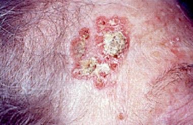
 Much controversy exists as to whether Bowen disease is associated with internal malignancies.1, 2, 3, 4 Many early papers reported such an association in 15-70% of cases. Some later reports supported an association of internal malignancies with Bowen disease that was associated with arsenic ingestion but not with Bowen disease from other causes. Most of the recent studies, with a larger number of cases and better scientific methods, have refuted any association; currently, Bowen disease is not believed to be a paraneoplastic condition.
Pathophysiology
Bowen disease is a form of intraepidermal carcinoma. It may ultimately become an invasive squamous cell carcinoma.
Frequency
United States
In 1991, a study from Minnesota reported the annual average rate of Bowen disease as 14 cases per 100,000 whites. In 1994, a study from Hawaii reported a rate 10 times that, 142 cases per 100,000 whites.5
Mortality/Morbidity
Prognosis of Bowen disease is favorable, with less than 5% of cases advancing to invasive SCC, and metastases are even more rare.
Race
Bowen disease most commonly is reported in whites.
Sex
The ratio is approximately equal between males and females.
Age
Bowen disease has its highest incidence in the older age groups.
History
Patients often present with an asymptomatic, slowly enlarging, erythematous, scaly patch or plaque. It may occur anywhere on the mucocutaneous surface. A delay in diagnosis often is encountered because the lesion is asymptomatic; early skin changes may be subtle and overlap with clinical features seen in many benign conditions such as tinea corporis, nummular eczema, and psoriasis.
Physical
Bowen disease presents as a single lesion in two thirds of cases. Lesions may appear on sun-exposed or covered skin. The head and neck are the most commonly affected anatomic locations, followed by the limbs. Lesions vary in size from a few millimeters to several centimeters in diameter. 
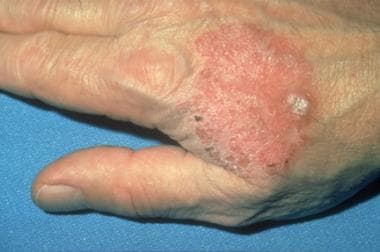
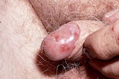
A sharply demarcated, irregular border usually is present. Lesions are erythematous, scaly patches or plaques that may become hyperkeratotic, crusted, fissured, or ulcerated. Rarely, the lesions are pigmented, especially in the genital region and the nails. Lesions in these locations may simulate melanoma.6 Bowen disease also may occur on mucous membranes. When it arises on the glans penis, it is referred to as erythroplasia of Queyrat. See Erythroplasia of Queyrat (Bowen Disease of the Glans Penis) for more information on this topic.
Causes
- Chronic UV radiation: Sun-exposed distribution of Bowen disease (over one half of lesions occur on the head, neck, and hands) implicates chronic sun damage as one factor in its formation.
- Arsenic exposure: The main sources of arsenic exposure include Fowler solution, a medication formerly used to treat psoriasis; Gay solution, a medication formerly used to treat asthma; contaminated well water; and certain pesticides.
- Human papilloma virus: Human papillomavirus type 16 is by far the most common subtype isolated from lesions of Bowen disease, although other subtypes, such as HPV 2, also have been found.
- Other possible causes include genetic factors, trauma, other chemical carcinogens, and x-ray radiation. See the Medscape Skin Cancer Resource Center for more information.
Actinic Keratosis
Basal Cell Carcinoma
Lichen Simplex Chronicus
Paget Disease, Mammary
Psoriasis, Plaque
Squamous Cell Carcinoma
Tinea Corporis
Other Problems to be Considered
Benign lichenoid keratosis
Eczema
Paget disease (extramammary)
Procedures
- Skin biopsy: Perform a shave or punch biopsy to confirm the diagnosis of Bowen disease. Incorporating follicular structures in the biopsy material is helpful. These procedures are typically performed in an office setting with the patient under local anesthesia. Pathologic analyses are best completed by dermatopathologists.
- Complete skin examination: Perform a total body skin examination on patients with Bowen disease on sun-exposed skin. Studies indicate a higher incidence of nonmelanoma skin cancers may exist in these patients.
Histologic Findings
Bowen disease is a full-thickness anaplasia of the epidermis, with loss of the normal maturation of its components. Keratinocytes are atypical and disorderly, often described as having a windblown appearance. Although the basal cell layer is intact, extension of keratinocyte atypia down the follicular epithelium is seen. Vacuolization, mitoses, individually keratinizing cells, and multinucleated cells are present in the epidermis. Large pale keratinocytes with abundant ground glass cytoplasm, so-called pagetoid cells, often are distributed haphazardly throughout the epidermis. Hyperkeratosis, parakeratosis, and acanthosis are seen to some degree in lesions of Bowen disease. The upper dermis has a moderate lymphocytic infiltrate.
Medical Care
- Topical therapy
- Administration of 5-fluorouracil topically under occlusion, following the use of a keratolytic or cryotherapy, or by iontophoresis (an electrogradient-driven chemical delivery system) has been used to effectively treat Bowen disease.7, 8, 9, 10
- Imiquimod 5% cream,11, 12 a topical immune response modifier, applied 3-7 d/wk appears to be a successful treatment option for Bowen disease in multiple body sites. Two reports indicate sustained clearance with at least 19 months disease-free follow-up after treatment of perianal Bowen disease with single-agent therapy using imiquimod 5% cream. Topical treatment for perianal Bowen disease may minimize the risk of scarring, poor wound healing, and functional impairment.
- Also note, however, that a cautionary report describes Bowen disease of the scalp treated with imiquimod that evolved into invasive SCC.13
- Consider x-ray or grenz-ray radiation therapy for poor surgical candidates or patients with multiple lesions.14
- In the last decade, photodynamic therapy (PDT) has shown promise in the treatment of superficial carcinomas, such as Bowen disease.15, 16 PDT involves the introduction of a photosensitizing agent into the body, which is retained preferentially by the tumor cells. Then, a light source is used to stimulate the photosensitizing agent, causing the release of toxins and leading to the destruction of the tumor.
Surgical Care
- Simple excision with conventional margins
- This surgery is the most common and preferred treatment for smaller lesions and those not in problematic areas, such as the face and digits.
- Although lesions are typically well demarcated, the actual extent of the disease may be well beyond the clinical margins. For this reason, the excision should be made at least 4 mm outside the clinical margin.
- Mohs micrographic surgery17
- This is an excellent method for larger lesions, recurrent lesions, or those in areas where tissue sparing is vital. Mohs micrographic surgery uses the systematic surgical removal of skin cancers with very small margins of normal tissue followed by frozen section examination of nearly 100% of the tissue margin.
- It offers the highest cure rate of all treatment modalities, and, because relatively thin layers are taken only in areas of proven tumor, it is a tissue-sparing procedure.
- Curettage and electrodesiccation, cryotherapy, and carbon dioxide laser ablation18
- These are blind surgical methods (no pathologic confirmation of removal) that are established treatment modalities for Bowen disease.
- As compared with excision and Mohs surgery, they are less likely to remove tumors that are present down in the adnexal structures.
The Medscape Dermatologic Surgery Resource Center may be of interest.
The goals of therapy are to reduce morbidity and to prevent complications.
Drug Category: Antineoplastic agents
Topical agents that may be used in the management of Bowen disease.
| Drug Name | 5-Fluorouracil (Efudex, Carac, Adrucil, Fluoroplex) |
| Description | 5-Fluorouracil administered topically under occlusion, following the use of keratolytic or cryotherapy, or by iontophoresis (an electrogradient-driven chemical delivery system), can be used. Interferes with DNA synthesis by blocking methylation of deoxyuridylic acid and inhibits thymidylate synthetase, which subsequently reduces cell proliferation. |
| Adult Dose | Only 5% strength recommended; apply bid, sparingly to cover lesions (minimum 3 wk); therapy may be required for 10-12 wk |
| Pediatric Dose | Administer as in adults |
| Contraindications | Documented hypersensitivity; potentially serious infections |
| Interactions | None reported |
| Pregnancy | X - Contraindicated; benefit does not outweigh risk
|
| Precautions | Incidence of inflammatory reactions may occur with occlusive dressings; porous gauze dressing may be applied for cosmetic reasons without increase in reaction |
| Drug Name | Imiquimod (Aldara) |
| Description | Precise mechanism of action for treatment of Bowen disease is unknown. May increase tumor infiltration of lymphocytes, dendritic cells, and macrophages. Indicated when surgical methods are not appropriate. |
| Adult Dose | Apply cream to treatment area (including 1 cm of skin surrounding tumor) 3-7 d/wk for up to 16 wk; leave on for at least 8 h, then wash area |
| Pediatric Dose | Not established |
| Contraindications | Documented hypersensitivity |
| Interactions | None reported |
| Pregnancy | C - Fetal risk revealed in studies in animals but not established or not studied in humans; may use if benefits outweigh risk to fetus
|
| Precautions | Mandatory follow-up to ensure treatment response; may cause redness, swelling, and sore development at application site; may cause itching or burning |
Further Outpatient Care
- Patients with a history of any type of skin cancer should be evaluated with a total body skin examination every 6-12 months.
Deterrence/Prevention
- Counsel at-risk individuals in the use of sunscreens, photoprotective clothing, and other measures to minimize further actinic damage. Frequent and routine screening should also be stressed to these patients.
Prognosis
- The prognosis for patients with Bowen disease is excellent.
Patient Education
- Patients with a history of Bowen disease should be counseled on safe sun behavior, to include avoiding the sun when it is most intense (between 10 am and 4 pm), wearing hats and other sun protective clothing, and using a dual-spectrum sunscreen with an SPF of 15 or higher.
- For excellent patient education resources, visit eMedicine's Cancer and Tumors Center and Burns Center. Also, see eMedicine's patient education articles Skin Cancer, Skin Biopsy, and Sunburn.
Medical/Legal Pitfalls
- A failure to recognize Bowen disease or to perform a biopsy in patients suspected of having Bowen disease leads to a delay in treatment. A high degree of suspicion is needed in both sun-exposed and non sun-exposed areas of the skin.
- Surgery for patients with Bowen disease may cause bleeding, scarring, infection, deformity, and nerve damage. Discussing these risks with the patient prior to surgery is important.
- Topical therapy of Bowen disease may result in recurrence or partial treatment, necessitating further biopsy and possible further surgical intervention with attendant risks as above.
Special Concerns
- Rule out an invasive SCC.
| Media file 1:
Squamous cell carcinoma in situ, Bowen disease. Courtesy of Hon Pak, MD. |
 | View Full Size Image | |
Media type: Photo
|
| Media file 3:
Erythroplasia of Queyrat, squamous cell carcinoma in situ of the glans penis. |
 | View Full Size Image | |
Media type: Photo
|
- Arbesman H, Ransohoff DF. Is Bowen's disease a predictor for the development of internal malignancy? A methodological critique of the literature. JAMA. Jan 23-30 1987;257(4):516-8. [Medline].
- Graham JH, Helwig EB. Bowen's disease and its relationship to systemic cancer. AMA Arch Derm. Aug 1959;80(2):133-59. [Medline].
- Jaeger AB, Gramkow A, Hjalgrim H, Melbye M, Frisch M. Bowen disease and risk of subsequent malignant neoplasms: a population-based cohort study of 1147 patients. Arch Dermatol. Jul 1999;135(7):790-3. [Medline].
- Poole S, Fenske NA. Cutaneous markers of internal malignancy. II. Paraneoplastic dermatoses and environmental carcinogens. J Am Acad Dermatol. Feb 1993;28(2 Pt 1):147-64. [Medline].
- Reizner GT, Chuang TY, Elpern DJ, Stone JL, Farmer ER. Bowen's disease (squamous cell carcinoma in situ) in Kauai, Hawaii. A population-based incidence report. J Am Acad Dermatol. Oct 1994;31(4):596-600. [Medline].
- Saxena A, Kasper DA, Campanelli CD, Lee JB, Humphreys TR, Webster GF. Pigmented Bowen's disease clinically mimicking melanoma of the nail. Dermatol Surg. Dec 2006;32(12):1522-5. [Medline].
- Bargman H, Hochman J. Topical treatment of Bowen's disease with 5-Fluorouracil. J Cutan Med Surg. Mar-Apr 2003;7(2):101-5. [Medline].
- Fulton JE Jr, Carter DM, Hurley HJ. Treatment of Bowen's disease with topical 5-fluorouracil under occlusion. Arch Dermatol. Feb 1968;97(2):178-80. [Medline].
- Sturm HM. Bowen's disease and 5-fluorouracil. J Am Acad Dermatol. Dec 1979;1(6):513-22. [Medline].
- Welch ML, Grabski WJ, McCollough ML, Skelton HG, Smith KJ, Menon PA, et al. 5-fluorouracil iontophoretic therapy for Bowen's disease. J Am Acad Dermatol. Jun 1997;36(6 Pt 1):956-8. [Medline].
- Mackenzie-Wood A, Kossard S, de Launey J, Wilkinson B, Owens ML. Imiquimod 5% cream in the treatment of Bowen's disease. J Am Acad Dermatol. Mar 2001;44(3):462-70. [Medline].
- van Egmond S, Hoedemaker C, Sinclair R. Successful treatment of perianal Bowen's disease with imiquimod. Int J Dermatol. Mar 2007;46(3):318-9. [Medline].
- Fernández-Vozmediano J, Armario-Hita J. Infiltrative squamous cell carcinoma on the scalp after treatment with 5% imiquimod cream. J Am Acad Dermatol. Apr 2005;52(4):716-7. [Medline].
- Dupree MT, Kiteley RA, Weismantle K, Panos R, Johnstone PA. Radiation therapy for Bowen's disease: lessons for lesions of the lower extremity. J Am Acad Dermatol. Sep 2001;45(3):401-4. [Medline].
- Braathen LR, Szeimies RM, Basset-Seguin N, Bissonnette R, Foley P, Pariser D, et al. Guidelines on the use of photodynamic therapy for nonmelanoma skin cancer: an international consensus. International Society for Photodynamic Therapy in Dermatology, 2005. J Am Acad Dermatol. Jan 2007;56(1):125-43. [Medline].
- Jones CM, Mang T, Cooper M, Wilson BD, Stoll HL Jr. Photodynamic therapy in the treatment of Bowen's disease. J Am Acad Dermatol. Dec 1992;27(6 Pt 1):979-82. [Medline].
- Leibovitch I, Huilgol SC, Selva D, Richards S, Paver R. Cutaneous squamous carcinoma in situ (Bowen's disease): treatment with Mohs micrographic surgery. J Am Acad Dermatol. Jun 2005;52(6):997-1002. [Medline].
- Tantikun N. Treatment of Bowen's disease of the digit with carbon dioxide laser. J Am Acad Dermatol. Dec 2000;43(6):1080-3. [Medline].
- Arnold HL, Odom RB, James WD. Bowen's disease. In: Arnold HL, Andrews GC, Odom RB, James WB, eds. Andrews' Diseases of the Skin. 8th ed. Philadelphia, Pa: WB Saunders; 1990:783-5.
- Cox NH, Eedy DJ, Morton CA. Guidelines for management of Bowen's disease: 2006 update. Br J Dermatol. Jan 2007;156(1):11-21. [Medline].
- Gard D. Nonpigmented premalignant lesions of the skin. Clin Plast Surg. Apr 1987;14(2):413-23. [Medline].
- Lee MM, Wick MM. Bowen's disease. Clin Dermatol. Jan-Mar 1993;11(1):43-6. [Medline].
- Ragi G, Turner MS, Klein LE, Stoll HL Jr. Pigmented Bowen's disease and review of 420 Bowen's disease lesions. J Dermatol Surg Oncol. Jul 1988;14(7):765-9. [Medline].
Bowen Disease excerpt Article Last Updated: Jul 15, 2008
|