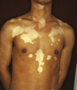
Ortonne J. Vitiligo and other disorders of Hypopigmentation. Bolognia J, Jorizzo J, Rapini R, eds. Dermatology. 2nd. Spain: Elsevier; 2008. Vol 1: 65.
McKee P, Calonje E, Granter S, eds. Disorders of Pigmentation. Pathology of the Skin with Clinical Correlations. 3rd ed. China: Elsevier Mosby; 2005. Vol 2: 993-7.
Moellmann G, Klein-Angerer S, Scollay DA, Nordlund JJ, Lerner AB. Extracellular granular material and degeneration of keratinocytes in the normally pigmented epidermis of patients with vitiligo. J Invest Dermatol. 1982 Nov. 79(5):321-30. [QxMD MEDLINE Link].
Matz H, Tur E. Vitiligo. Curr Probl Dermatol. 2007. 35:78-102. [QxMD MEDLINE Link].
Ezzedine K, Silverberg N. A Practical Approach to the Diagnosis and Treatment of Vitiligo in Children. Pediatrics. 2016 Jul. 138 (1):[QxMD MEDLINE Link].
van Geel N, Ongenae K, Naeyaert JM. Surgical techniques for vitiligo: a review. Dermatology. 2001. 202(2):162-6. [QxMD MEDLINE Link].
Rusfianti M, Wirohadidjodjo YW. Dermatosurgical techniques for repigmentation of vitiligo. Int J Dermatol. 2006 Apr. 45(4):411-7. [QxMD MEDLINE Link].
Alikhan A, Felsten LM, Daly M, Petronic-Rosic V. Vitiligo: a comprehensive overview Part I. Introduction, epidemiology, quality of life, diagnosis, differential diagnosis, associations, histopathology, etiology, and work-up. J Am Acad Dermatol. 2011 Sep. 65 (3):473-91. [QxMD MEDLINE Link].
Schallreuter KU, Wood JM, Pittelkow MR, et al. Regulation of melanin biosynthesis in the human epidermis by tetrahydrobiopterin. Science. 1994 Mar 11. 263(5152):1444-6. [QxMD MEDLINE Link].
Ongenae K, Van Geel N, Naeyaert JM. Evidence for an autoimmune pathogenesis of vitiligo. Pigment Cell Res. 2003 Apr. 16(2):90-100. [QxMD MEDLINE Link].
Zhang BX, Lin M, Qi XY, Zhang RX, Wei ZD, Zhu J, et al. Characterization of circulating CD8+T cells expressing skin homing and cytotoxic molecules in active non-segmental vitiligo. Eur J Dermatol. 2013 Jun 19. [QxMD MEDLINE Link].
Oiso N, Suzuki T, Fukai K, Katayama I, Kawada A. Nonsegmental vitiligo and autoimmune mechanism. Dermatol Res Pract. 2011. 2011:518090. [QxMD MEDLINE Link].
Toussaint S, Kamino H. Noninfectous papular and squamous diseases. Elder D, Elenitas R, Jaworsky D, Johnson B Jr. Lever's Histopathology of the Skin. Philadelphia, Pa: Lippincot-Raven; 1997. 154-5.
Spritz RA. The genetics of generalized vitiligo. Curr Dir Autoimmun. 2008. 10:244-57. [QxMD MEDLINE Link].
Zhang XJ, Chen JJ, Liu JB. The genetic concept of vitiligo. J Dermatol Sci. 2005 Sep. 39 (3):137-46. [QxMD MEDLINE Link].
Jin Y, Birlea SA, Fain PR, et al. Genome-Wide Analysis Identifies a Quantitative Trait Locus in the MHC Class II Region Associated with Generalized Vitiligo Age of Onset. J Invest Dermatol. 2011 Jun. 131(6):1308-12. [QxMD MEDLINE Link].
Halder R, Taliaferro S. Vitiligo. Wolff K, Goldsmith L, Katz S, Gilchrest B, Paller A, Lefell D, eds. Fitzpatrick's Dermatology in General Medicine. 7th ed. New York, NY: McGraw-Hill; 2008. Vol 1: 72.
Dev A, Vinay K, Bishnoi A, Kumaran MS, Dogra S, Parsad D. Dermatoscopic assessment of treatment response in patients undergoing autologous non-cultured epidermal cell suspension for the treatment of stable vitiligo: A prospective study. Dermatol Ther. 2021 Sep. 34 (5):e15099. [QxMD MEDLINE Link].
van Geel N, Speeckaert R, Taieb A, Picardo M, Böhm M, Gawkrodger DJ, et al. Koebner's phenomenon in vitiligo: European position paper. Pigment Cell Melanoma Res. 2011 Jun. 24 (3):564-73. [QxMD MEDLINE Link].
Lee DY, Kim CR, Park JH, Lee JH. The incidence of leukotrichia in segmental vitiligo: implication of poor response to medical treatment. Int J Dermatol. 2011 Aug. 50 (8):925-7. [QxMD MEDLINE Link].
Chandrashekar L. Dermatoscopy of blue vitiligo. Clin Exp Dermatol. 2009 Jul. 34 (5):e125-6. [QxMD MEDLINE Link].
Hann S-K. Clinical variants of vitiligo. Lotti T, Hercogova J, eds. Vitiligo: Problems and Solutions. New York, NY: Marcel Dekker; 2004. 159-73.
Ezzedine K, Diallo A, Leaute-Labreze C, et al. Multivariate analysis of factors associated with early-onset segmental and nonsegmental vitiligo: a prospective observational study of 213 patients. Br J Dermatol. 2011 Jul. 165(1):44-9. [QxMD MEDLINE Link].
Yang Y, Lin X, Fu W, Luo X, Kang K. An approach to the correlation between vitiligo and autoimmune thyroiditis in Chinese children. Clin Exp Dermatol. 2009 Oct 23. [QxMD MEDLINE Link].
Rashtak S, Pittelkow MR. Skin involvement in systemic autoimmune diseases. Curr Dir Autoimmun. 2008. 10:344-58. [QxMD MEDLINE Link].
Pajvani U, Ahmad N, Wiley A, et al. The relationship between family medical history and childhood vitiligo. J Am Acad Dermatol. 2006 Aug. 55(2):238-44. [QxMD MEDLINE Link].
Aydogan K, Turan OF, Onart S, Karadogan SK, Tunali S. Audiological abnormalities in patients with vitiligo. Clin Exp Dermatol. 2006 Jan. 31(1):110-3. [QxMD MEDLINE Link].
Ardiç FN, Aktan S, Kara CO, Sanli B. High-frequency hearing and reflex latency in patients with pigment disorder. Am J Otolaryngol. 1998 Nov-Dec. 19(6):365-9. [QxMD MEDLINE Link].
Gul U, Kilic A, Tulunay O, Kaygusuz G. Vitiligo associated with malignant melanoma and lupus erythematosus. J Dermatol. 2007 Feb. 34(2):142-5. [QxMD MEDLINE Link].
Nordlund JJ, Kirkwood JM, Forget BM, Milton G, Albert DM, Lerner AB. Vitiligo in patients with metastatic melanoma: a good prognostic sign. J Am Acad Dermatol. 1983 Nov. 9 (5):689-96. [QxMD MEDLINE Link].
Dahir AM, Thomsen SF. Comorbidities in vitiligo: comprehensive review. Int J Dermatol. 2018 May 28. [QxMD MEDLINE Link].
Migayron L, Boniface K, Seneschal J. Vitiligo, From Physiopathology to Emerging Treatments: A Review. Dermatol Ther (Heidelb). 2020 Sep 19. [QxMD MEDLINE Link].
Machado RD, de Morais MC, da Conceição EC, Vaz BG, Chaves AR, Rezende KR. Crude plant extract versus single compounds for vitiligo treatment: Ex vivo intestinal permeability assessment on Brosimum gaudichaudii Trécul. J Pharm Biomed Anal. 2020 Sep 7. 191:113593. [QxMD MEDLINE Link].
Salloum A, Bazzi N, Maalouf D, Habre M. Microneedling in vitiligo: A systematic review. Dermatol Ther. 2020 Sep 17. e14297. [QxMD MEDLINE Link].
Sach TH, Thomas KS, Batchelor JM, et al. An economic evaluation of the randomised controlled trial of topical corticosteroid and home-based narrowband UVB for active and limited vitiligo (The HI-Light Trial). Br J Dermatol. 2020 Sep 12. [QxMD MEDLINE Link].
Bae JM, Jeong KH, Choi CW, Park JH, Lee HJ, Kim HJ, et al. Development of evidence-based consensus on critical issues in the management of patients with vitiligo: A modified Delphi study. Photodermatol Photoimmunol Photomed. 2020 Aug 9. [QxMD MEDLINE Link].
Ohguchi R, Kato H, Furuhashi T, Nakamura M, Nishida E, Watanabe S, et al. Risk factors and treatment responses in patients with vitiligo in Japan-A retrospective large-scale study. Kaohsiung J Med Sci. 2015 May. 31 (5):260-4. [QxMD MEDLINE Link].
Schallreuter KU, Bahadoran P, Picardo M, et al. Vitiligo pathogenesis: autoimmune disease, genetic defect, excessive reactive oxygen species, calcium imbalance, or what else?. Exp Dermatol. 2008 Feb. 17(2):139-40; discussion 141-60. [QxMD MEDLINE Link].
Bae JM, Jung HM, Hong BY, Lee JH, Choi WJ, Lee JH, et al. Phototherapy for Vitiligo: A Systematic Review and Meta-analysis. JAMA Dermatol. 2017 Mar 29. [QxMD MEDLINE Link].
Bae JM, Yoo HJ, Kim H, Lee JH, Kim GM. Combination therapy with 308-nm excimer laser, topical tacrolimus, and short-term systemic corticosteroids for segmental vitiligo: A retrospective study of 159 patients. J Am Acad Dermatol. 2015 May 6. [QxMD MEDLINE Link].
Do JE, Shin JY, Kim DY, Hann SK, Oh SH. The effect of 308 nm excimer laser on segmental vitiligo: a retrospective study of 80 patients with segmental vitiligo. Photodermatol Photoimmunol Photomed. 2011 Jun. 27(3):147-51. [QxMD MEDLINE Link].
Saraceno R, Nistico SP, Capriotti E, Chimenti S. Monochromatic excimer light 308 nm in monotherapy and combined with topical khellin 4% in the treatment of vitiligo: a controlled study. Dermatol Ther. 2009 Jul-Aug. 22(4):391-4. [QxMD MEDLINE Link].
Esfandiarpour I, Ekhlasi A, Farajzadeh S, Shamsadini S. The efficacy of pimecrolimus 1% cream plus narrow-band ultraviolet B in the treatment of vitiligo: a double-blind, placebo-controlled clinical trial. J Dermatolog Treat. 2009. 20(1):14-8. [QxMD MEDLINE Link].
Njoo MD, Westerhof W. Therapeutic guidelines for vitiligo. Lotti T, Hercogova J, eds. Vitiligo: Problems and Solutions. New York, NY: Marcel Dekker; 2004. 235-52.
Birlea SA, Costin GE, Norris DA. Cellular and molecular mechanisms involved in the action of vitamin D analogs targeting vitiligo depigmentation. Curr Drug Targets. 2008 Apr. 9(4):345-59. [QxMD MEDLINE Link].
Ermis O, Alpsoy E, Cetin L, Yilmaz E. Is the efficacy of psoralen plus ultraviolet A therapy for vitiligo enhanced by concurrent topical calcipotriol? A placebo-controlled double-blind study. Br J Dermatol. 2001 Sep. 145 (3):472-5. [QxMD MEDLINE Link].
Khullar G, Kanwar AJ, Singh S, Parsad D. Comparison of efficacy and safety profile of topical calcipotriol ointment in combination with NB-UVB vs. NB-UVB alone in the treatment of vitiligo: a 24-week prospective right-left comparative clinical trial. J Eur Acad Dermatol Venereol. 2015 May. 29 (5):925-32. [QxMD MEDLINE Link].
Akdeniz N, Yavuz IH, Gunes Bilgili S, Ozaydın Yavuz G, Calka O. Comparison of efficacy of narrow band UVB therapies with UVB alone, in combination with calcipotriol, and with betamethasone and calcipotriol in vitiligo. J Dermatolog Treat. 2014 Jun. 25 (3):196-9. [QxMD MEDLINE Link].
Grimes PE, Hamzavi I, Lebwohl M, Ortonne JP, Lim HW. The efficacy of afamelanotide and narrowband UV-B phototherapy for repigmentation of vitiligo. JAMA Dermatol. 2013 Jan. 149 (1):68-73. [QxMD MEDLINE Link].
Lim HW, Grimes PE, Agbai O, Hamzavi I, Henderson M, Haddican M, et al. Afamelanotide and narrowband UV-B phototherapy for the treatment of vitiligo: a randomized multicenter trial. JAMA Dermatol. 2015 Jan. 151 (1):42-50. [QxMD MEDLINE Link].
Graham A, Westerhof W, Thody AJ. The expression of alpha-MSH by melanocytes is reduced in vitiligo. Ann N Y Acad Sci. 1999 Oct 20. 885:470-3. [QxMD MEDLINE Link].
Dillon AB, Sideris A, Hadi A, Elbuluk N. Advances in Vitiligo: An Update on Medical and Surgical Treatments. J Clin Aesthet Dermatol. 2017 Jan. 10 (1):15-28. [QxMD MEDLINE Link].
Kim KI, Jo JW, Lee JH, Kim CD, Yoon TJ. Induction of pigmentation by a small molecule tyrosine kinase inhibitor nilotinib. Biochem Biophys Res Commun. 2018 Jun 28. [QxMD MEDLINE Link].
Bunker CB, Manson J. Vitiligo remitting with tocilizumab. J Eur Acad Dermatol Venereol. 2018 Jun 10. [QxMD MEDLINE Link].
White C, Miller R. A Literature Review Investigating the Use of Topical Janus Kinase Inhibitors for the Treatment of Vitiligo. J Clin Aesthet Dermatol. 2022 Apr. 15 (4):20-25. [QxMD MEDLINE Link]. [Full Text].
Focht M. FDA Approves Topical Ruxolitinib (Opzelura) for Nonsegmental Vitiligo. Medscape Medical News. Available at https://www.medscape.com/viewarticle/977464. 2022 July 19; Accessed: July 19, 2022.
Jancin B. Vitiligo: First-ever RCT is smashing success. MDedge.com/dermatology. Available at https://www.mdedge.com/dermatology/article/211169/pigmentation-disorders/vitiligo-first-ever-rct-smashing-success. October 29, 2019; Accessed: October 31, 2019.
Berbert Ferreira S, Berbert Ferreira R, Neves Neto AC, Assef SMC, Scheinberg M. Topical Tofacitinib: A Janus Kinase Inhibitor for the Treatment of Vitiligo in an Adolescent Patient. Case Rep Dermatol. 2021 Jan-Apr. 13 (1):190-194. [QxMD MEDLINE Link].
Mobasher P, Guerra R, Li SJ, Frangos J, Ganesan AK, Huang V. Open-label pilot study of tofacitinib 2% for the treatment of refractory vitiligo. Br J Dermatol. 2020 Apr. 182 (4):1047-1049. [QxMD MEDLINE Link].
Grau C, Silverberg NB. Vitiligo patients seeking depigmentation therapy: a case report and guidelines for psychological screening. Cutis. 2013 May. 91(5):248-52. [QxMD MEDLINE Link].
Chimento SM, Newland M, Ricotti C, Nistico S, Romanelli P. A pilot study to determine the safety and efficacy of monochromatic excimer light in the treatment of vitiligo. J Drugs Dermatol. 2008 Mar. 7(3):258-63. [QxMD MEDLINE Link].
AlGhamdi KM, Kumar A. Depigmentation therapies for normal skin in vitiligo universalis. J Eur Acad Dermatol Venereol. 2011 Jul. 25 (7):749-57. [QxMD MEDLINE Link].
Ju HJ, Bae JM, Lee RW, Kim SH, Parsad D, Pourang A, et al. Surgical Interventions for Patients With Vitiligo: A Systematic Review and Meta-analysis. JAMA Dermatol. 2021 Mar 1. 157 (3):307-316. [QxMD MEDLINE Link].
van Geel N, Wallaeys E, Goh BK, De Mil M, Lambert J. Long-term results of noncultured epidermal cellular grafting in vitiligo, halo naevi, piebaldism and naevus depigmentosus. Br J Dermatol. 2010 Dec. 163(6):1186-93. [QxMD MEDLINE Link].
Gupta S, Relhan V, Garg VK, Sahoo B. Autologous noncultured melanocyte-keratinocyte transplantation in stable vitiligo: A randomized comparative study of recipient site preparation by two techniques. Indian J Dermatol Venereol Leprol. 2018 Jul 9. [QxMD MEDLINE Link].
Gou D, Currimbhoy S, Pandya AG. Suction blister grafting for vitiligo: efficacy and clinical predictive factors. Dermatol Surg. 2015 May. 41 (5):633-9. [QxMD MEDLINE Link].
Bae JM, Lee JH, Kwon HS, Kim J, Kim DS. Motorized 0.8 mm micro-punch grafting for refractory vitiligo: A retrospective study of 230 cases. J Am Acad Dermatol. 2018 Jun 15. [QxMD MEDLINE Link].
Falabella R. Surgical approaches for stable vitiligo. Dermatol Surg. 2005 Oct. 31(10):1277-84. [QxMD MEDLINE Link].
McGovern TW, Bolognia J, Leffell DJ. Flip-top pigment transplantation: a novel transplantation procedure for the treatment of depigmentation. Arch Dermatol. 1999 Nov. 135(11):1305-7. [QxMD MEDLINE Link].
Kovacs SO. Vitiligo. J Am Acad Dermatol. 1998 May. 38(5 Pt 1):647-66; quiz 667-8. [QxMD MEDLINE Link].
Salman A, Kurt E, Topcuoglu V, Demircay Z. Social Anxiety and Quality of Life in Vitiligo and Acne Patients with Facial Involvement: A Cross-Sectional Controlled Study. Am J Clin Dermatol. 2016 Jun. 17 (3):305-11. [QxMD MEDLINE Link].
Brown MM, Chamlin SL, Smidt AC. Quality of life in pediatric dermatology. Dermatol Clin. 2013 Apr. 31 (2):211-21. [QxMD MEDLINE Link].
Bonotis K, Pantelis K, Karaoulanis S, Katsimaglis C, Papaliaga M, Zafiriou E, et al. Investigation of factors associated with health-related quality of life and psychological distress in vitiligo. J Dtsch Dermatol Ges. 2016 Jan. 14 (1):45-9. [QxMD MEDLINE Link].
Porter J, Beuf AH, Nordlund JJ, Lerner AB. Psychological reaction to chronic skin disorders: a study of patients with vitiligo. Gen Hosp Psychiatry. 1979 Apr. 1 (1):73-7. [QxMD MEDLINE Link].
Silverberg JI, Silverberg NB. Association between vitiligo extent and distribution and quality-of-life impairment. JAMA Dermatol. 2013 Feb. 149 (2):159-64. [QxMD MEDLINE Link].
Nogueira LS, Zancanaro PC, Azambuja RD. [Vitiligo and emotions]. An Bras Dermatol. 2009 Jan-Feb. 84 (1):41-5. [QxMD MEDLINE Link].
Schmid-Ott G, Künsebeck HW, Jecht E, Shimshoni R, Lazaroff I, Schallmayer S, et al. Stigmatization experience, coping and sense of coherence in vitiligo patients. J Eur Acad Dermatol Venereol. 2007 Apr. 21 (4):456-61. [QxMD MEDLINE Link].
Silverberg JI, Silverberg NB. Quality of life impairment in children and adolescents with vitiligo. Pediatr Dermatol. 2014 May-Jun. 31 (3):309-18. [QxMD MEDLINE Link].
Eleftheriadou V, Atkar R, Batchelor J, et al. British Association of Dermatologists guidelines for the management of people with vitiligo 2021. Br J Dermatol. 2022 Jan. 186 (1):18-29. [QxMD MEDLINE Link]. [Full Text].


