Practice Essentials
Tinea versicolor is a common, benign, superficial cutaneous fungal infection usually characterized by hypopigmented or hyperpigmented macules and patches on the chest and the back. In patients with a predisposition, tinea versicolor may chronically recur. The fungal infection is localized to the stratum corneum. Note the image below.
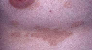 In patients with lighter skin color, lesions frequently are light tan or salmon colored.
Signs and symptoms
Please see Physical Examination for a full discussion.
Most individuals with tinea versicolor report cosmetically disturbing, abnormal pigmentation. The involved skin regions are usually the trunk, the back, the abdomen, and the proximal extremities. The face, the scalp, and the genitalia are less commonly involved. In patients with fair skin, the color of each lesion varies from almost white to reddish-brown or fawn colored. In darker skin types, involved areas can have varying degrees of either hypopigmentation or hyperpigmentation. A fine, dustlike scale covers the lesions.
Tinea versicolor patients often report that the involved skin lesions fail to tan in the summer and cause the affected areas to become more apparent. Conversely, affected areas may become subtler in winter months as background tan fades.
Occasionally, a tinea versicolor patient also reports mild pruritus. In most instances, the condition is asymptomatic.
Greater than 20% of tinea versicolor patients report a family history of the condition. This subset of patients records a higher rate of recurrence and longer duration of disease. [1]
Diagnostics
Please see Workup for a full discussion.
While the clinical presentation of tinea versicolor is distinctive, examination under a Wood lamp often, but not always, produces a coppery-orange fluorescence. Confirmation can be obtained through potassium hydroxide preparation and periodic acid-Schiff (PAS) or methenamine silver staining.
Management
Please also see Treatment and Medication.
Topical antifungals are the first-line treatment for tinea versicolor. Oral systemic antifungals may also be employed. Prophylactic treatment may be required to prevent recurrence as tinea versicolor is often resistant to eradication.
Pathophysiology
Tinea versicolor is caused by the dimorphic, lipophilic organisms in the genus Malassezia, formerly known as Pityrosporum. Fourteen species are recognized within this classification of yeasts, of which Malassezia globosa, Malassezia sympodialis, and Malassezia furfur are the predominant species isolated in tinea versicolor. [2, 3, 4, 5, 6, 7, 8, 9] Malassezia is extremely difficult to propagate in laboratory culture and is culturable only in media enriched with C12- to C14-sized fatty acids. Malassezia is naturally found on the skin surfaces of many animals, including humans. Indeed, it can be isolated in 18% of infants and 90-100% of adults. [10]
The organism can be found on healthy skin and on skin regions demonstrating cutaneous disease. In patients with clinical disease, the organism is found in both the yeast (spore) stage and the filamentous (hyphal) form. Factors that lead to the conversion of the saprophytic yeast to the parasitic, mycelial morphologic form include a genetic predisposition; warm, humid environments; immunosuppression; malnutrition; pregnancy; and Cushing disease. Human peptide cathelicidin LL-37 plays a role in skin defense against this organism.
Even though Malassezia is a component of the normal flora, it can also be an opportunistic pathogen. The organism is considered to be a factor in other cutaneous diseases, including Pityrosporum folliculitis, confluent and reticulate papillomatosis, seborrheic dermatitis, psoriasis, and some forms of atopic dermatitis. Malassezia species have also been shown to be a pulmonary pathogen in patients with immunosuppression due to stem cell transplantation. [11]
Etiology
Most cases of tinea versicolor occur in healthy individuals with no immunologic deficiencies. Nevertheless, several factors predispose some people to develop this condition. These factors include genetic predisposition; warm, humid environments; immunosuppression; malnutrition; application of oily preparations; corticosteroid usage; and Cushing disease. [12, 13, 14] The use of bath oils and skin lubricants may increase the risk of developing tinea versicolor. [15]
The reason why this organism causes tinea versicolor in some individuals while remains as normal flora in others is not entirely known. Several factors, such as the organism's nutritional requirements and the host's immune response to the organism, are significant.
The organism is lipophilic, and lipids are essential for growth in vitro and in vivo. Furthermore, the mycelial stage can be induced in vitro by the addition of cholesterol and cholesterol esters to the appropriate medium. Because the organism more rapidly colonizes humans during puberty when skin lipids are increased more than that of adolescent levels and tinea versicolor is manifested in sebum-rich areas (eg, chest, back), individual variations in skin surface lipids are hypothesized to play a major role in disease pathogenesis. However, patients with tinea versicolor and control subjects do not demonstrate any quantitative or qualitative differences in skin surface lipids. Skin surface lipids are significant for the normal presence of M furfur on human skin, but they probably play little role in the pathogenesis of tinea versicolor.
Evidence has been accumulating to suggest that amino acids, rather than lipids, are critical for the appearance of the diseased state. In vitro, the amino acid asparagine stimulates the growth of the organism, while another amino acid, glycine, induces hyphal formation. In vivo, the amino acid levels have been shown to be increased in the uninvolved skin of patients with tinea versicolor in two separate studies. The gene MGL_3741 has been reported in a study to play a role in amino acid biosynthesis pathways in M globosa, which are an important source of carbon and nitrogen for yeast. This gene has been proposed as potentially increasing the pathogenicity of M globosa. [16]
Another significant causative factor is the patient's immune system. Although sensitization against M furfur antigens is routinely present in the general population (as proven by lymphocyte transformation studies), lymphocyte function on stimulation with the organism has been shown to be impaired in patients who are affected. This outcome is similar to the situation of sensitization with Candida albicans. In short, cell-mediated immunity plays some role in disease causation.
Oxidative stress as shown by expression of reduced glutathione contributes to the pathogenesis of this condition. [17]
In steroid-associated atrophying tinea versicolor, the Malassezia infecting the epidermis may impair the barrier function of skin, thus allowing improved penetration of topical corticosteroids. This may lead to enhanced corticosteroid-induced atrophy of the affected skin. [18, 19, 20] However, as atrophying tinea versicolor has been reported in patients who have not used topical steroids, it has been proposed that a T-cell–mediated immune response to the Malassezia organism may be responsible for the atrophy noted. This theory suggests that Th1 cytokines recruit histiocytes to the site of epidermal infection. These histiocytes serve as a source of elastases and they up-regulate the metalloproteinase activity, ultimately leading to atrophy of the affected epidermis. [19, 21, 22]
A study from 2020 found a statistically significant relationship between Helicobacter pylori infection and tinea versicolor, proposing H pylori infection as an etiologic factor for this fungal infection, although this has not been verified in larger studies and the controls population with telogen effluvium was not clearly demographically matched. [23]
Epidemiology
Frequency
United States
Tinea versicolor occurs more frequently in areas with higher temperatures and higher relative humidities. The national prevalence of this condition is 2-8% of the population. The exact incidence in the United States is difficult to assess because many individuals who are affected may not seek medical attention.
International
Tinea versicolor occurs worldwide, with prevalences reported to be as high as 50% in the humid, hot environment of Western Samoa and as low as 1.1% in the colder temperatures of Sweden.
Race
Although the alteration in skin pigmentation is more apparent in darker-skinned individuals, the incidence of tinea versicolor appears to be the same in all races.
Sex
Several studies have addressed the frequency of tinea versicolor based on sex, and no dominance of either sex is apparent.
Age
In the United States, tinea versicolor is most common in persons aged 15-24 years, when the sebaceous glands are more active. The occurrence of tinea versicolor before puberty or after age 65 years is uncommon. [24] In more tropical countries, age frequency varies; most cases involve people aged 10-19 years who live in warmer, humid countries, such as Liberia and India.
Prognosis
Tinea versicolor is a benign skin disease that causes scaly macules or papules on the skin. As the name implies (versi means several), the condition can lead to discoloration of the skin, with colors ranging from white to red to brown. The condition is not considered contagious because the causative fungal pathogen is a normal inhabitant of the skin. Treatment leads to cessation of scaling within a few days, but discoloration may last for weeks to months. If scale cannot be provoked and new lesions are not developing, then there is no need to repeat treatment and the patient can be reassured that ongoing infection in unlikely.
Although tinea versicolor is recurrent for some patients and, therefore, a chronic disease, the condition remains treatable with the available remedies (see Medical Care and Medication). Thus, the prognosis is excellent and new treatments continue to be developed. [25]
Patient Education
Patients need to realize that tinea versicolor is caused by a fungus that is normally present on the skin surface; thus, it is not considered a contagious disease. Sequelae from the disease are not permanent, and any pigmentary alterations resolve entirely 1-2 months after treatment is initiated. Treatment is needed to remedy the condition and for prophylaxis to prevent recurrences.
-
In patients with lighter skin color, lesions frequently are light tan or salmon colored.
-
Hyperpigmented macules forming some confluent patches on the abdomen. While scale is not readily apparent, it is easily provoked with light scratching. On dark skin, affected areas may be hypopigmented or hyperpigmented.


