Continually Updated Clinical Reference
 
 
  All Sources     eMedicine     Medscape     Drug Reference     MEDLINE
 
eMedicine - Perforating Folliculitis : Article by

Quick Find
Authors & Editors
Introduction
Clinical
Differentials
Workup
Treatment
Follow-up
Miscellaneous
Multimedia
References

Related Articles
Acneiform Eruptions

Dermatologic Manifestations of Renal Disease

Elastosis Perforans Serpiginosum

Folliculitis

Gram-Negative Folliculitis

Keratosis Pilaris

Kyrle Disease

Pityrosporum Folliculitis

Reactive Perforating Collagenosis


Patient Education
Click here for patient education.


Author: James W Patterson, MD, Director of Dermatopathology, Professor of Pathology and Dermatology, Departments of Pathology and Dermatology, University of Virginia Medical Center

James W Patterson is a member of the following medical societies: American Academy of Dermatology, American College of Physicians, American Medical Association, American Society of Dermatopathology, Medical Society of Virginia, Royal Society of Medicine, Society for Investigative Dermatology, and United States and Canadian Academy of Pathology

Editors: Timothy McCalmont, MD, Director, UCSF Dermatopathology Service, Professor of Clinical Pathology and Dermatology, Departments of Pathology and Dermatology, University of California at San Francisco; Michael J Wells, MD, Associate Professor, Department of Dermatology, Texas Tech University Health Sciences Center; Rosalie Elenitsas, MD, Associate Professor of Dermatology, University of Pennsylvania School of Medicine; Director, Penn Cutaneous Pathology Services, Department of Dermatology, University of Pennsylvania Health System; Joel M Gelfand, MD, MSCE, Medical Director, Clinical Studies Unit, Assistant Professor, Department of Dermatology, Associate Scholar, Center for Clinical Epidemiology and Biostatistics, University of Pennsylvania; Dirk M Elston, MD, Director, Department of Dermatology, Geisinger Medical Center

Author and Editor Disclosure

Synonyms and related keywords: acquired perforating dermatosis, acquired perforating dermatitis, Kyrle disease, Kyrle's disease, hyperkeratosis follicularis et parafollicularis in cutem penetrans, perforating disorder of renal failure, elastosis perforans serpiginosa

Background

In classic perforating folliculitis, keratotic follicular papules develop, particularly over extensor surfaces. Microscopically, the disorder is characterized by disruption of the infundibular portion of the follicular wall, with transepidermal (transfollicular) elimination of connective tissue elements and cellular debris.

The condition may present as an isolated finding, apparently unrelated to other disease states, but also can be associated with chronic renal failure. Perforating folliculitis is closely related, if not identical, to the acquired perforating dermatosis that occurs with chronic renal disease. Kyrle disease (hyperkeratosis follicularis et parafollicularis in cutem penetrans) probably simply represents an exaggerated form of perforating folliculitis. In addition, another disorder of transepidermal elimination, elastosis perforans serpiginosa, occasionally displays involvement of follicular units.

Pathophysiology

As in Kyrle disease, the concept of an extrinsic keratin plug penetrating the epidermis generally has been discredited. Abnormally premature keratinization at the expense of proliferation is a possible explanation, as proposed by Carter and Constantine and Tappeiner et al in Kyrle disease. A role for fibronectin has been postulated. In addition, a primary alteration of connective tissue or deposition of foreign material within the superficial dermis, with subsequent engulfment and elimination by proliferative follicular epithelium, also is conceivable as a mechanism. Such a response to experimental implantation of foreign material has been described.

In addition, evidence suggests a pathologic role for excessively coiled hairs. Mehregan first proposed that curled hairs within follicular canals may act as springs, penetrating the lateral follicular wall, thereby initiating the process of transepidermal elimination. Support for this concept has been provided by an ultrastructural study of acquired perforating dermatosis that showed hair shaft fragments within transepidermal channels, even in patients in whom follicular involvement was not demonstrable on routine light microscopy. Factors that may promote coiling of hairs include follicular hyperkeratosis (occasional perforated follicles can be identified in keratosis pilaris) or contact dermatitis (eg, resulting from formaldehyde in clothing). Finally, trauma, such as scratching of pruritic skin, may well play a significant role in lesional development, possibly by setting in motion one or more of the pathologic events described above.

Frequency

United States

Incidence of perforating folliculitis in the United States is not known precisely, although the disorder is not uncommon. In Detroit, Michigan, 50 cases were reported during a 2-year period in the early 1970s, although this observation was followed by a declining incidence of new cases.

International

International incidence is not known but probably is comparable to the incidence observed in the United States.

Mortality/Morbidity

Morbidity is associated with the appearance of lesions and the pruritus that occasionally accompanies them. Although cutaneous disease is insignificant, substantial morbidity or mortality rates can be seen in association with the primary underlying diseases, such as diabetes mellitus or chronic renal failure.

Race

Although generally no ethnic predilection has been identified, 1 study found a higher incidence of Kyrle disease in chronic renal failure among African American individuals.

Sex

Perforating folliculitis occurs equally in males and females; no sex predilection has been reported.

Age

The condition is more common in the second through fourth decades of life.


History

In perforating folliculitis, papules typically are concentrated on hair-bearing portions of the extremities and buttocks. Often, lesions are asymptomatic, although pruritus may be a striking feature, especially in patients with renal insufficiency. Lesions may wax and wane and persist for months or years. Spontaneous remission can occur, and remission following renal transplantation has been documented in 1 patient who developed lesions secondary to renal failure.

Physical

Typical primary lesions consist of scaly papules, often folliculocentric, with small central keratotic plugs and varying degrees of erythema (see Media File 1). Compression of papules may yield keratin debris and a coiled hair or hair fragments. An initial eruption of follicular pustules, followed by typical plugged papules and subsequent development of prurigo-nodularis–like lesions was reported in 1 study. The Köbner phenomenon usually is not readily demonstrable, but a linear configuration occasionally can be observed. A case of progressive generalized perforating folliculitis has been reported to be associated with erythroderma, keratoderma of the palms and soles, alopecia of the scalp and eyebrows, and nail changes. Accompanying jaundice has been observed in patients with underlying liver disease.

Causes

A number of reported cases appear to be idiopathic, but specific associations also have been observed. Although some associations could be coincidental, the association with chronic renal failure (including both dialysis-dependent and nondialysis patients) is relatively common, suggesting a pathogenetic link. Perforating folliculitis also is observed relatively commonly in association with diabetes mellitus. Less common associations include sclerosing cholangitis, hypertension, atherosclerotic cardiovascular disease, acanthosis nigricans, psoriasis, and phrynoderma. A single case report described an association with Poland syndrome (unilateral absence of the pectoralis major muscle and ipsilateral symbrachydactyly), but this patient also had diabetes mellitus, hyperuricemia, and dilated cardiomyopathy.


Acneiform Eruptions
Dermatologic Manifestations of Renal Disease
Elastosis Perforans Serpiginosum
Folliculitis
Gram-Negative Folliculitis
Keratosis Pilaris
Kyrle Disease
Pityrosporum Folliculitis
Reactive Perforating Collagenosis


Lab Studies

  • Perform renal function studies, including urinalysis, serum creatinine, and creatinine clearance, to evaluate for possible underlying renal disease, if warranted by history and/or physical findings.
  • Test blood glucose levels to evaluate for possible diabetes mellitus.

Imaging Studies

  • Imaging is not applicable to the evaluation of a patient with this diagnosis.

Procedures

  • Skin biopsy
    • Biopsy is indicated when uncertainty about the nature of an eruption exists, ie, whether or not it is a folliculitis. In particular, consultants in other specialties, such as nephrologists who are following patients for chronic renal failure, may wish to exclude vasculitis, an infectious process, or a drug eruption. Biopsy also helps distinguish perforating folliculitis from other inflammatory disorders of the hair follicle, such as irritant folliculitis, acneiform folliculitis, or infectious folliculitis (eg staphylococcal, Pseudomonas-induced, or Pityrosporum-induced folliculitis).
    • Since histopathologic findings in perforating folliculitis are focal, they can be missed if a skin biopsy is sectioned incompletely. As a result, a pathology report may be returned with a nonspecific diagnosis. To avoid this, a small punch biopsy that completely encompasses the lesion is recommended. The clinician should indicate on the accession form that perforating folliculitis is suspected and that multiple sections may be necessary to demonstrate diagnostic changes.

Histologic Findings

The involved hair follicle shows focal disruption of its lateral wall, with a transepithelial channel and, often, a parakeratotic luminal plug. Connective tissue elements, including collagen and elastin, and varying numbers of inflammatory cells can be found within this transfollicular channel and within the follicular lumen, admixed with parakeratotic keratin. In some cases, a hair shaft or hair fragment can be demonstrated within the follicular lumen and/or within the transfollicular channel. The dermis surrounding the follicle typically shows sparse inflammation and focal fibrosis.


Medical Care

Since some cases of perforating folliculitis are associated with systemic disorders (eg, diabetes mellitus, chronic renal failure), direct therapeutic efforts towards the underlying condition in these patients. Soothing antipruritic lotions may be helpful for patients in whom itching is a significant problem. UV-B irradiation also may be of general benefit in the control or amelioration of pruritus.

Assessing treatments for perforating folliculitis is difficult. Although the condition is not uncommon, controlled therapeutic studies are not available. Unsuccessful treatments have included oral beta-carotene, keratolytics, antiacne therapies, and topical corticosteroids. A case reported by Zachariae and Sogaard featured progressive generalized perforating folliculitis combined with erythroderma, keratoderma, and alopecia and was resistant to corticosteroids, adrenocorticotropic hormone, psoralen plus UV-A, cytostatic agents, and aromatic retinoids. A case of phrynoderma with perforating folliculitis, reported by Neill et al, responded to 50,000 U/d of beta-carotene for 1 month, followed by 2000 U/d. Hurwitz found that a combination of antistaphylococcal therapy, phototherapy, and a topical corticosteroid lotion was helpful in controlling the condition in patients with chronic renal failure.

Tretinoin 0.1% cream has cleared some lesions but has not prevented the development of others. Despite the single negative result with aromatic retinoids, these agents may play a role in the treatment of some patients with perforating folliculitis; however, experience with these agents is not sufficient to allow recommendation of a particular dosing schedule.

A recent study of 5 patients showed good clinical responses with narrowband UV-B therapy. Patients were treated 2-3 times weekly. The initial dose was 400 mJ/cm2, with increases to a maximum of 1500 mJ/cm2. Lesions completely resolved after 10-15 exposures, although some recurrences were observed.

Surgical Care

Surgical care is not required for perforating folliculitis lesions per se, except when indicated for the performance of a diagnostic skin biopsy and subsequent wound management.

Surgery in the form of renal transplantation may be an option for selected patients in whom perforating folliculitis is associated with chronic renal failure. Of course, such a procedure would be based on the overall medical needs of the patient and is not performed simply to treat the perforating folliculitis (although improvement of perforating folliculitis has been reported following renal transplantation).

Consultations

  • Dermatologist: Practitioners may wish to consult a dermatologist when uncertainty regarding a papular eruption exists in a patient. Dermatologists often are able to determine clinically whether such an eruption is likely to be a folliculitis, and they can appreciate subtle lesional changes that suggest a perforating folliculitis. Dermatologists also can suggest or perform the proper type of biopsy procedure for this type of folliculitis.
  • Internist: Specialists in internal medicine should be consulted for management of patients in whom perforating folliculitis may be associated with an underlying medical condition, such as chronic renal failure, diabetes mellitus, or atherosclerotic cardiovascular disease.
  • General surgeon: Consultation with a surgeon may be indicated for patients whose perforating folliculitis is associated with chronic renal failure. Renal transplantation may be an option.


Further Inpatient Care

  • Inpatient care is not required for this cutaneous condition.

Further Outpatient Care

  • Further outpatient dermatologic care may be required for ongoing management of the perforating folliculitis when lesions persist, spread, or become resistant to therapy.
  • Follow-up care may be necessary for complications related to therapy.
  • Outpatient internal medicine care may be indicated for management of any underlying medical conditions, such as chronic renal failure, hypertension, diabetes mellitus, or atherosclerotic cardiovascular disease.

Complications

  • Potential complications include secondary infection of lesions (resulting from trauma or other factors), spread of lesions despite therapy, or adverse effects of treatments, such as irritation from topical retinoids or atrophy and telangiectasia from chronic topical corticosteroid use.
  • Complications related to any underlying medical conditions may occur.

Prognosis

  • Skin lesions can improve or clear, either spontaneously or with therapy. Hopefully, further therapeutic experience will permit development of specific treatment guidelines.
  • Reports of improvement of lesions with stabilization of renal damage (in patients with chronic renal failure) are encouraging and underscore the important role of internal medicine or surgery consultants in treating these patients.

Patient Education

  • Inform patients about the nature of the disease and its relation to any underlying medical condition that may be present.
  • Instruct patients to avoid rubbing or scratching the lesions, since this may result in their spread or in the development of secondary infection.
  • Fully inform patients about the treatment being used, including its proper use or application and possible adverse effects.


Medical/Legal Pitfalls

  • Failure to exclude significant underlying diseases


Media file 1:  
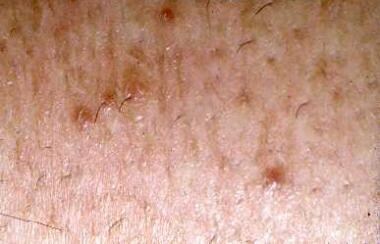
Typical appearance of lesions of perforating folliculitis consisting of keratotic follicular papules.
Click to see larger pictureClick to see detailView Full Size Image
Media type:  Photo


  • Bilezikçi B, Seçkin D, Demirhan B. Acquired perforating dermatosis in patients with chronic renal failure: a possible pathogenetic role for fibronectin. J Eur Acad Dermatol Venereol. Mar 2003;17(2):230-2. [Medline].
  • Carter VH, Constantine VS. Kyrle''s disease. I. Clinical findings in five cases and review of literature. Arch Dermatol. Jun 1968;97(6):624-32. [Medline].
  • Chang P, Fernandez V. Acquired perforating disease: report of nine cases. Int J Dermatol. Dec 1993;32(12):874-6. [Medline].
  • Fistarol SK, Itin PH. Acquired perforating dermatosis in a patient with Poland syndrome. Dermatology. 2003;207(4):390-4. [Medline].
  • Headley CM, Wall B. ESRD-associated cutaneous manifestations in a hemodialysis population. Nephrol Nurs J. Dec 2002;29(6):525-7, 531-9; quiz 540-1. [Medline].
  • Hong SB, Park JH, Ihm CG, Kim NI. Acquired perforating dermatosis in patients with chronic renal failure and diabetes mellitus. J Korean Med Sci. Apr 2004;19(2):283-8. [Medline].
  • Hurwitz RM. The evolution of perforating folliculitis in patients with chronic renal failure. Am J Dermatopathol. Jun 1985;7(3):231-9. [Medline].
  • Kahana M, Trau H, Dolev E, et al. Perforating folliculitis in association with primary sclerosing cholangitis. Am J Dermatopathol. Jun 1985;7(3):271-6. [Medline].
  • Mahajan S, Koranne RV, Sardana K, et al. Perforating folliculitis with jaundice in an Indian male: a rare case with sclerosing cholangitis. Br J Dermatol. Mar 2004;150(3):614-6. [Medline].
  • Mehregan AH, Coskey RJ. Perforating folliculitis. Arch Dermatol. Apr 1968;97(4):394-9. [Medline].
  • Neill SM, Pembroke AC, du Vivier AW, Salisbury JR. Phrynoderma and perforating folliculitis due to vitamin A deficiency in a diabetic. J R Soc Med. Mar 1988;81(3):171-2. [Medline].
  • Ohe S, Danno K, Sasaki H, et al. Treatment of acquired perforating dermatosis with narrowband ultraviolet B. J Am Acad Dermatol. Jun 2004;50(6):892-4. [Medline].
  • Patterson JW, Graff GE, Eubanks SW. Perforating folliculitis and psoriasis. J Am Acad Dermatol. Sep 1982;7(3):369-76. [Medline].
  • Patterson JW. The perforating disorders. J Am Acad Dermatol. Apr 1984;10(4):561-81. [Medline].
  • Patterson JW, Brown PC. Ultrastructural changes in acquired perforating dermatosis. Int J Dermatol. Mar 1992;31(3):201-5. [Medline].
  • Pavlović MD, Zecević RD, Stamenković M, et al. Trauma-induced perforating folliculitis. Eur J Dermatol. Nov-Dec 2003;13(6):592. [Medline].
  • Pavlović MD, Zecević RD, Stamenković M, et al. Trauma-induced perforating folliculitis. Eur J Dermatol. Nov-Dec 2003;13(6):592. [Medline].
  • Sehgal VN, Jain S, Thappa DM, et al. Perforating dermatoses: a review and report of four cases. J Dermatol. Jun 1993;20(6):329-40. [Medline].
  • Tappeiner J, Wolff K, Schreiner E. [Kyrle''s disease]. Hautarzt. Jan 1969;20(1):296-310. [Medline].
  • Zachariae H, Sogaard H. Progressive generalized perforating folliculitis. Dermatologica. 1984;168(3):131-7. [Medline].

Perforating Folliculitis excerpt

Article Last Updated: May 17, 2006