Practice Essentials
Mycobacterium marinum is an atypical Mycobacterium found in salt water and fresh water. The bacterium was first isolated from tubercles during a saltwater fish necropsy at a Philadelphia aquarium in 1926. The first human case was reported in Sweden in 1951. [1] M marinum is the most common atypical Mycobacterium to cause infection in humans. Infection occurs following inoculation of a skin abrasion or puncture and manifests as a localized granuloma or sporotrichotic lymphangitis (see the image below).
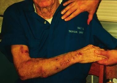 Photograph of Mycobacterium marinum infection lesions.
Also known as fish tank granuloma, diagnosis and treatment of the disease are often delayed because of a lack of suspicion for mycobacterial involvement, ie, versus more common bacterial pathogens. Due to the increased use of immunosuppressants for transplant recipients and tumor necrosis factor (TNF) inhibitors for a variety of conditions, infections with mycobacteria other than tuberculosis (MOTT) are increasing.
Prognosis
Once identified and appropriately treated, M marinum infection can typically be successfully eradicated, usually with no major sequelae. M marinum skin infection typically remains localized and does not cause significant morbidity in patients who are immunocompetent. Cases reported in patients who are severely immunocompromised document disseminated infection via lymphatics and can involve the bone marrow and viscera, with rare reports of death secondary to the infection. [2]
Complications
Complications can include the following:
-
Persistent ulceration
-
Osteomyelitis, bony erosion
-
Bursitis
-
Tenosynovitis
-
Arthritis
-
Disseminated infection
Note that there have been 3 reported cases of M marinum pulmonary infections. One of these cases was in the setting of anorexia nervosa. [3]
Treatment
See Treatment.
In a case of M marinum left upper extremity disseminated infection in a 61-year-old woman, the patient was treated with ethambutol 1200 mg daily, rifampin 600 mg daily, and oral clarithromycin 500 mg twice daily. Trimethoprim-sulfamethoxazole treatment was unsuccessful. She showed slow improvement, and surgical debridement was planned. The patient was not actively immunosuppressed, but she did have a history of Sjogren syndrome, breast cancer, and chemotherapy. [4]
Consultations
A variety of specialists may be involved in the diagnosis and treatment, such as dermatologists, rheumatologists, and infectious disease physicians.
Prevention
People who work near or in saltwater should take precautions to avoid abrasions, trauma, or bites from fish and marine animals. People who work with aquariums should wear gloves if they are cleaning tanks or expect to encounter trauma to their hands or feet. If bites or abrasions occur, cleanse the skin, apply an antibacterial preparation, and dress with an appropriate bandage.
Pathophysiology
M marinum is a slow-growing species that resides in both freshwater and saltwater environments, with optimal growth at 30-32°C. It is carried by many fish species and can result in human infection via inoculation of the skin by a fish bite, exposure of an open wound to contaminated water, contact with an aquarium, or contact with marine animals such as fish or turtles. [2] Exposure to M marinum via swimming pools is rare because most pools are chlorinated. [5]
The pathogen is classified as a photochromagen in Runyon group 1, which means that it produces yellow pigment when cultured and exposed to light. Culture growth occurs over 7-21 days and is optimal at 25-32°C (77-89.6°F) given the organism is adapted to infect ectotherms, such as fish. When endotherms, such as humans, are infected the infection favors the cooler extremities more than central sites. Systemic infection, usually of an immunocompromised host, has been reported. This indicates that the organism is capable of adapting to grow in conditions closer to 37°C. [6]
After inoculation into the host tissues via an abrasion or other wound, the mycobacteria are phagocytosed by macrophages. Inside the macrophage, they are able to interrupt the formation of the phagolysosome, which would normally kill the organisms. The mycobacteria, however, are able escape the lysosome and can move intracellularly and extracellularly via actin-based motility. This may contribute to cell-to-cell spread.
Tumor necrosis factor (TNF) is important for the immune response against mycobacteria. Studies have demonstrated that in the absence of TNF, macrophages engulf but do not destroy the mycobacteria. Instead, the mycobacteria survive and grow, finally killing the macrophage. [7] The importance of TNF is also supported by a number of reports of infection occurring in patients treated with TNF inhibitors, and these medications should be stopped during the course of antibiotic therapy. If not, the lesions may rapidly extend. [8, 9, 10]
Studies have revealed 2 pathophysiologically and genetically (ie, via amplified restriction-based polymorphism analysis) distinct populations of M marinum. One group can infect humans and causes acutely lethal disease in fish, while a second group cannot infect humans and causes chronic progressive disease in fish.
Special concerns
Utility of M marinum as an immunotherapy agent to elicit an antituberculosis response is currently being explored. [11] There is specific scientific interest in M marinum because of its genetic relatedness to Mycobacterium tuberculosis and because experimental infection of M marinum in fish mimics tuberculosis pathogenesis. [10]
Epidemiology
Infections caused by M marinum are uncommon but well described in the literature. The estimated annual incidence in the United States is 0.05-0.27 case per 100,000 adult patients. [12, 10] Of the more than 160 cases described, most are case reports of cutaneous infection; some report concomitant osteomyelitis, tenosynovitis, arthritis, and/or disseminated infection. Nosocomial infection has never been described.
Infection occurs worldwide, most commonly in individuals with occupational and recreational exposure to nonchlorinated fresh water or saltwater. [13]
No racial or sexual predilections have been noted for M marinum skin infection. M marinum infection has been reported in persons of every age group; however, it appears to be rare in the pediatric population. [14, 15]
-
Photograph of Mycobacterium marinum infection lesions.


