Practice Essentials
Milia are very common, benign, keratin-filled cysts (see the image below).
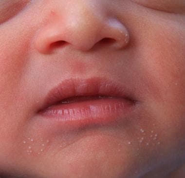 Milia in a week-old infant.
Primary milia are typically seen in infants but also may occur in children and adults. Secondary milia develop after trauma to the skin, such as after burns (eg, sunburns), dermabrasion, or in blistering disorders. Milia en plaque is a rare inflammatory condition characterized by plaques of milia in the periauricular area. Multiple eruptive milia is a condition characterized by the sudden development of crops of milia over the course of weeks to months.
Milia are asymptomatic. In children and adults, they usually arise around the eye. Eruptive milia, as the name suggests, have a rapid onset, often within a few weeks.
No systemic complications have been reported.
Pathophysiology
Milia are tiny epidermoid cysts. The cysts may be derived from the pilosebaceous follicle. Primary milia arise on facial skin bearing vellus hair follicles. Secondary milia result from damage to the pilosebaceous unit.
Prognosis
Milia seen in infancy tend to spontaneously disappear within the first few weeks of life. Milia in older children and adults tend to persist.
Secondary milia arising from blisters rarely resolve.
Patients or their parents can be taught how to treat milia with a needle.
Workup
No investigations are needed for simple milia. The clinical appearance is diagnostic. Investigation of the underlying disease is necessary in persons with secondary milia.
Performing a skin biopsy is necessary only if the diagnosis is in doubt. If milia en plaque is suspected, performing a biopsy is prudent to exclude follicular mucinosis and multiple trichoepitheliomata. In an elderly person with sun-damaged skin, Favre-Racouchot syndrome (nodular elastosis of the skin) needs to be excluded.
Treatment
No topical or systemic medications are effective on primary and secondary milia. Single case reports have demonstrated the success of topical tretinoin [20] and oral etretinate, [21] and minocycline in treating patients with milia en plaque.
Epidemiology
Primary milia in newborns are so common that they can be considered normal (occurring in approximately half of all infants). Milia formation may be delayed in premature infants, with milia appearing several weeks after birth. [32] Multiple eruptive milia and milia en plaque are rare entities. No racial predilection is recognized for milia. Sexual prevalence is equal for primary and secondary milia. Eruptive milia and milia en plaque occur more frequently in women. Milia occur in persons of all ages but are typically found in infants.
-
Milia in a week-old infant.