Practice Essentials
Maffucci syndrome is characterized by enchondromas with venous malformations with or without spindle cell hemangiomas. It was first reported by Maffucci in 1881 after a 40-year-old woman died from complications following amputation of an arm. The patient had frequent and severe hemorrhage from a vascular tumor for which she was admitted to the hospital. In view of the profuse bleeding, an amputation was performed and the patient died from infection. Maffucci reported a thorough autopsy that described all the main points of the syndrome named after him. In 1941, Carleton et al proposed the eponym Maffucci syndrome.
Maffucci syndrome is a rare mosaic genetic disorder that affects both males and females. It is associated with heterozygous somatic mutations in the isocitrate dehydrogenase 1 and 2 (IDH1/IDH2) genes. [1, 2] In 2021, an L309I mutation in the ELKS/RAB6-interacting/CAST family member 2 (ERC2) gene was also identified as being associated with Maffucci syndrome. [3]
Maffucci syndrome is characterized by benign enlargements of cartilage (enchondromas), bone deformities, and venous malformations. No racial or sexual predilection is apparent in Maffucci syndrome. No familial pattern of inheritance has been shown. Maffucci syndrome manifests early in life, usually around age 4-5 years, with 25% of cases being congenital. Patients apparently are of average intelligence, and no associated mental or psychiatric abnormalities seem to be present. About 160 cases of Maffucci syndrome have been published in the English literature. [4, 5, 6, 1] Note the image below.
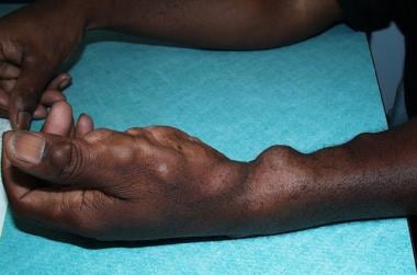 Characteristic venous malformations on the patient's right upper extremity. These vascular malformations are benign and asymptomatic.
Causes
Maffucci syndrome has no familial pattern of inheritance and appears sporadically. It has been shown to be associated with heterozygous somatic mutations in the isocitrate dehydrogenase 1 and 2 (IDH1/IDH2) genes. [1, 2]
Complications
Neoplastic changes occur in enchondromas. Chondrosarcoma is the most common neoplasm in this syndrome, affecting about 30% of patients (see Pathophysiology).
Enchondromas can cause a fracture, leading to further complications, such as shortened or unequal length limbs.
Varied but rare complications and associations of Maffucci syndrome have been reported in the literature, including giant tubular adenoma of the breast, [7] pedal synovial sarcoma, [8] nasopharynx clival enchondroma, [9] and intrahepatic cholangiocarcinoma. [10]
Prognosis
Patients with Maffucci syndrome usually lead reasonably normal lives with a normal life expectancy if no malignant transformation occurs. Although the skeletal malformations can sometimes be crippling, patients have managed to perform activities of daily living rather well.
Treatment
No medical care is needed in Maffucci syndrome patients who are asymptomatic. Patients do need careful follow-up care to evaluate any changes in the skin and bone lesions.
Physical activity is not limited for Maffucci syndrome. Some patients may have difficulty ambulating because of the bone abnormalities.
Patient education
Patients can obtain further information from the following source: The National Organization for Rare Disorders, Inc (NORD), PO Box 1968, 55 Kenosia Avenue, Danbury, CT 06813-1968, (203) 744-0100 or (800) 999-6673. Contact NORD by e-mail at orphan@rarediseases.org.
Pathophysiology
Heterozygous mutations in IDH1 and IDH2 are associated with both Maffucci syndrome and Ollier disease. These mutations permit the accumulation of D-2-hydroxyglutarate, which is associated with tumor formation. An additional mutation associated with Maffucci syndrome, in ERC2, was identified by Cheng et al. This mutation is likely responsible for hemangioma formation in Maffucci syndrome by causing increased endothelial intracellular calcium concentrations. Notably, ERC2 mutations have not been identified in Ollier disease patients, likely accounting for the lack of hemangiomas in that syndrome. [3]
Maffucci syndrome affects the skin and the skeletal systems. Superficial and deep vascular lesions (venous malformations) often protrude as soft nodules or tumors, usually on the distal extremities, but they can appear anywhere. They can be bilateral or unilateral, but they usually are asymmetric. Venous-lymphatic malformations and hemangioendotheliomas can occur but are much less common. [11, 12, 13, 14] Enchondromas are benign cartilaginous tumors with the potential for malignant transformation. They can appear anywhere, but they usually are found on the phalanges and the long bones. These bone abnormalities usually are asymmetric and can cause secondary fractures. Approximately 30-37% of enchondromas develop into a chondrosarcoma.
The venous malformations in Maffucci syndrome manifest as blue subcutaneous nodules that can be emptied by pressure. Thrombi often form within vessels and develop into phleboliths. Under microscopic examination, these phleboliths appear as calcified vessels.
Enchondromas develop from the mesodermal dysplasia associated with Maffucci syndrome. As the bones grow, some cartilage material is left behind and grows irregularly, developing into the characteristic bone deformities. Bone irregularities in Maffucci syndrome include shortened length of the long bones, unequal leg length, pathologic fractures, and malunion of fractures. [15]
In Maffucci syndrome, neoplastic changes occur in enchondromas. Chondrosarcoma is the most common neoplasm in this syndrome, affecting about 30% of patients. The average age for neoplastic change in Maffucci syndrome patients is 40 years. Vascular neoplasms have occurred in 4 reported cases: 2 hemangiosarcomas and 2 lymphangiosarcomas. Other malignancies associated with Maffucci syndrome include pancreatic and hepatic adenocarcinoma, ovarian tumors, brain gliomas, astrocytomas, and other types of sarcomas. [6]
Epidemiology
Maffucci syndrome is rare. Fewer than 100 cases of Maffucci syndrome have been reported in the United States with about 160 total case reports in the English literature. No increased frequency of Maffucci syndrome occurs because of race. Maffucci syndrome appears to be sporadically inherited. No sexual bias is present. Lesions of Maffucci syndrome are first noted usually by age 4-5 years.
-
Characteristic venous malformations on the patient's right upper extremity. These vascular malformations are benign and asymptomatic.
-
Enchondroma on the left elbow.
-
Radiograph of a patient's hands showing enchondromas and phleboliths. Areas of translucency represent enchondromas, and opaque spots represent phleboliths.
-
Venous malformations on a patient's right hand.


