Background
Granuloma annulare (GA) is a benign inflammatory dermatosis. T. Colcott Fox first described granuloma annulare in 1895; however, not until 1902 did Radcliffe-Crocker label it as granuloma annulare.
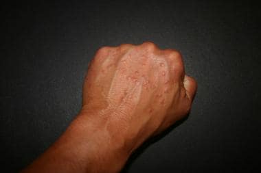
Granuloma annulare is relatively common disease that occurs in all age groups, but it is rare in infancy. [1, 2] Granuloma annulare is characterized clinically by dermal papules and annular plaques (see the image below). The precise cause is unknown. Histological examination reveals foci of degenerative collagen associated with palisaded granulomatous inflammation.
 Granuloma annulare. By Mierlo at English Wikipedia - Transferred from en.wikipedia to Commons by Stevenfruitsmaak using CommonsHelper., Public Domain, https://commons.wikimedia.org/w/index.php?curid=4560836.
The following clinical variants are recognized:
-
Localized granuloma annulare: This is the most common form. Localized granuloma annulare is characterized by skin-colored to violaceous lesions up to 5 cm in diameter. Usually, the epidermis has attenuated surface markings. Annular rings with solitary firm papules or nodules may be present. Localized granuloma annulare has a predilection for the feet, ankles, lower limbs, and wrists.
-
Generalized granuloma annulare: This form occurs predominantly in adults. The trunk is usually involved, as well as the neck, extremities, face, scalp, palms, and soles. Lesions range from widespread papules to annular plaques to large, discolored patches with a variety of coloration from yellow to violaceous.
-
Subcutaneous granuloma annulare [3] : This form occurs predominantly in children. Subcutaneous granuloma annulare is characterized by firm or hard asymptomatic nodules in the deep dermis or subcutaneous tissues, with individual lesions measuring from 5 mm to 4 cm in diameter. They are prevalent on the anterotibial plateau, ankles, dorsal feet, buttocks, hands, scalp, and eyelids.
-
Perforating granuloma annulare [4] : This form is very rare. Perforating granuloma annulare is usually localized to the dorsal hands and fingers or may be generalized on the trunk and extremities. A variety of superficial umbilicated papules develop, with or without a discharge, that heal with scarring.
-
Arcuate dermal erythema: This is an uncommon form of granuloma annulare that manifests as infiltrated erythematous patches that may form large, hyperpigmented rings with central clearing.
Some authorities consider actinic granuloma (AG) to be a subset of granuloma annulare, but others view actinic annulare as a separate, but related, entity. [5]
Pathophysiology
Proposed pathogenic mechanisms for granuloma annulare include cell-mediated immunity (type IV), immune complex vasculitis, and an abnormality of tissue monocytes. Some other possible mechanisms include primary degeneration of connective tissue leading to granulomatous inflammation, lymphocyte-mediated immune reaction with macrophage activation, and cytokine-mediated degradation of connective tissue. [1]
Etiology
The etiology of granuloma annulare is usually unknown, and the pathogenetic mechanisms are poorly understood, with a vast majority of granuloma annulare cases occurring in patients who are otherwise healthy. The range of predisposing events and associated diseases is diverse, and granuloma annulare is thought to represent a reaction pattern with many different initiating factors. [1]
Granuloma annulare has been hypothesized to be associated with tuberculosis, insect bites, trauma, sun exposure, thyroiditis, vaccinations, and viral infections, including HIV, Epstein-Barr virus, hepatitis B virus, hepatitis C virus, and herpes zoster virus. However, these suggested etiologic factors remain unproven.
Familial cases of granuloma annulare observed in identical twins and siblings in several generations, along with an association of granuloma annulare with HLA phenotypes, suggest the possibility of a hereditary component in some cases. The HLA-B8 level has been reported to be increased in localized granuloma annulare; HLA-A29 and HLA-BW35 levels are reported to be increased in generalized granuloma annulare.
Some reports associate chronic stress with granuloma annulare as a trigger of the disease. Granuloma annulare also has some predilection for the sun-exposed areas and photodamaged skin. Photosensitive granuloma annulare has been found in association with HIV infection. Finally, some cases of granuloma annulare or granuloma annulare–like reactions have been reported after gold therapy and treatment with allopurinol, diclofenac, quinidine, calcitonin, amlodipine, ACE inhibitors, daclizumab, [6] checkpoint inhibitors, [7, 8, 9] and calcium channel blockers.
Relationship to systemic diseases
Granuloma annulare has been associated primarily with type I diabetes mellitus, but it is only rarely associated with type II diabetes mellitus and thyroid disease, based on an increased number of granuloma annulare patients with these diseases in small case series. [10]
Small case series have reported granuloma annulare to occur in association with malignancy, AIDS, and herpes zoster lesions. Although no definite patterns relating granuloma annulare and systemic disease have been thoroughly established, it has been suggested that an atypical histologic (vasculopathy or extravascular neutrophilia) or clinical presentation (unusual appearance or location) may indicate an associated disease. In the case of malignancy, a 2003 study by Li et al reviewed classic cases in the literature and could find no definite relationship between granuloma annulare and malignant neoplasms. [11]
Relationship with malignant diseases
Certain malignancies are accompanied by different mucocutaneous paraneoplastic syndromes. Lesions that mimic granuloma annulare or are histologically confirmed as granuloma annulare have occurred in association with the following:
-
Acute myelogenous leukemia
-
Chronic lymphocytic leukemia
-
Myelomonocytic leukemia
-
Large granular lymphocytic leukemia
-
Follicular lymphoma
-
Lennert lymphoma
-
Solid tumors - Breast tumors, cervical cancer, colon cancer, lung cancer, prostate cancer, testicular tumors, thyroid cancer
Epidemiology
The frequency of granuloma annulare is in the general population is unknown. Granuloma annulare does not favor a particular race, ethnic group, or geographical area.
Localized granuloma annulare is the most common among the various subtypes. Of all patients with granuloma annulare, 9-15% have the generalized variant. Perforating granuloma annulare has been reported to have a prevalence of 5% among granuloma annulare subtypes; further, reports suggest that this variant may be more common in the Hawaiian Islands.
Sex
Women are affected by granuloma annulare twice as often as men.
Age
Localized granuloma annulare is most commonly found in children and in adults younger than 30 years. Generalized granuloma annulare demonstrates a bimodal age distribution, occurring in patients younger than 10 years and in patients aged 30-60 years. Although subcutaneous granuloma annulare can occur in adults, it is predominantly a disease of otherwise healthy children, who are typically aged 2-10 years. Similarly, perforating granuloma annulare most often affects children.
Prognosis
Spontaneous resolution of localized granuloma annulare has occurred within 2 years in 50% of cases, although lesions may last weeks to decades. Recurrence, often at the same site, is noted in 40% of cases.
Generalized granuloma annulare has a more chronic course, with rare spontaneous resolution, poor response to treatment, and frequent relapses.
Subcutaneous granuloma annulare lesions often spontaneously regress. Local or distant recurrences have been reported in 20-75% of cases in different studies.
Patient Education
Patients and families should be reassured about the typically benign nature and course of granuloma annulare.
-
Granuloma annulare. By Mierlo at English Wikipedia - Transferred from en.wikipedia to Commons by Stevenfruitsmaak using CommonsHelper., Public Domain, https://commons.wikimedia.org/w/index.php?curid=4560836.


